Case History:
A 65 year old man presents to the emergency room with acute onset back pain. Of note, the man was diagnosed with Burkitt’s lymphoma two months prior and had recently received a course of chemotherapy. During the workup for his back pain, a chest CT is obtained and reveals a 2 cm pulmonary nodule in the left upper lobe with a surrounding “groundglass halo” highly suspicious for invasive fungal involvement. A fine needle aspiration (FNA) of the nodule is performed and tissue is sent for histopathologic examination as well as bacterial and fungal culture.


Laboratory Identification:
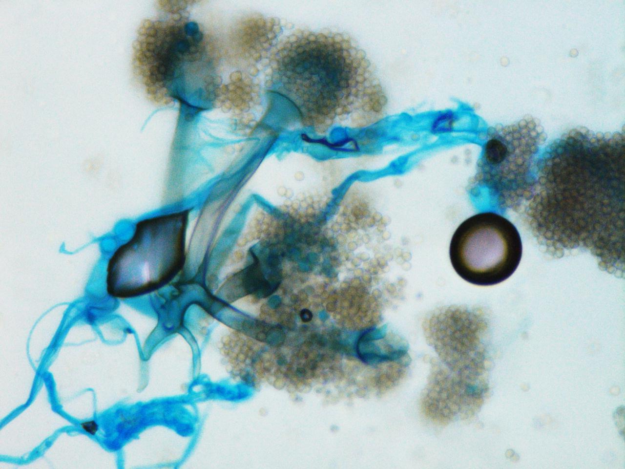
One rapidly growing white colony was identified that became grey over time. The colony was a “lid-lifter” that began pushing at the lid after only a few days. Microscopically, the organisms had broad hyphae with single and branching sporangiophores. At the ends of the sporangiophores there were pyriform, or pear-shaped, sporangia sitting atop funnel-shaped apophyses. Rhizoids were found to be internodal, or arising from the hyphae between the sporangiophores. Based on this morphology, the fungal organisms were identified as Lichtheimia corymbifera complex (formally Absidia corymbifera).
Discussion:
Lichtheimia corymbifera is an organism within the phylum Zygomycota and is one of two pathogenic species in the genus Lichtheimia. This organism is known as an uncommon cause of Zygomycosis and is only implicated in approximately 5% of cases. As in most cases of Zygomycosis, exemplified in our patient, Lichtheimia corymbifera most often affects immunocompromised patients. It is ubiquitous in the environment and is associated with decaying plant matter and soil. Disease is caused by inhalation of spores.
Important points for laboratory identification:
Lichtheimia
- Growth at 35-37°C (capable of growth up to 50°C)
- Inhibited by media containing cycloheximide
- Internodal rhizoids
- Pyriform sporangia
- Apophysis
Compared to other common Zygomycetes:
Mucor
- No rhizoids
- Round sporangia
- No apophysis
Rhizopus
- Nodal rhizoids (directly opposite of the sporangiophores)
- Round sporangia
- No apophysis
-Britni Bryant, MD is a 2nd year anatomic and clinical pathology resident at the University of Vermont Medical Center.

-Christi Wojewoda, MD, is the Director of Clinical Microbiology at the University of Vermont Medical Center and an Assistant Professor at the University of Vermont.
