Thyroid Tales (First Edition)

I’ve found that our cytologists have a love-hate relationship with thyroids. Pathologists do too. Or it could be that we see so many goiters (50%) and follicular lesions or atypia of undetermined significance (35%) that the rare papillary thyroid carcinoma is a gem in our eyes. Minimally-invasive thyroid FNAs are instrumental in the management of thyroid nodules. It’s important to note that due to lack of architecture and assessment of capsular invasion, cytologic diagnoses may be limited, and prior to referring the patient for a potentially unnecessary surgery, various molecular tests can be utilized. The ongoing evolution of molecular testing on thyroid FNAs help classify indeterminate and suspicious cytology diagnoses (Bethesda Categories III and IV), examining the risk of malignancy or detecting the presence of genetic alterations, which help guide surgical intervention versus surveillance. This post (the first edition) features a series of our classic Bethesda Category VI specimens, which bypassed the need for risk classification and defaulted in surgical intervention based on guidelines at the time of diagnosis. It is worth mentioning that many of these cases occurred prior to the implementation of the Thyroid Imaging Reporting and Data System (TI-RADS), so to preserve the accuracy of patient history, a TI-RADS score will not be assumed.

Case 1

Okay, I know I said Bethesda VI, but let’s kick this series off with a Bethesda Category IV case. Thankfully, the patient decided to undergo a partial thyroidectomy, yielding a beautiful tissue follow-up. A 59-year-old male with newly diagnosed melanoma of the neck underwent imaging for staging purposes. A left thyroid nodule was identified measuring 3.0 centimeters. The patient presented for an ultrasound-guided fine needle aspiration.

Images 1-3: Thyroid, Left Lobe, FNA 1: DQ-stained smear; 2: Pap-stained smear; 3. H&E Cell Block section (400X).

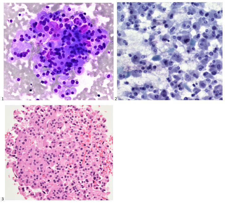
Abundant oncocytic Hürthle cells, some with mild atypia, were identified, suggestive of Hürthle cell neoplasm (Images 1-3). With a lack of lymphocytes, we did not feel comfortable suggesting Hashimoto’s (lymphocytic) thyroiditis. Immunostains performed on cell block sections show the tumor cells are positive for TTF-1, focally positive for thyroglobulin and AE1/AE3 (rare), and negative for calcitonin. The morphology and immunohistochemical profile support the above diagnosis.

The patient underwent a left lobectomy and isthmusectomy. Pathology showed a 3.2 cm Hürthle cell carcinoma (Images 4-5) in the left lobe of the thyroid (encapsulated with a foci of capsular invasion without vascular invasion) as well as a 0.3 cm micropapillary carcinoma. Since Hürthle cell carcinoma does not typically concentrate radioiodine, the patient would not be responsive to treatment with radioactive iodine. Therefore, there would be less benefit derived from treating the smaller right lobed nodules (which don’t meet biopsy criteria) from remnant ablation. The patient had a clinically limited stage thyroid cancer. The patient is monitored with neck ultrasounds rather than serum thyroglobulin testing (due to the remaining right lobe).

Images 4-5: Thyroid, Left Lobe with Isthmus, Excision: H&E section (600X).

Cytology diagnosis: Hürthle cell neoplasm.

Pathology diagnosis: Hürthle cell carcinoma.


Case 2

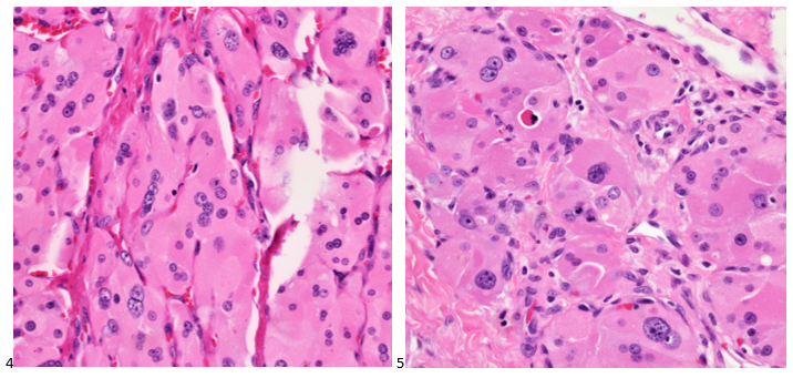
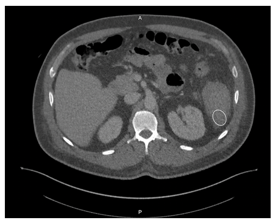
A 53-year-old female with no prior history presented with fatigue and a self-palpated right thyroid nodule and normal thyroid function tests. She reported an extensive family history of hypothyroidism. On thyroid ultrasound, the right upper pole thyroid nodule measured 2.0 x 2.0 x 1.8 cm and was mostly solid and hypoechoic with microcalcifications. Pre-intervention serum calcitonin measured 1634 pg/mL. The patient underwent an FNA of the thyroid nodule and the smears are depicted below.

Images 6-7: Thyroid, Right Lobe, Upper Pole, FNA: 6: DQ-stained smear; 7: Pap-stained smear.

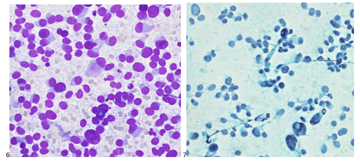
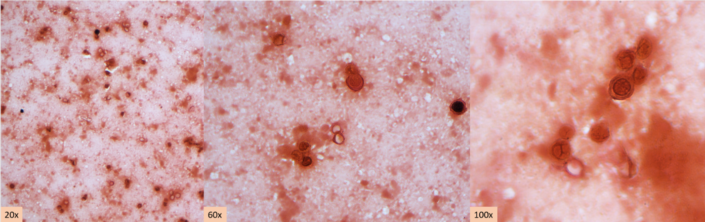
Cells appear plasmacytoid and appear both isolated and in clusters (Image 6). The nuclei are eccentrically placed, and the chromatin has a salt and pepper appearance akin to a neuroendocrine tumor (Image 7-8). Also identified were pink granules and intranuclear pseudoinclusions (Images 6-7). We performed immunohistochemical stains on paraffin sections of the cell block. Tumor cells show positive staining for calcitonin, chromogranin, mCEA, and TTF-1, while negative staining for thyroglobulin and CD45.

Following the diagnosis, the patient had a CT scan for staging purposes. Multiple lymph nodes in the right cervical chain were identified. the patient at a clinical stage IVA diagnosis. In the interim, the patient had a total thyroidectomy which revealed medullary thyroid carcinoma of the right lobe measuring 2.2 cm, a micropapillary carcinoma of the left lobe measuring 0.1 cm (Image 8). Lymphovascular invasion was not identified, the inked surgical resection margins are free of carcinoma, and metastatic medullary carcinoma was identified in 6 of the 77 lymph nodes removed during the central compartment lymph node dissection, and bilateral cervical lymphadenectomies. The calcitonin level dropped to 23 pg/mL postoperatively.  Genetic testing was performed to assess for Multiple Endocrine Neoplasia Type 2 (MEN2), and although her result was indeterminate, a RET mutation was not identified.

Image 8: Thyroid, Excision: H&E section (600X).

Case 3

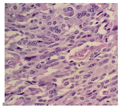
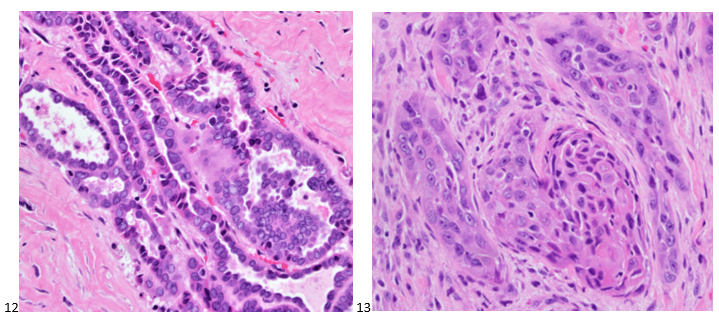
A 67-year-old male with no pertinent medical history presented to the endocrinology clinic after his primary care physician identified a large lump in the patient’s neck. A 7.0 cm hypoechoic right thyroid mass with macrocalcifications was noted on ultrasound imaging. The patient was referred to diagnostic imaging for a thyroid FNA. The smears and cell block section are depicted below. While the papillary formation of Image 9 is not evident on the pap-stained slide (Image 10), the nuclear grooves and pseudoinclusions along with irregular nuclear membranes and powdery chromatin are highlighted. A separate needle pass was collected for molecular testing, which revealed a BRAF V600E mutation in the tumor cells.

Images 9-11: Thyroid, Right Lobe, FNA 9: DQ-stained smear; 10: Pap-stained smear; 11. H&E Cell Block section (600X).

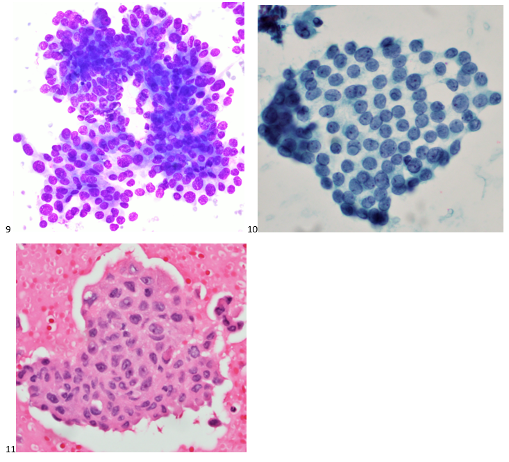
Two weeks after the FNA diagnosis, the patient was scheduled for a partial thyroidectomy of the right lobe. While 60% of the mass demonstrated well-differentiated papillary thyroid carcinoma (Image 12), 40% of the tumor contained poorly differentiated thyroid carcinoma with squamous features (Image 13). No sarcomatous components or giant tumor cells were identified. Carcinoma with squamous features invaded into the surrounding tissue, strap muscle, thymus, and right paratracheal lymph node. Interestingly, the right-sided levels 3 and 4 lymph nodes contained predominantly well-differentiated papillary thyroid carcinoma with rare foci of poorly differentiated thyroid carcinoma with squamous differentiation.

Images 12-13: Thyroid, Right Lobe, Excision: H&E section (600X).

Five months after the excision, the patient developed a left-sided pleural effusion. A diagnostic thoracentesis was performed and metastatic thyroid carcinoma was identified. Immunostains performed on the cell block slides with adequate controls show that the tumor cells are positive for PAX8, and negative for TTF-1, and thyroglobulin. The findings support the diagnosis. While patients with papillary thyroid carcinoma tend to have better disease-free survival rates, the poorly differentiated tumor was difficult to control and eventually resulted in widespread metastasis.

Cytology diagnosis: Papillary thyroid carcinoma.

Pathology diagnosis: Poorly differentiated thyroid carcinoma with squamous features in a background of well-differentiated papillary thyroid carcinoma.


Case 4

A 73-year-old female presented with a rapidly growing and painful thyroid mass that measured 8 cm on imaging. Originating from the right lobe, multiple needle passes targeted various areas of the mass via ultrasound-guidance. The smears and cell block section are presented below. Smears (Images 14-15) feature pleomorphic nuclei in a background of inflammation and necrosis. The cell block section (Image 16) demonstrates increased mitotic figures and neutrophils.

Images 14-16: Thyroid, Right Lobe, FNA: 14: DQ-stained smear; 15: Pap-stained smear; 16. H&E Cell Block section (600X).

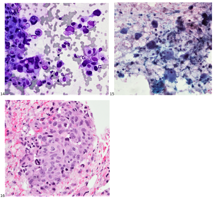
We performed immunocytochemical stains on paraffin sections of the cell block. Tumor cells how positive staining for p53, focal staining for cyclin D1, and negative staining for AE1/AE3, thyroglobulin, and BCL-2. Rare tumor cells show staining for TTF-1. The proliferation index by Ki-67 immunostaining is approximately 70%.

While not a standard procedure for thyroid specimens, core biopsies (Image 17) were also obtained from this mass.

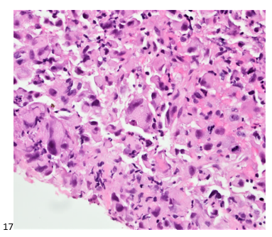
Molecular testing on the core biopsy sample identified a high mutation burden, with the tissue harboring both TP53-inactivating and TERT promoter mutations. Imaging demonstrated widespread metastasis, and this patient did not survive the extensiveness of her disease.

Cytology Diagnosis: Undifferentiated (anaplastic) thyroid carcinoma.

Pathology Diagnosis: High-grade carcinoma consistent with anaplastic carcinoma (interchangeable diagnoses).


That’s enough for our classic thyroid cases. Stay tuned for the second edition featuring thyroid FNAs with unsuspecting findings!

-Taryn Waraksa-Deutsch, MS, SCT(ASCP)CM, CT(IAC), has worked as a cytotechnologist at Fox Chase Cancer Center, in Philadelphia, Pennsylvania, since earning her master’s degree from Thomas Jefferson University in 2014. She is an ASCP board-certified Specialist in Cytotechnology with an additional certification by the International Academy of Cytology (IAC). She is also a 2020 ASCP 40 Under Forty Honoree.

Microbiology Case Study: A 25 Year Old Male with Abdominal Pain and Fever

Case History

A 25 year old male with severe right lower quadrant abdominal pain, fever and history of congenital neutropenia and thrombocytopenia presented to the emergency department. The patient reported experiencing similar episodes over the previous six months, sometimes requiring hospitalization. His symptoms would typically improve following several days of antibiotic therapy and were thought to be secondary to typhlitis. He reported that his current symptomology was more severe than any of the previous episodes. In the emergency department, the patient was found to be tachycardic and severely hypotensive, prompting admission for sepsis and empiric antimicrobial therapy with piperacillin/tazobactam and vancomycin. Imaging at the time revealed severe terminal ileitis with mild pancolitis, markedly worsened from a study obtained months prior during a previous episode. After failing to improve with conservative management, an exploratory laparotomy with total abdominal colectomy and end ileostomy was completed to achieve source control.

Laboratory workup

Anaerobic bottles from blood cultures collected at the time of hospital admission flagged positive in under 24 hours. Gram staining from the positive bottle broth revealed large, gram variable bacilli (Image 1A). Multiplex PCR performed on the positive blood culture broth failed to reveal the identity of the organism. After 24 hours, close observation was required to visualize bacterial growth on anaerobic blood culture media, which appeared as a thin, translucent film (Image 1B). To provide better visualization for this case, a loop was utilized to scrape the growth so a path could be observed within the biomass (Images 1B and 1C, dotted lines). The bacterium recovered was definitively identified as Clostridium septicum by MALDI-TOF MS, prompting continued coverage with piperacillin/tazobactam until discharge.

Image 1. A) Representative Gram stain from the positive anaerobic blood culture bottle. Staining reveals large, gram variable rods. Given the size and morphology of the cells, a gram positive organism was suspected. B) Growth of the organism on CDC anaerobic blood agar after 72 hours. A lack of distinct colonies was noted, and the organism grew as a thin film across the surface of the plate which was difficult to visualize. The hatched oval denotes where a loop was pulled across the plate surface to help visualize the bacterial growth. A closer, enhanced image is presented in C.

Discussion

Clostridium septicum is an anaerobic, Gram-positive spore-forming bacillus.1,2 Colony morphology on solid media evolves over time, with a characteristic “medusa head” morphology at under 8 hours, a “sand grain” appearance at approximately 24 hours, and visible swarming after 48 hours (Image 1B and 1C).2 The organism is found in the soil and the gastrointestinal tracts of both humans and animals with the potential to cause a variety of diseases. It is especially problematic as an infectious agent of livestock and other ruminants, poultry, and horses.1 Clinical manifestations of infection with C. septicum in humans includes necrotizing enterocolitis, bacteremia, and clostridial myonecrosis.2 Prompt identification of infection with Clostridium septicum is essential due to the rapidly fatal nature of untreated bacteremia with this organism. The mainstay of treatment is early initiation of antibiotic therapy and aggressive surgical debridement of affected tissue. The drug of choice for medical management is penicillin, although metronidazole and carbapenems may be used in patients with penicillin allergies.

C. perfringens and C. septicum are the two clostridial species most associated with bacteremia in neutropenic patients.5 Clostridium septicum bacteremia can allow for the hematogenous dissemination of infection and can be rapidly fatal.3 Infection of the bowels of a susceptible host and compromise to the bowel wall integrity leads to entry of bacteria into the circulation. In severe cases, this in turn can lead to seeding of distal sites with progression to sequelae including clostridial myonecrosis (gas gangrene). While classical traumatic myonecrosis associated with an infected wound becomes contaminated with clostridial spores from the external environment (most commonly C. perfringens), atraumatic (or spontaneous) myonecrosis is associated with C. septicum (among other clostridial species) and results from hematogenous seeding of distal sites during bacteremia. Virulence and tissue necrosis are mediated by a variety of exotoxins produced by the organism,1 with alpha-toxin being the primary virulence determinant.4

Importantly, Clostridium septicum bacteremia is strongly associated with both hematogenous and gastrointestinal cancers. For this reason, detection of this pathogen in the blood may warrant further patient evaluation for potential sources of underlying pathology.2 In this patient’s case, typhlitis and neutropenic enterocolitis were precipitating factors leading to his C. septicum bacteremia as this patient presented without evidence of gastrointestinal malignancy. Following the procedure, the patient stabilized, and his condition continued to improve through recovery. His antibiotics were eventually discontinued, and he was discharged.

References

  1. Alves MLF, Ferreira MRA, Donassolo RA, Rodrigues RR, Conceição FR. Clostridium septicum: A review in the light of alpha-toxin and development of vaccines. Vaccine. 2021;39(35):4949-4956. doi:10.1016/j.vaccine.2021.07.019
  2. Mallozzi MJG, Clark AE. Trusting your gut: Diagnosis and management of clostridium septicum infections. Clinical Microbiology Newsletter. 2016;38(23):187-191. doi:10.1016/j.clinmicnews.2016.11.001
  3. Koransky JR, Stargel MD, Dowell VR Jr. Clostridium septicum bacteremia. Its clinical significance. Am J Med. 1979;66(1):63-66. doi:10.1016/0002-9343(79)90483-2
  4. Kennedy CL, Krejany EO, Young LF, et al. The alpha-toxin of Clostridium septicum is essential for virulence. Mol Microbiol. 2005;57(5):1357-1366. doi:10.1111/j.1365-2958.2005.04774.x
  5. Bennet JE, Dolin R, Blaser MJ, Onderdonk AB, Garrett WS. Diseases Caused by Clostridium. In Mandell, Douglas, and Bennett’s principles and practice of infectious diseases (2nd ed., pp. 2960-2968). Elsevier.

-Amanda Means is a PGY-1 resident in Anatomic and Clinical Pathology at the University of Texas Southwestern Medical Center in Dallas, Texas.  Dr. Means did her undergraduate studies at Sam Houston St University in Huntsville, TX and received her medical degree from the University of Texas Health Sciences Center in San Antonio.


-Clare McCormick-Baw, MD, PhD is an Assistant Professor of Clinical Microbiology at UT Southwestern in Dallas, Texas. She has a passion for teaching about laboratory medicine in general and the best uses of the microbiology lab in particular.

-Andrew Clark, PhD, D(ABMM) is an Assistant Professor at UT Southwestern Medical Center in the Department of Pathology, and Associate Director of the Clements University Hospital microbiology laboratory. He completed a CPEP-accredited postdoctoral fellowship in Medical and Public Health Microbiology at National Institutes of Health, and is interested in antimicrobial susceptibility and anaerobe pathophysiology.

Containment and Contagion: How Far Do We Go?

One of my favorite older horror movies is The Shining. The acting, the music, and the pace of the story create a good long scare for the audience. In one scene, the mother and child are playing in an outdoor maze constructed of tall bushes. Later in the film they would be running for their lives in that selfsame maze, but I do not want to give away any spoilers if you haven’t watched it. The maze sometimes reminds me of some laboratory departments that I have seen, and I have seen how winding hallways and multiple doorways create confusion for lab employees and others in the facility.

The International Organization for Standardization (ISO) states in its standard Medical Laboratories — Requirements for Safety (ISO 15190:2020) that clean and dirty areas need complete separation from floor to ceiling. For example, the break room must be a completely separate room from the space where lab work is performed. While not all laboratories are ISO-certified, this is clearly an infection control and safety best practice. The Occupational Safety and Health Administration (OSHA) has been known to enforce this when they cite labs for incomplete separation of clean and dirty areas.

The Centers for Disease Control (CDC) published its resource, Biosafety in Microbiological and Biomedical Laboratories (BMBL-6th Edition) in 2020, and it requires a hand hygiene sink near the exit of laboratories so that hands can be washed before exiting the department. That means door handles inside the department must be considered clean and not touched with gloved hands.

Think about those requirements and apply them to your lab space(s). Do they work? Do they make sense to you? If you work in a Biosafety (BSL) 2 or 3 laboratory, you should understand the basics of pathogen containment. After all, the Biosafety Level of your laboratory is determined by the infectivity of pathogens present, the severity of disease they could cause, their transmissibility, and the nature of the work conducted. Based on risk and task assessments, labs should utilize the personal protective equipment (PPE) appropriate for the tasks performed, and that PPE should never be brought outside of the laboratory (another OSHA regulation).

One of the most common questions I receive from lab safety professionals is how to improve PPE compliance. The answer is, we need to make it easy for our staff to do the right thing. That means providing education so they understand the consequences of unsafe behaviors, and it means putting practices in place that make sense and are easy to follow.

In my travels I have seen labs that require that gloves must be removed before touching lab telephones and keyboards. I have seen specimen transport policies that allow for the use of wearing one glove for holding specimens and keeping the other hand clean to touch doors, and I have also seen staff who are told to keep a glove on to touch all lab door handles. If your staff are required to figure out that crazy maze of practices, they are not ever all going to get it right, and someone is going to acquire an infection.

If you require all gloves to be removed and hands to be cleaned before exiting the lab (as you should), are there sinks and waste cans at the exits? If lab coats must be hung up before going into the break room or rest room (as they should be), do you provide coat hooks near those doors? If staff must leave one BSL2 lab and walk through a clean hallway to deliver specimens to another lab, what do they wear? Do they wear their PPE (violating OSHA’s regulation) and do they touch door handles with gloves that other staff will use without gloves when leaving the department for the day?

Lab Acquired Infections (LAIs) are vastly under-reported, and many time people do not even realize how they got infected. There are probably many lab practices (like the ones mentioned above) that lead to these. Could this happen in your workplace? How can you prevent it?

Start, as always, with a risk assessment. Determine the hazards in your workplace, create hazard mitigation steps, and determine if any residual risks still exist. Take a good look at your physical layout. Can a “clean” hallway be designated as “dirty?” Would something as simple as adding a door to close off a hallway make it so safety regulations can be followed? Those risk assessments and diagrams used as a show-and-tell for facility leadership can often be what gets you the approval or funds you might need to make those changes. Correcting those unsafe practices and those long lab winding hallways and exits will certainly make it easy for staff to find the way in and out while containing the pathogens where they should be.

Dan Scungio, MT(ASCP), SLS, CQA (ASQ) has over 25 years experience as a certified medical technologist. Today he is the Laboratory Safety Officer for Sentara Healthcare, a system of seven hospitals and over 20 laboratories and draw sites in the Tidewater area of Virginia. He is also known as Dan the Lab Safety Man, a lab safety consultant, educator, and trainer.

Microbiology Case Study: Infected Hangnail in a 13 Year Old Female

Case History

A 13 year old female presented to the emergency department with a painful left thumb swelling. Eight days ago, she picked out a hangnail from the medial side of her left thumb, resulting in a worsening pain and swelling. She went to urgent care five days ago and was prescribed Augmentin with no improvement. Physical examination showed a tender circumferential swelling of the right thumb past the flexion joint, with paronychia extending 1 cm in length with fluctuance extending an additional 0.5 cm. The wound was incised, and the drainage was sent to the microbiology laboratory for culture.

The culture grew Gram negative bacilli after 24 hours of incubation (Figure 1). The culture grew a moderate quantity of small nonhemolytic colonies that showed “pitting” appearance on blood (Figure 2) and chocolate (Figure 3) agar plates. The colonies notably smelled musty/bleachy. There was no growth on MacConkey agar.

Identification by Matrix-Assisted Laser Desorption Ionization – Time of Flight (MALDI-ToF) revealed Eikenella corrodens.

Discussion

Eikenella corrodens is a facultatively anaerobic, gram negative, oxidase positive, catalase negative, nonmotile, non-spore-forming, rod. It belongs to the HACEK group and is in the family Neisseriaceae. It is a part of the normal flora of the mouth and upper respiratory tract.

E. corrodens is an opportunistic pathogen that can lead to various infections including a variety of head and neck infections (ocular, mastoid, submandibular, thyroid), pleuropulmonary infections, skin and soft tissue infections, etc.). It can cause subcutaneous abscesses, cellulitis of soft tissues, and bacteremia in intravenous drugs users. E. corrodens also causes infections of the hand (i.e., ostomyelitis, cellulitis) because of chronic nail biting, human bite, or clenched-fist injury. The bacterium can gain access to the central nervous system by way of periodontal, middle ear, or sinus infections and cause meningitis, brain or spinal abscesses, subdural empyema, and osteomyelitis. In some predisposed individuals, endocarditis and bacteremia can also occur.

Under aerobic conditions, it grows slowly in blood and chocolate agar at 35–37℃ and does not grow on MacConkey Agar. It forms small, convex, round or irregular, gray, and non-hemolytic colonies. Most isolates may “pit” the agar although both pitting and non-pitting strains have similar biochemical characteristics. Most strains produce a musty or bleach-like odor.

E. corrodens is generally sensitive to ampicillin, amoxicillin, second- and third generation cephalosporins, tetracyclines, azithromycin, sulfamethoxazole, and fluoroquinolones. They are resistant to clindamycin, vancomycin, erythromycin, metronidazole, aminoglycosides, and penicillinase-resistant penicillins, with penicillin susceptibility varying from strain to strain. In clinical microbiology laboratories, the routine antimicrobial susceptibility testing for E. corrodens is not usually performed since it is generally responsive to beta-lactams. If necessary, susceptibility testing can be performed following the CLSI guidelines for fastidious organisms.

The disease spectrum of E. corrodens is increasing in complexity, and it is important to study its clinical characteristics for timely diagnosis, and effective treatment.

References

  1. Chapter 9, Color Atlas and Textbook of Diagnostic Microbiology, Seventh Edition.
  2. Li, L, Shi, Y-B, Weng, X-B. Eikenella corrodens infections in human: Reports of six cases and review of literatures. J Clin Lab Anal. 2022; 36:e24230. doi:10.1002/jcla.24230
  3. Mühlhauser M. Eikenella corrodens. Rev Chilena Infectol. 2013. doi:10.4067/S0716-10182013000200007

-Fahad Sheikh is a 2nd year AP/CP pathology resident in the Department of Pathology at Montefiore Medical Center in Bronx, NY.

-Phyu Thwe, Ph.D, D(ABMM), MLS(ASCP)CM is Associate Director of Infectious Disease Testing Laboratory at Montefiore Medical Center, Bronx, NY. She completed her medical and public health microbiology fellowship in University of Texas Medical Branch (UTMB), Galveston, TX. Her interests includes appropriate test utilization, diagnostic stewardship, development of molecular infectious disease testing, and extrapulmonary tuberculosis.

The Importance of Patient Identification in Forensics

Body identification is one of the core responsibilities of a forensic pathologist yet is also probably the most common one to be overlooked. Most of the deceased people who come to our office are visually identifiable, and the identity may already have been confirmed if they were transported to the hospital. In some situations, though, the body may be disfigured by fire, decomposition, injury, or there may only be partial remains recovered. In incidents with multiple fatalities, we need to be sure the correct remains are returned to the correct family. Particularly when foul play is involved, there may be intentional attempts to conceal the decedent’s identity or disguise them as another person.

There are two different levels of identification: “positive” identification (the gold standard) and “presumptive” identification.

The three generally accepted forms of “positive” identification are DNA comparison, fingerprint comparison, and radiograph comparison. While television shows have DNA “matches” coming back in the time it takes for a commercial break, DNA identification can pose challenges. A pre-existing specimen from the decedent or close family members is needed for comparison, which means you need to already have some suspicion of who they are. If they’ve been previously arrested or charged with a felony (the laws vary slightly by state), their DNA may have been uploaded to the Combined DNA Index System (CODIS), and a match may potentially be obtained blindly by uploading the decedent’s sample. By comparison, one can collect fingerprints from a decedent and submit them to the Automated Fingerprint Identification System (AFIS) for relatively rapid identification. Many people have been fingerprinted in their lifetime, whether for relatively minor arrests, employment, or background checks. However, there are still limitations. The hands (or at least fingertips) need to be intact, with printable skin. For mummified remains, the tissue can be rehydrated by soaking in sodium carbonate or sodium hydroxide to obtain legible prints. “Degloved” remains, where the skin has sloughed from the hands due to decomposition, can be fingerprinted by inserting one’s gloved hand into the sloughed skin.

Radiographs, like DNA, are limited by the need to have a pre-existing sample from the decedent (meaning you need to know who they might be). Radiographs are invaluable when trying to identify someone with no usable fingerprints, or no fingerprints on file. A variety of locations can be used for comparison, including the dentition, frontal sinuses, vertebral processes, healed fractures, or orthopedic implants. Serial numbers on implanted devices can also be traced back to the decedent, although not all implantable devices have such markings.

“Presumptive” identifications are based on many other common sense factors including context, visual identification, tattoos, belongings, and clothing. Depending on the context of the case, a presumptive identification may suffice. For a decomposed body in a secure apartment occupied by a single, elderly person the neighbors haven’t seen in days, monogrammed dentures within the mouth may be sufficient. But in a fire with three charred female victims, aged 20-25, it’s much more important to confirm the identifications by a positive method. As I mentioned earlier any situation involving foul play may provide motivation for to conceal a victim’s identity, and so all homicide victims must be positively identified. It’s often taken for granted that the tag on our patient’s toe is accurate, but we need to approach our autopsies with the same level of diligence a laboratorian has when evaluating the label on a blood tube. Knowing who your patient is, and who your sample comes from, is the first critical step for any pathologist.

The mummified remains of a young adult were found in an abandoned house; while the fingertips were initially too dessicated to yield fingerprints, rehydration revealed excellent ridge details. Fingerprints were then uploaded to AFIS, and the decedent was identified within an hour.
For skeletal remains with intact teeth, dental radiographs of the remains can be used for identification; however, for edentulous patients, a different strategy must be used.

-Alison Krywanczyk, MD, FASCP, is currently a Deputy Medical Examiner at the Cuyahoga County Medical Examiner’s Office.

Microbiology Case Report: 47 Year Old with Left Flank Pain

Case Description

A 47 year old male presented to the emergency department with a two-week history of progressively worsening left-sided flank and abdominal pain with radiation to his back. He also endorsed associated nausea, vomiting, dizziness, lack of appetite, and a 1-month history of intermittent fever, chills, night sweats, cough, and chest pain. Patient history was also notable for travel throughout the Amazon basin in Peru and Colombia, then to Panama where he felt ill while hiking in the rainforest. He also had sustained injuries to his left flank and head while traveling.

Given his symptomology, further evaluation and management were undertaken. CBC showed a drop in hemoglobin from 10.3 g/dL to 8.5 g/dL, and imaging revealed splenomegaly with a caudally located subcapsular hematoma (Figure 1). Small hemoperitoneum on the pelvis with small non-loculated fluid along the bilateral paracolic gutters was also observed. The patient received two units of packed red blood cells to stabilize his hemoglobin levels.

Laboratory Diagnosis

Malaria smears were ordered given his history travel and evidence of anemia. Plasmodium vivax was identified on thin smear at a parasitemia of 0.1%. The Infectious Disease service was consulted and the patient received IV artesunate followed by oral artemether/lumefantrine and was also started on primaquine for hypnozoite eradication as he was not G6PD deficient. The malaria-associated splenic rupture was well managed with conservative treatment, and no surgical intervention was needed.

Figure 1Computer Tomography scan of the abdomen reveals a caudally located subcapsular hematoma (white circle).
Figure 2. Peripheral blood smears (Giemsa stain, 1000x magnification, oil immersion) from the presented patient. A) An enlarged red blood cell with an irregular membrane containing a ring form (black arrowhead) and a mature trophozoite (white arrowhead) of P. vivax. B) An enlarged red blood cell with dot-like staining and a ring form, consistent with P. vivax (black arrowhead). C) P. vivax gametocyte (black arrowhead) and mature trophozoite (white arrowhead). Infected cells are enlarged and the mature trophozoite expands to nearly fill the red blood cell.

Discussion

Malaria is a severe and sometimes fatal tropical disease caused by Plasmodium sp. The World Health Organization (WHO) estimates 300-500 million humans suffer from malaria each year, of whom >1 million die.1 Five species of Plasmodium cause human malaria: P. falciparumP. malariaeP. vivaxP. ovale, and P. knowlesi. Patients with malaria initially present with a combination of non-specific symptoms such as fever, chills, headache, and general malaise. Moreover, it can cause various complications, including severe anemia, neurologic defects, nephrotic syndrome, hepatosplenomegaly, and even splenic rupture.

Splenic rupture is an infrequent but life-threatening complication of malaria. Due to a paucity of reported cases, the incidence of pathological splenic rupture in natural vector-transmitted malaria is poorly defined. Additionally, cases of splenic rupture in the setting of malaria are likely to be underreported due to a lack of diagnostic methods, especially in endemic regions.2 One case series determined splenic rupture during malaria was fatal 22% of the collected cases, and it was likely to be the most frequent life-threatening issue of Plasmodium vivax infection reported worldwide.3 Similarly, P. vivax tends to cause more pronounced splenomegaly and a higher incidence of splenic rupture when compared to other Plasmodium species as noted in experimental and clinical studies.2.4 However, the underlying mechanism is incompletely established.

The cause of splenic rupture in malaria infections is thought to be a complication of the macroscopic consequences of different microscopic infiltrative phenomena. Specificities of clotting regulation in the spleen red pulp and acute spleen-specific pooling/unpooling of platelets may lead to increase in intrasplenic tension, making the spleen prone to rupture.3 Although malaria-related splenic rupture is essentially a ‘pathological rupture’ in the absence of overt trauma, minor trauma can also be a triggering event, which possibly contributed to the splenic rupture in this case.

Malaria patients with splenic rupture may present with abdominal pain, hypotension, tachycardia, nausea, and vomiting, which can be difficult to distinguish from signs and symptoms of malaria. Referred left shoulder pain (Kehr sign) caused by diaphragmatic irritation from peri-splenic effusion may also occur. The physical examination can show left upper quadrant or diffuse abdominal tenderness, abdominal rigidity or guarding, and splenomegaly. Radiological studies (i.e., ultrasound or CT scan) play a critical role in diagnosing malaria-associated splenic rupture, and the main findings may include splenic enlargement, splenic tear, subcapsular/intrasplenic hematoma, peri-splenic effusion, and hemoperitoneum. Non-radiological workup by contrast (e.g., decreased hemoglobin/hematocrit level) is generally non-specific.

Management of malaria-associated splenic rupture generally follows the same principles as traumatic injuries. Early diagnosis and appropriate management are essential for a favorable prognosis. It is suggested that conservative treatment can be considered in patients with stable hemodynamics. However, the risk of delayed hemorrhage still exists, which makes strict monitoring crucial. Some data suggest surgical intervention should be considered a first-line treatment if tears involve the splenic hilum.5 Although it may be challenging to decide on surgery versus conservative strategy in clinical practice, there are several definitive surgical indications including signs of peritonitis and unstable hemodynamic status despite aggressive resuscitation. Of note, splenectomy may increase the risk of severe infection caused by encapsulated bacteria and complicated malaria; therefore, spleen-preserving procedures should be attempted in selected patients, such as frequent travelers to malarious regions.6

References

  1. Guerra, C.A., et al., The limits and intensity of Plasmodium falciparum transmission: implications for malaria control and elimination worldwide. PLoS Med, 2008. 5(2): p. e38.
  2. Zingman, B.S. and B.L. Viner, Splenic complications in malaria: case report and review. Clin Infect Dis, 1993. 16(2): p. 223-32.
  3. Imbert, P., C. Rapp, and PA Buffet, Pathological rupture of the spleen in malaria: analysis of 55 cases (1958-2008). Travel Med Infect Dis, 2009. 7(3): p. 147-59.
  4. Schmidt, L.H., Plasmodium falciparum and Plasmodium vivax infections in the owl monkey (Aotus trivirgatus). I. The courses of untreated infections. Am J Trop Med Hyg, 1978. 27(4): p. 671-702.
  5. Fuks, D., et al., Spontaneous splenic rupture during a febrile crisis of Plasmodium falciparum malaria. J Chir (Paris), 2005. 142(6): p. 403-5.
  6. Jimenez, B.C., et al., Spontaneous splenic rupture due to Plasmodium vivax in a traveler: case report and review. J Travel Med, 2007. 14(3): p. 188-91.

-Bo Zhang, MD is an AP/CP resident at UT Southwestern Medical Center in Dallas, Texas. He is a first year resident interested in different aspects of surgical pathology.

-Andrew Clark, PhD, D(ABMM) is an Assistant Professor at UT Southwestern Medical Center in the Department of Pathology, and Associate Director of the Clements University Hospital microbiology laboratory. He completed a CPEP-accredited postdoctoral fellowship in Medical and Public Health Microbiology at National Institutes of Health, and is interested in antimicrobial susceptibility and anaerobe pathophysiology.

-Clare McCormick-Baw, MD, PhD is an Assistant Professor of Clinical Microbiology at UT Southwestern in Dallas, Texas. She has a passion for teaching about laboratory medicine in general and the best uses of the microbiology lab in particular.

Bandemia: What is it and Why is it Important?

Recently our lab was asked by physicians to start reporting band counts of ≧10% as critical values. While we have always reported bands when a manual differential is performed, we have also heard for years about other labs that have stopped reporting bands. Mayo Clinic stopped reporting bands in the 1990’s- other nationally and internationally known hospitals and community hospitals alike have followed suit. CAP proficiency surveys for cell ID do not separate segmented neutrophils and band neutrophils for cell IDs. Our hematology analyzer reports Immature granulocytes (IG) and we have learned about the benefits of IG over the band count. Thus, when our pathologists asked us to add a critical value for bandemia, we wondered if we were moving ‘backwards’.

Bandemia is defined as elevated band neutrophils in the peripheral blood. Neutrophils are produced to help fight infection. With infection, there is an increase in WBCs being released by the bone marrow into the peripheral blood. Bands are considered mature neutrophils and can fight infection, and in an effort to keep up with demand, some of these infection fighting cells which are released are bands. However, we are reminded that this is a nonspecific clinical finding. Bands can be elevated in many situations including inflammation, autoimmune disease, metabolic abnormalities, pregnancy, and treatment with granulocyte colony stimulating factors. Band counts of ≧10% have been used clinically as an indicator of serious bacterial illness. A finding of bandemia can help providers decide what steps need to be taken in evaluation of a patient, and in conjunction with clinical findings, can help in making a differential diagnosis.

The trouble with bands is that they are notoriously difficult to measure accurately and precisely. Bands are somewhat controversial and there are conflicting opinions on the utility of bands. Should bands be reported or included with neutrophils? A left shift reflects bone marrow response to bacterial infection, and this has been quantified as band count or immature granulocyte count. Is the IG a better parameter than the band count?

If your Hematology analyzer reports the absolute neutrophil count (ANC) with an automated differential, this includes all mature neutrophils. The theory that supports this is that neutrophilic bands and segmented neutrophils are both mature cells, and both fight infection. A true left shift would therefore be the presence of immature granulocytes (metamyelocytes, myelocytes and promyelocytes). Yet, we also now have analyzers that offer a 6-part differential that includes the immature granulocyte (IG) count. And we know that an automated differential is based on counting thousands of cells, and a manual differential is based on counting 100 cells. So, which is better? We can theorize that the IG is a better indicator of left shift because the automated diff count looks at more cells than a 100-cell manual diff.

Let’s say that we have a patient with a WBC of 20 x 103/μL. Left shift is defined as an absolute IG count >0.1 x 10³/μL. Automated diffs can count >30,000 cells depending on the WBC count. If the auto diff counts 32,000 cells and finds 1% IG, this means that the analyzer identified 320 IG. Absolute counts are calculated as % x the WBC, in this case 0.01 x 20,000=.2 x 103/μL. This meets the definition of a left shift. If we do a manual diff and count 100 cells and see 1 meta, the absolute IG would be .2 x 103/μL, again meeting the criteria for a left shift. However, if we did not see any metas or other IG on the manual diff, the absolute IG would be 0. Thus, performing a manual diff, the difference in seeing 1 cell or 0 would make the difference of reporting a suspected infection versus no suspicion of infection.

Table 1: Left shift? comparison of absolute IG when 1 or no IGs are seen on manual diff

With the advent of the IG count on automated differentials, labs have moved away from reporting bands. I have attended conferences and heard presentations about “banning” bands. A few years ago, I wrote a blog called “Beyond Bands: The Immature Granulocyte Count”, describing the benefits of using the IG count over bands in manual diffs. The above example would support “Ban the bands” arguments. Using the IG count from analyzers can take advantage of analyzing many more cells and give us statistically more precise values.

Results from auto diff can get to patients’ chart faster than a manual diff result, leading to faster treatment. To report bands, a manual differential must be done. We must wait for a slide to be made, dry, and a diff to be analyzed either under the microscope or on CellaVision. Bands are subjective, relying on technologist interpretation.

So, why are we suddenly being asked to make bandemia a critical value?

Physicians asked for this change and have cited cases where patients were seen in the emergency department (ED) and subsequently released, later to return, or experiencing negative outcomes. These patients had bands reported on differentials. Other area hospitals are reporting bands and have critical values for bandemia. Because patients often are seen at more than one area hospital, and doctors may have privileges at more than one of these, for consistency, this makes sense. But is also makes sense clinically.

Recent data has drawn renewed attention to bands as a reliable predictor of severity of patient condition. A number of research papers have been published that indicate that bands may indeed be important for patient care. A 2012 study investigated bandemia in patients with normal white blood cell counts. This cohort study found that patients with normal WBC counts with moderate (11%-18%) or high (>20%) band counts had increased odds of having positive blood cultures and in-hospital mortality. (Drees, 2012)

In 2019 a study showed that there was an “increasing risk for death with increasing bandemia, irrespective of leukocyte count. (Davis, 2019) A 2021 study done at Rhode Island Hospital showed a strong correlation between increasing percentages of bands on an initial emergency room CBC and the likelihood of significant positive blood cultures and in-hospital mortality. This was noted even at band levels below 10%. (Hseuh, 2021) S. Davis, MD, from the Department of Emergency Medicine, George Washington University School of Medicine and Health Sciences, in Washington, DC wrote that “While emergency physicians may find reassurance in a normal leukocyte count, the balance of evidence strongly suggests a more prudent approach would be to wait for the bands.” (S. Davis, 2021) In other words, wait for that manual differential. He stated that emergency room physicians get results from automated CBCs before the manual diff and do not see or are aware of any internal laboratory flags on these specimens. Physicians should be aware of reporting processes to avoid early discharge of otherwise well-appearing patients before band counts are reported. Last year, trends in bandemia and clinical trajectory among patients was reviewed in a retrospective chart review at George Washington University Hospital. They noted that “Bandemia clearance and trending, in conjunction with other existing clinical tools, may be of use as a marker of improvement in sepsis. Conversely, worsening bandemia may be predictive of a deteriorating clinical status and possibly a higher mortality.” They also noted that following trends of band levels in patients with sepsis or septic shock may help to predict a clinical course and overall prognosis. (Prasanna, 2022) Additionally, a band count greater than 10% is one of the American College of Chest Physicians/Society of Critical Care Medicine’s systemic inflammatory response syndrome (SIRS) criteria used to diagnose sepsis. (Chakraborty, 2022) These are just a few of the many articles that support a clinical utility of reporting the band count.

When we first learned that we would be reporting bandemia as a critical value, we realized that we would need to get everyone on the same page. We do most of our diffs on CellaVision, so, in theory, that should be easy, but, and a big but, is that bands are notoriously subjective. Different technologists may have been taught or have used their own definitions of bands. Variation can occur depending on slide quality, tech training, definition of bands used, and number of cells counted. So, how do we make this work? We need to be sure that all our technologists are reporting bands using the same criteria, so we are not reporting differing or confusing information to physicians. The concern lies in a physician making a significant clinical decision based on apparent changes in band counts that are not real but only reflect predictable statistical factors and unpredictable technologist variability.

In our laboratory, we approached the implementation of this new critical value as an educational opportunity. Differentials that had been performed on our CellaVision were reviewed, and it was apparent that bands were not being categorized consistently among techs. (See Figure1) We started with reviewing the definition of bands with all technologists and writing updated detailed procedures that includes these definitions. We use CAP’s definition of bands, which is the definition used in most textbooks and references for over 60 years. (See Table 1) Many examples of both bands and segmented neutrophils were added to our reference library on CellaVision. These included textbook perfect bands and some that may be more subjective. These reference cells can be used by techs to compare cells when making decisions as to in which category they belong. It was also stressed to techs that they need to look at cells carefully, and in a view that is large enough to see both detail and differentiation.

Figure1. Cells reported as segmented neutrophils on Differential. How many bands do you see?
Table 2. CAP Definition of bands vs segmented neutrophils
Figure 2. Are these bands or segmented neutrophils?

Patient samples that had bands reported were located on CellaVision and multiple slides were made from these samples to be used as competency slides. In developing the differential evaluation tool, Rumke’s data showed that for a differential with a reported 12% bands, a second differential would have to have greater than 23% bands or fewer than 3% bands before the difference could be considered statistically significant. But this can be significant to our patients and patient care. In this example, diffs with <10% bands would not indicate bandemia, and diffs over 10% would initiate a critical bandemia call. And this could happen on the same slide depending on who did the diff, or on sequential samples on the same patient over a short period of time. These competency slides were assigned to techs to collect statistics on the mean and SD of the bands reported on each slide. Retraining and coaching will be provided as necessary. Follow up competency slides will be assigned, and statistics will be recalculated. The goal is to decrease variability in our band counts and to show that we have done so. This ongoing quality project has involved writing procedures, offering continuing education, assigning and reviewing competency slides, coaching technologists and reviewing slides with them and calculating statistics. Our goal is consistency and reporting meaningful results to our physicians. 

As we saw in the studies cited above, % bands and trends are both important when evaluating clinical correlations. The chart below shows examples of how this might affect patient care. If a patient on presenting at the ED had a 19% band count, this would be called as a critical, and the patient would be further evaluated, and depending on clinical symptoms and medical history would likely have blood cultures drawn and be admitted to the hospital. If a second tech did the diff and reported 5% bands, the patient may be sent home without further evaluation. On subsequent CBCs, we could be giving confusing results to the physician if we are not consistent in our reporting. With multiple techs doing differentials on different shifts, it could look like this patient is getting better,

getting worse, or it could look like the patient is responding to therapy, and then the next day they had a setback. While we understand that bands will probably always be somewhat subjective, we need to narrow this down. By adhering to one definition, our goal is to report consistent and accurate results.

Table 3. Various results on differentials on the same patient over the course of 3 days, showing technologist dependent results.

ED physicians are looking for an early marker that can be used to identify septic patients as early as possible. Bandemia may be used as this marker. We therefore need to be as objective as possible when reporting bands. “Ultimately the band count is only one factor amongst several others which will be used in assessing the patient’s clinical state and in determining any subsequent medical management. Yet, identifying bands is important, and emphasizes the key role that our laboratory professionals play in identifying causes for concern” Dr Edgar Alonsozana, Mercy Medical Center, Baltimore, Md.

I welcome your comments about how your laboratories report bands and if bandemia is a critical value in your facilities!

References

P.Joanne Combleet, Clinical utility of the band count, Clinics in Laboratory Medicine, 2002;22:101-136

Al-Gwaiz, Layla A. and H H Babay. “The Diagnostic Value of Absolute Neutrophil Count, Band Count and Morphologic Changes of Neutrophils in Predicting Bacterial Infections.” Medical Principles and Practice 16 (2007)

S. Davis, R. Shesser, K. Authelet, A. Pourmand. “Bandemia” without leukocytosis: A potential Emergency Department diagnostic pitfall. The American Journal of Emergency Medicine,Volume 37, Issue 10, 2019

Harada T, Harada Y, Morinaga K, Hirosawa T, Shimizu T. Bandemia as an Early Predictive Marker of Bacteremia: A Retrospective Cohort Study. Int J Environ Res Public Health. 2022 Feb 17;19(4):2275.

Christine DeFranco DO and Terrance McGovern DO MPH
St. Joseph’s Regional Medical Center, Paterson, NJ. Isolated Bandemia: What Should We Do with It? Critical Care, Oct. 2016,

Harmening, Denise. Clinical hematology and Fundamentals of Hemostasis, 4th ed. 1997

Prasanna N, DelPrete B, Ho G, et al. The utility of bandemia in prognostication and prediction of mortality in sepsis. Journal of the Intensive Care Society. 2022;0(0).

https://www.mlo-online.com/home/article/13007276/answering-your-questions

Takayuki Honda, Takeshi Uehara, Go Matsumoto, Shinpei Arai, Mitsutoshi Sugano, Neutrophil left shift and white blood    cell count as markers of bacterial infection,Clinica Chimica Acta, Volume 457, 2016

Drees M, Kanapathippillai N, Zubrow MT. Bandemia with normal white blood cell counts associated with infection. Am J Med. 2012 Nov;125(11):1124.e9-1124.e15.

Leon Hsueh, Janine Molino, Leonard Mermel,

Elevated bands as a predictor of bloodstream infection and in-hospital mortality,The American Journal of Emergency Medicine,2021

https://www.ncbi.nlm.nih.gov/books/NBK547669/

Socha-small

-Becky Socha, MS, MLS(ASCP)CMBBCM graduated from Merrimack College in N. Andover, Massachusetts with a BS in Medical Technology and completed her MS in Clinical Laboratory Sciences at the University of Massachusetts, Lowell. She has worked as a Medical Technologist for over 40 years and has taught as an adjunct faculty member at Merrimack College, UMass Lowell and Stevenson University for over 20 years.  She has worked in all areas of the clinical laboratory, but has a special interest in Hematology and Blood Banking. She currently works at Mercy Medical Center in Baltimore, Md. When she’s not busy being a mad scientist, she can be found outside riding her bicycle.

Microbiology Case Study: A Middle-Aged Adult with Pulmonary Blastomycosis

Case History

A middle-aged patient with a complex past medical history presented to the Emergency Department with the primary complaints of shortness of breath and cough. The patient had recently been admitted for community-acquired pneumonia and discharged with antibiotics 10 days ago. However, the patient continued to have a dry cough, a fever of 102 degrees F, and shortness of breath. On the new admission, they were noted to be hypotensive and labs showed elevated WBC (14.7), lactate of 7.3, proBNP of 852, and procalcitonin of 4.62. Bacterial blood cultures showed no growth. A chest x-ray showed worsening left lower lobe consolidation with extension to the left upper lobe, compared to the x-ray at the previous admission. A bronchoscopy was done and bronchoalveolar lavage (BAL) was submitted for Gram stain, aerobic bacterial culture, and fungal culture. The Gram stain and bacterial culture were negative. However, the fluorochrome stain showed many large budding yeast forms, most suggestive of Blastomyces dermatitidis. Lung tissue sample culture (Image 1), Gram stain (Image 2) and histopathology (Image 3) confirmed the identification as Blastomyces dermatitidis. The patient was expired before the results were released. Gross images of lungs are shown in image 4.

Image 1. Fungal growth on the Sabouraud dextrose plate observed after 2 days.
Image 2. Gram stain of left lower lobe of lung tissue sample demonstrating budding yeast.
Image 3. GMS and H&E stains of left lower lobe of lung sections demonstrating budding yeast.
Image 4. Gross pictures of lungs showing left lower lobe hepatization and miliary pattern consolidation in the right lung.

Discussion

Blastomyces dermatitidis is a dimorphic fungus found in soil and decaying wood. Often found in areas close to a water source such as a lake, river, or stream.1 The fungus is endemic to parts of the United States, including parts of the Appalachian Mountains, the Great Lakes, and the Ohio and Mississippi River Valley.1 Human infection occurs when airborne conidia are inhaled. Blastomycosis (aka Gilchrist’s disease) may cause a broad range of clinical presentations. The most affected organs are the lungs and the skin.2 Osseous, genitourinary, central nervous system (CNS), and disseminated blastomycosis can also be seen, but at a lower frequency.2 Unlike opportunistic fungal pathogens such as H. capsulatum, C. immitis, and C. neoformans, B. dermatitidis no more likely to cause disease in immunocompromised population compared to the non-immunocompromised populatin.3 However, the disease is more severe, with higher morbidity and mortality, and more likely to be disseminated in immunocompromised patients.3

Although growth of fungi took 2 days in our case, Blastomyces is a slow growing fungi in that mycelial forms mature in 14-21 days. In suspicious cases, cultures should be hold up to 8 weeks. Furthermore, Blastomyces should be cultured as soon as possible since it does not survive well in samples. Morphologically, Blastomyces colony appears as a mold, which is white, prickly, and cottony at 25-30 °C However, at 35-37 °C, the colony appears as a yeast, which is tan, wrinkled, and waxy. Microscopically, at 37 °C, Blastomyces appear as round to oval cells with refractile and thick cell walls and measure 8 – 15 uM in diameter. Each yeast cell produces only one, broad-based (4-5 uM), bud. At 25-30 °C, septate hyphae form with short or long conidiophores. Round to pear-shaped conidia attach to the apex, resembling lollipops (Image 5).4

Image 5. Spherical, oval, or pyriform conidia rising from aerial hyphae, mold phase.

History of recent travel to the endemic areas or already living in there usually triggers the suspicion with clinical findings. There are different auxiliary diagnostic modalities for B. dermatitidis including culture, cytology smear, histopathology, urine antigen test, serum antigen test, serum antibody test, and molecular techniques. Serum antibody test exhibits high degree of cross-reactivity with other endemic mycoses.5 Serum and urine antigen testing has a sensitivity of 89 % and specificity of 79 % and it is useful in diagnosis, monitoring treatment, and detecting recurrence.5,6,7 Direct visualization of the distinctive yeast form with broad-based budding on a cytology smear of respiratory secretions or tissue samples using fluorochrome stain is considered as presumptive diagnosis and enough for initiation of antifungal therapy. However, a negative result in smear cannot exclude the diagnosis due to lack of sensitivity. Polymerase chain reaction (PCR) based assays have been developed to detect B. dermatitidis.8 However, although they are promising, utility has not been confirmed and there are no FDA approved assays.8

Only histopathology and culture can provide a definitive diagnosis. Although H&E may be enough in selected cases, periodic acid-Schiff (PAS) and Grocott’s methenamine silver (GMS) stains are useful to highlight the yeast form in tissue sections. In culture, seeing the characteristic morphologic and microscopic appearance is very useful for diagnosis. However, confirmatory testing with chemiluminescent DNA probe may still be necessary.9

Depending on the severity and site of the infection fluconazole, itraconazole, voriconazole, or amphotericin B can be used in treatment.10

References

  1. Castillo CG, Kauffman CA, Miceli MH. Blastomycosis. Infect Dis Clin North Am. 2016;30(1):247-264.
  2. Mazi PB, Rauseo AM, Spec A. Blastomycosis. Infect Dis Clin North Am. 2021;35(2):515-530.
  3. McBride JA, Gauthier GM, Klein BS. Clinical Manifestations and Treatment of Blastomycosis. Clin Chest Med. 2017;38(3):435-449.
  4. Maresca B, Kobayashi GS. Dimorphism in Histoplasma capsulatum and Blastomyces dermatitidis. Contrib Microbiol. 2000;5:201-216.
  5. Wheat LJ. Antigen detection, serology, and molecular diagnosis of invasive mycoses in the immunocompromised host. Transpl Infect Dis. 2006;8(3):128-139.
  6. Mongkolrattanothai K, Peev M, Wheat LJ, Marcinak J. Urine antigen detection of blastomycosis in pediatric patients. Pediatr Infect Dis J. 2006;25(11):1076-1078.
  7. Tarr M, Marcinak J, Mongkolrattanothai K, et al. Blastomyces antigen detection for monitoring progression of blastomycosis in a pregnant adolescent. Infect Dis Obstet Gynecol. 2007;2007:89059.
  8. Bariola JR, Hage CA, Durkin M, et al. Detection of Blastomyces dermatitidis antigen in patients with newly diagnosed blastomycosis. Diagn Microbiol Infect Dis. 2011;69(2):187-191.
  9. Saccente M, Woods GL. Clinical and laboratory update on blastomycosis. Clin Microbiol Rev. 2010;23(2):367-381.
  10. Chapman SW, Dismukes WE, Proia LA, et al. Clinical practice guidelines for the management of blastomycosis: 2008 update by the Infectious Diseases Society of America. Clin Infect Dis. 2008;46(12):1801-1812.

-Kadir Isidan, MS, MD is a pathology resident at University of Chicago (NorthShore). His academic interests include gastrointestinal pathology and cytopathology.

-Paige M.K. Larkin, PhD, D(ABMM), M(ASCP)CM is the Director of Molecular Microbiology and Associate Director of Clinical Microbiology at NorthShore University HealthSystem in Evanston, IL. Her interests include mycology, mycobacteriology, point-of-care testing, and molecular diagnostics, especially next generation sequencing.

Guess Who’s coming to the Lab?

When we enter the laboratory, we know of the dangers that can be encountered. Our training tells us there could be microbes and other potential pathogens in the samples we are about to analyze. We also learned how to protect ourselves; how our behavior while in the lab has consequences. We even know how to dress properly and what engineering controls we have at our disposal to keep us safe. We put on our personal protective equipment (PPE) before we start to work and remove it before leaving the lab. For some, these behaviors are automatic, actions that are done almost without even thinking. But is this the same for all who enter the lab? Do visitors who comes into the department know what they are really walking into or how to keep themselves safe in an environment that may be foreign to them? One common question asked by lab staff regarding visitors is “do they have to adhere to the lab safety policies and if so, why?”.  

On a recent safety audit, I visited a lab that happened to be getting a new chemistry analyzer installed. I noticed the vendor team, which consisted of 5 individuals, were not wearing any PPE. There were backpacks, open water bottles, and cell phones sitting on the counters and floors. The new instrument was not hidden in a back corner of the lab far away from the daily work. It was close to the area where the lab process, spins, and runs patient samples. Members of the vendor team were lying on the floor and crawling around. How does that scene make you feel?

Vendors and service representatives are regular visitors in your lab. A laboratory can have a representative on site a dozen times before you even begin to use that piece of equipment.  Once it is installed, you can bet you will see them multiple times for preventative maintenance and service calls. How does your lab welcome these guests? Do you let them in and have them get right to work? If they are there to repair an analyzer   you are likely eager to have them get started, but do you ask them to wear a lab coat?  Did they bring one of their own that was kept in their backpack?  If so, do you think that coat is clean or was it used in a different lab, packed up, and brought to your lab? Vendor compliance is a safety issue for many labs because these visitors are not lab employees, yet they are in your department and may be putting themselves and your team at risk. Often vendors are seen with drinks in labs, using cell phones or touching instruments without gloves – behaviors lab folk are told not to follow. So why is it tolerated? It shouldn’t be, and you have the right to speak up and ask them to adhere to your lab policies.

What about other potential laboratory visitors? Do pathologists come in to look at a patient slide in Hematology? Do they just sit down at your bench and look at the slide without gloves or a lab coat? Is lab staff allowed to scan a smear without PPE? Probably not, and no one else should be allowed too either. The microscope has most likely been touched with dirty gloves, and no one else should touch the same scope without gloves. Even lab doorknobs are a consideration. Staff should wash hands before leaving the department. That means no one should use contaminated gloves to open the door.

Speaking up about these safety issues to lab visitors can feel uncomfortable. A conversation with a physician about safe practices in the lab can be daunting, but the cost of not speaking up can be high. Take the opportunity to show you care about visitors and want to keep them protected. Sometimes you know who is coming to the lab, and you feel confident they have been trained and will use the best safety practices. At other times, though, those guests may be unexpected and lacking in safety knowledge. Make sure to treat them with respect, give them the safety training and tools they need so they can leave both happy and healthy.

-Jason P. Nagy, PhD, MLS(ASCP)CM is a Lab Safety Coordinator for Sentara Healthcare, a hospital system with laboratories throughout Virginia and North Carolina. He is an experienced Technical Specialist with a background in biotechnology, molecular biology, clinical labs, and most recently, a focus in laboratory safety.

Microbiology Case Study: A 40 Year Old with Polysubstance Use Disorder

Case History

A 40-year-old woman with past medical history of polysubstance use disorder (cocaine, IVDU-heroin) presented with shortness of breath on exertion, weight loss, and weakness. Cardiac ultrasound showed aortic valve endocarditis with mild thickening of the aortic valve, vegetation and severe aortic regurgitation without stenosis. The patient was taken to the operating room for aortic valve replacement. A tissue biopsy was sent for surgical pathology workup and microbiology cultures. H&E staining of the right and left coronary cusp and noncoronary cusp and showed focal giant cell reaction, and acute inflamed granulation tissue consistent with vegetation/infective endocarditis. The Gram stain of the tissue specimen revealed gram positive cocci but no bacterial growth was seen. Fungal and mycobacterial cultures were also negative. Broad Range Bacterial PCR and Sequencing (BRBPS) of the heart valve detected Streptococcus mutans.

H&E staining of the heart valve revealed focal giant cell reaction (black arrow), and acute inflamed granulation tissue consistent with vegetation/infective endocarditis (left image). The Gram stain of the tissue specimen revealed gram positive cocci (right image). (H&E diagnosis credits: Dr. Irena Manukyan from George Washington University.)

Discussion

The first step of BRBPS is performing polymerase chain reaction (PCR) targeting the 16S ribosomal RNA (rRNA), a conserved gene across many bacterial species including mycobacteria. If PCR is negative, then sequencing is not performed but if PCR is positive, sequencing such as Sanger sequencing or Next generation sequencing will be followed. Subsequent bioinformatics analysis of the sequencing results will allow identification of the specific bacteria.1,2 In the patient here, the PCR for 16S rRNA was positive and sequencing revealed S. mutans.

S. mutans is part of the viridans-group of Streptococcus and can be identified as a gram positive cocci with growth that is alpha-hemolytic, optochin resistant, and bile insoluble. Viridans group Streptococcus invade the bloodstream following dental treatment or dental procedures and can be associated with high risk for infectious endocarditis in patients with underlying heart disorders. S. mutans first gained medical attention due to its role in dental caries.3 S. mutans is considered part of the normal flora of the oropharynx, more specifically the dental plaque, and can form biofilms on the hard surface of the tooth. Bacterial strains that cause endocarditis have been shown to be able to bind to type I, III, and IV collagen, which are major components of the host cardiovascular tissues.4 The bacteria also have virulence factors that can cause strong adhesion to human endothelial cells. Additionally, aggregation and interaction with fibrinogen, platelets, and completement allow this bacteria to successfully cause cardiovascular diseases.5  

In infective endocarditis, 20% of the cases are negative by conventional methods such microbiology cultures.6 Several studies have shown that in specimens from patients with endocarditis, 16s rRNA sequencing can correctly detect the organism that was grown in culture but also successfully detect organisms in specimens where there was no growth, allowing clinical teams to accurately treat patients, similar to our case here.7,8 For prosthetic heart valve tissue, the sensitivity of sequencing versus culture is 93% and 35%, respectively. A study describing periprosthetic joint infections using elbow joint fluids showed that sequencing was positive in 47 specimens but 8% of those were negative by culture.10 Additionally, 16s rRNA sequencing has been useful in identifying bacterial tick-borne organisms not typically grown in culture such as BorreliaAnaplasmsaEhrlichia, and Rickettsia].11

The BRBPS test is validated for and ideally be tested on specimens from sterile sources (joint fluid, blood, heart valves, CSF) and ideal specimens are those where bacterial organisms can be visualized using microscopy. Limitations of BRBPS is that this test only detects bacterial organisms and will not detect viruses, fungi, and parasites. False-positive results are possible if the specimen is contaminated with patient flora. False-negative results may occur due to sequence variability affecting how the primers bind, presence of PCR inhibitors, or the quantity of nucleic acid material below the limit of detection. Clinical tests utilizing sequencing approaches may be costly at the moment in time; those who order sequencing tests need to understand the purpose of different sequencing tests so the most appropriate test is ordered. For example, if fungal pathogens are suspected, a sequencing test based on amplifying 28S rRNA or ribosomal ITS genes for the conserved genes found in fungal pathogens is appropriate.12 Such tests where you selectively enrich for specific targets such as 16s or ITS are ‘targeted NGS tests’ which have higher sensitivity for detection of microorganisms in sample types with large amounts of DNA. In comparison, metagenomics is a more agnostic approach and allows for detection of all nucleic acid in the specimen (including both host and microbial reads). While metagenomics can successfully detect pathogens involved in an infection, it can also detect the microbiome present in the same specimen. Hence, the one limitation is the background noise from human nucleic acid and the microflora [13-14. Another type of sequencing is whole genome sequencing where the microbial genome of a particular organism is sequenced and assembled. WGS aids in identification, typing, and determining the microbial susceptibility. WGS is helpful in identifying outbreaks or for epidemiological purposes, but the limitation is that a pure culture is needed. WGS requires a pure culture so this is a limitation for organisms that are non-viable in culture [13-14].

References

  1. Rosey AL, Abachin E, Quesnes G, Cadilhac C, Pejin Z, Glorion C, Berche P, Ferroni A. Development of a broad range 16S rDNA real-time PCR for the diagnosis of septic arthritis in children. J Microbiol Methods. 2007 Jan;68(1):88-93. doi: 10.1016/j.mimet.2006.06.010. Epub 2006 Aug 14. PMID: 16904782.
  2. Broad Range Bacterial PCR and Sequencing, Varies. Mayo Clinic Laboratories. https://www.mayocliniclabs.com/test-catalog/Overview/65058
  3. Loesche WJ. 1986. Role of Streptococcus mutans in human dental decay. Microbiol Rev 50:353–380.
  4. Nomura R, Naka S, Nemoto H, Inagaki S, Taniguchi K, Ooshima T, Nakano K. 2013. Potential involvement of collagen-binding proteins of Streptococcus mutans in infective endocarditis. Oral Dis 19:387–393. doi: 10.1111/odi.12016.
  5. Otsugu, M., Nomoura R., Matayoshi, S., Teramoto, N., Nakanoa, K. Contribution of Streptococcus mutans Strains with Collagen-Binding Proteins in the Presence of Serum to the Pathogenesis of Infective Endocarditis. Infect Immun. 2017 Dec; 85(12): e00401-17.
  6. Baddour LM at al; American Heart Association Committee on Rheumatic Fever, Endocarditis, and Kawasaki Disease of the Council on Cardiovascular Disease in the Young, Council on Clinical Cardiology, Council on Cardiovascular Surgery and Anesthesia, and Stroke Council. Infective Endocarditis in Adults: Diagnosis, Antimicrobial Therapy, and Management of Complications: A Scientific Statement for Healthcare Professionals from the American Heart Association. Circulation. 2015 Oct 13;132(15)
  7. Peeters, B., Herijgers, P., Beuselinck, K., Verhaegen, J., Peetermans, W.E., Herregods, M.-C., Desmet, S., Lagrou, K. Addd diagnostic value and impact on antimicrobial therapy of 16s rRNA PCR and amplicon sequencing on resected heart valves in infective endocarditis: a prospective cohort study. Clinical Microbiology and Infection. 2017 Nov; 23(11): 888.e1-888.e5
  8. Premru, M.M, Zupanc, T.L., Klokocovnik, T., Sabljic, E.R., Cerar, T. Broad-Range 16s rRNA PCR on Heart Valves in Infective Endocarditis. J Heart Valve Dis. 2016 Mar; 25(2):221-22
  9. Miller, R. J.H., Chow, B., Pillai, D., Church, D. Development and evaluation of a novel fast broad-range 16S ribosomal DNA PCR and sequencing assay for diagnosis of bacterial infective endocarditis: multi-year experience in a large Canadian healthcare zone and a literature review. BMC Infect Dis. 2016 April 12:16:146
  10. Flurin, L.; Wolf, M.J.; Greenwood-Quaintance, K.E.; Sanchez-Sotelo, J.; Patel, R. Targeted next generation sequencing for elbow periprosthetic joint infection diagnosis. Diagn. Microbiol. Infect. Dis. 2021, 101, 115448.
  11. Kingry, L.; Sheldon, S.; Oatman, S.; Pritt, B.; Anacker, M.; Bjork, J.; Neitzel, D.; Strain, A.; Berry, J.; Sloan, L.; et al. Targeted Metagenomics for Clinical Detection and Discovery of Bacterial Tick-Borne Pathogens. J. Clin. Microbiol. 2020, 58, e00147-20.
  12. Yeo, S.F.; Wong, B. Current status of nonculture methods for diagnosis of invasive fungal infections. Clin. Microbiol. Rev. 2002, 15, 465–484.
  13. Mitchell SL, Simner PJ. Next-Generation Sequencing in Clinical Microbiology: Are We There Yet? Clin Lab Med. 2019 Sep;39(3):405-418
  14. Hilt, E.E., Ferrieri, P. Next Generation and Other Sequencing Technologies in Diagnostic Microbiology and Infectious Diseases. Genes (Basel). 2022 Aug 31;13(9):1566. doi: 10.3390/genes13091566.

Rami Abdulbaki, MD is a Pathology Resident (PGY-4) at The George Washington University Hospital. His academic interest includes hematopathology and molecular pathology.

-Maikel Benitez Barzaga, MD is a Pathology Resident (PGY-2) at The George Washington University Hospital. His academic interest include hematology, microbiology, molecular and surgical pathology.

Rebecca Yee, PhD, D(ABMM), M(ASCP)CM is the Chief of Microbiology, Director of Clinical Microbiology and Molecular Microbiology Laboratory at the George Washington University Hospital. Her interests include bacteriology, antimicrobial resistance, and development of infectious disease diagnostics.