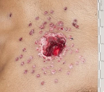
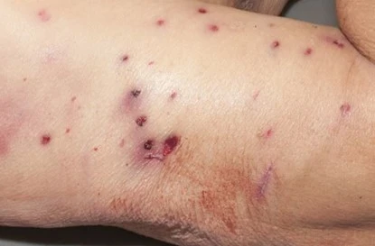
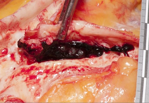
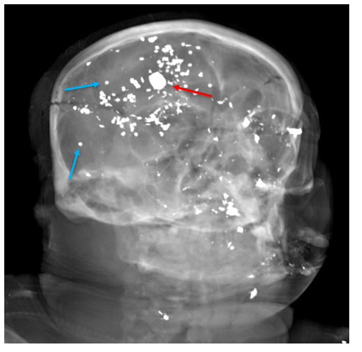
Emergency services were called to a fire in a small apartment building, in which the structure was completely engulfed. Most of the occupants had been evacuated – however, once the fire was extinguished, the charred remains of an adult woman were found in the debris.
At the autopsy of severely fire-damaged human remains, two key questions must be answered: 1) who is the decedent?, and 2) were they alive when the fire started?
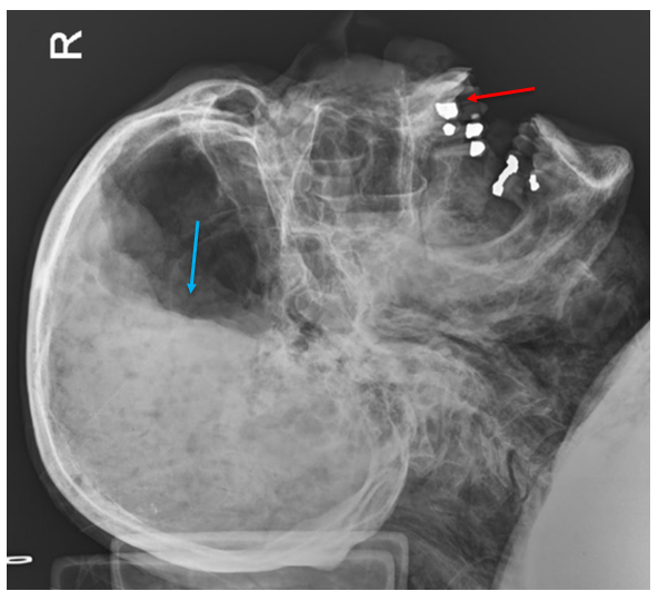
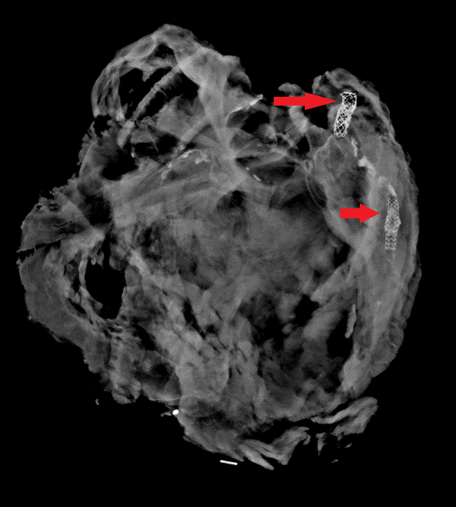
Question #1 is particularly relevant in this case, as many people lived in the building. Presumptive identification based on the tenant list may seem reasonable at first, but this victim could represent a visitor, contractor, or subletter. When facial identification isn’t possible, radiographic identification can be done with dental x-rays or x-rays of other bones which may have unique features from healed trauma or degeneration. Additional methods of positive identification could include fingerprints (if still intact), or DNA comparison to first degree relatives.
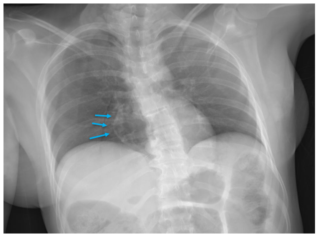
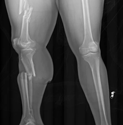
Question #2 is of importance because fire can be used in an attempt to disguise the identity of a victim of violent crime and destroy evidence. Cutaneous evidence of trauma may be disguised by burns, so full body x-rays are taken of every fire-damaged body. X-rays can also reveal retained bullets, knife tips, or fractures unlikely to have been caused by the fire.
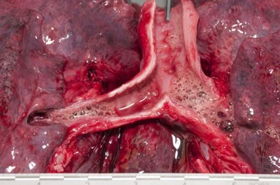
When deciding if a fire victim was alive when the fire started, we first examine the upper and lower airways for soot. Most fire victims do not die from cutaneous burns, but from smoke inhalation – including carbon monoxide (CO) toxicity, which is often apparent by cherry red discoloration of the blood and viscera. Postmortem carboxyhemoglobin measurements in house fire victims are typically greater than 50%. There are exceptions to this rule, of course. Rarely, someone who was clearly alive when the fire began will have minimal or no soot in their airways and a negligible carbon monoxide concentration. This can happen in a “flash fire”, such as one ignited by gasoline or oxygen tanks, in which thermal injury to the upper airway may cause rapid occlusion by laryngospasm or edema. People with underlying heart or lung conditions will be more susceptible to the effects of carboxyhemoglobin, and may not survive long enough to obtain a level above 50%. Fires also produce other toxic products of combustion such as cyanide, and can lower ambient oxygen saturations to result in asphyxiation by lack of ambient oxygen (even without CO).
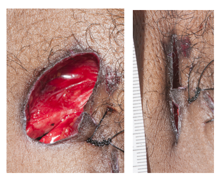
Forensic pathologists need to be aware of the artifacts that fires can create. Pugilistic posturing of fire victims (limb flexion) is due to heat-related contraction of muscle fibers. Epidural hematomas can result from boiling blood and bone marrow within the calvarium extravasating into the epidural space. The heat can induce fractures in exposed bone once the surrounding soft tissue is consumed or fully charred. Finally, the heat can split apart skin and soft tissue, resulting in sharp-force-like defects which occur parallel to the orientation of muscle fibers (rather than across them, which is more suspicious for penetrating trauma).
Of utmost importance in fire-related deaths, however, is scene investigation. The manner of death in fire fatalities is related to the origin of the fire. Most fire deaths are accidental, as the fire is unintentionally sparked by some electrical malfunction or unattended flame. However if the fire started intentionally, the manner of death can be homicide (if started by another) or suicide (started by the victim). It is therefore crucial to review the final fire investigation report before finalizing the autopsy report and death certificate.
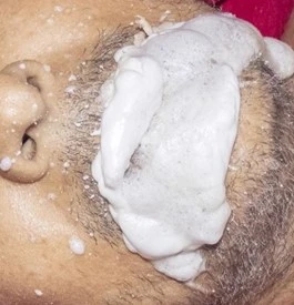
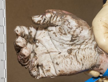
This image shows dark black soot lining the main and lobar bronchi; this indicates the victim was breathing during the fire.Heat-related epidural hematomas have a brown, amorphous appearance rather than the bright red color of traumatic epidural hematomas.The scalp has been consumed by fire, and the exposed bone is calcined and brittle with fractures of the outer table.
-Alison Krywanczyk, MD, FASCP, is currently a Deputy Medical Examiner at the Cuyahoga County Medical Examiner’s Office.
One of the most common questions I’m asked by family members is “do you know when they died?” If death occurs in the hospital, or is witnessed, the time of death isn’t controversial. It’s common though in forensics that people may not be found for hours, days, weeks, or more. Forensics television shows usually depict an investigator measuring body temperature at the scene, and then confidently declaring they’ve been dead for 44 hours. Unfortunately, there aren’t any existing methods that actually give that level of precision – but there is a way we can systematically approach the question.
When determining time of death (TOD), it’s most important to keep in mind that it will be an estimate. The estimate starts with the “window of death” – the time between when the decedent was last known alive and when their body was found. The smaller this window, the greater accuracy is possible.
Once the window is known, one can assess postmortem changes of the body. Livor mortis is the gravity-dependent settling of blood within vessels, which can appear as soon as twenty minutes after death. Sparing of lividity will be present in areas of pressure, such as parts of the body pressed against the floor or with tight clothing. Livor is initially blanchable, but after 8 to 12 hours blood extravasates from vessels and it becomes “fixed”. Clearly though, this only allows one to differentiate between ‘less than’ or ‘greater than’ 8 to 12 hours.
Rigor mortis (stiffening of the body after death) occurs because of postmortem ATP depletion. Muscle fibers require a supply of ATP to both contract and relax – once ATP levels are sufficiently low, muscle will remain contracted until the fibers are broken down by decompositional changes. Generally speaking, rigor starts to develop within an hour of death, peaks from 12 to 24 hours, and dissipates by 36 hours. However, these are average intervals. The onset of rigor is hastened by vigorous physical activity, seizures, electrocution, or increased body temperature, which preemptively deplete ATP. Rigor is also harder to detect in people with low muscle mass (e.g. infants), and can’t be assessed in frozen bodies with those with extensive thermal damage.
Cooling of the body after death, known as algor mortis, is similarly prone to interfering elements. One can find many formulas for estimating the time of death based on the temperature of the body – unfortunately, none of them are particularly useful because of the assumptions that must be made. Change in temperature after death is affected by numerous variables, including body habitus, clothing, wind, actual body temperature at the time of death (not many people are constantly at 98.6℉), sepsis, terminal seizures, and many others. If the environment is warmer than the body, the temperature can even increase after death.
I’ll briefly mention vitreous potassium measurement, which is probably the most recently discovered (and debunked) “holy grail” of time of death. Similar to algor and rigor mortis, vitreous potassium does a reasonably decent job predicting time of death in a controlled experiment – but in this line of work, people don’t tend to die in controlled environments.
At the end of the day, time of death is best estimated by thorough scene investigation, correlated with the evidence the body provides. Newspapers or mail not retrieved from the mailbox, expiration dates on perishable groceries, last refills of prescriptions, and unreturned text messages or phone calls can all narrow down the window of death.
As stated earlier, the longer the interval between death and discovery of the body, the more difficult time of death determination becomes. In advanced decomposition, there is no rigor, livor, or algor remaining to assess (there may even be scant residual soft tissue). In one such situation, despite months of a potential “window of death”, dates on unopened bills and crossed-off calendar dates helped us place the time of death within one or two days. It’s not as flashy as multivariate equations for temperature or potassium levels, but it’s far more accurate and scientifically defensible.
Image 1. The quilting pattern of this decedent’s mattress is visible in the livor mortis on his back.Image 2. This decedent’s right arm is defying gravity due to rigor – he was initially face down, and his arm musculature became temporarily fixed in this position. Rigor can be forcibly broken if needed, but will also break down as decomposition proceeds.
-Alison Krywanczyk, MD, FASCP, is currently a Deputy Medical Examiner at the Cuyahoga County Medical Examiner’s Office.
When I was about to complete residency in anatomic and clinical pathology, I was speaking with a colleague and mentioned I was pursuing dual fellowships in forensic and cardiovascular pathology. He furrowed his brow and asked, “What are you going to do with that?”
I was slightly surprised by this response, but he’s not the only person who would react that way. Many people (even pathologists) think of forensic pathology as gunshot wounds and motor vehicle accidents. While those deaths do come to our office, the majority of autopsies performed in the forensic setting are still due to natural causes, with heart disease making up a significant proportion. My interest in cardiovascular pathology was piqued when, as a medical student, I observed an autopsy on a healthy adolescent athlete who collapsed during a cross country race. The pathologist identified a congenital anomaly in his coronary arteries, in which the left coronary artery arose from the opposite cusp and traveled between the aortic and pulmonary arteries. This meant the coronary artery was susceptible to compression by the two surrounding, larger arteries, leading to ischemia and potential lethal arrhythmia whenever his heart rate became elevated. In another case, a relatively healthy young man had suddenly collapsed shortly after taking his first dose of prescribed azithromycin for a sinus infection. While the autopsy was macroscopically unremarkable, postmortem genetic testing revealed a likely pathogenic variant in a gene associated with long QT syndrome. In the context of the azithromycin (a drug known to prolong the QT interval), a lethal arrhythmia was triggered. His family was unaware of this heritable channelopathy, and they were urged to see a cardiologist themselves for a risk assessment.
These experiences made me see how our ability to detect and identify subtle cardiac disease at autopsy could have profound impacts on the emotional and physical well-being of families. It’s not news that pathology is facing a shortage of recruits, and both forensics and cardiovascular pathology are particularly feeling the squeeze. Unsurprisingly, these are both fields to which residents have very little exposure. Many residents don’t rotate through forensics until their 3rd year (after they’ve already chosen a specialty) and few academic centers have a specialized cardiovascular pathology service. The required number of autopsies to complete residency has now been decreased from 50 to 30, meaning residents see even less cardiovascular pathology during training. I can anecdotally add that myself and several other forensic pathologists I’ve met were occasionally discouraged from entering the field by academic mentors, who considered it a waste of potential. As a profession, we need to recognize the public health impact and academic worth of forensic autopsies and encourage residents’ exposure to the field. Not only is a well-trained forensic pathologist needed to accurately interpret injuries at autopsy, they are the front line in recognizing natural diseases that went undiagnosed prior to death. Additional cardiovascular training helps us to recognize potentially heritable cardiovascular disease; this not only helps families understand why and how their loved one died, but it also affords them the opportunity to obtain screening and interventional measures. It isn’t just natural deaths, either; people who died from any cause could have early signs of heritable disease, and overlooking them could mean disastrous consequences for the family. I would strongly encourage any pathology trainee with an interest in public and preventative health, molecular pathology, and non-neoplastic disease to consider combined training in forensics and cardiovascular pathology. The National Association of Medical Examiners offers free membership to trainees, and the Society for Cardiovascular Pathology offers a one-on-one mentorship program to introduce new members to the field – you will be a welcome addition to either or both groups! If you have specific questions you’d like to ask, I’m available at akrywanczyk@cuyahogacounty.us.
-Alison Krywanczyk, MD, FASCP, is currently a Deputy Medical Examiner at the Cuyahoga County Medical Examiner’s Office.
Image 1. Looks like this medical lab science blogger made quite the … shady… joke. CSI: Miami’s Lt. Horatio Caine (played by David Caruso) donned his shades at pivotal plot times. (Source: CBS)
Okay-okay, I couldn’t resist that. How many times have you
just wanted a CSI-style joke on here?
No? Just me? That’s fine…
Hello again everybody! Welcome back! Last month I talked a
bit about “Just
Culture,” a sort of bridge between the values we tout as clinical leaders
in our laboratories and the medical culture’s evolving and value-informed
paradigm shift. There was a little in there about the lessons paralleled in LMU
and the benefits of interdisciplinary teamwork. This month, on the subject of
interdisciplinary collaboration, I’d like to talk about our colleagues who often
are secluded or in more remote areas in our hospitals, offices, and academic
centers. Not here to stereotype; I’m talking about our friends in forensic
pathology!
Before I get there, let me go back a bit. I’ve already written several times about the stereotypes that surround our field of lab medicine and there are two times when that is glaringly present: when you’re a medical student or when you’re in forensics. I got the chance to meet someone who falls into both categories.
I’ve just finished up my OB/GYN rotation. But before my last
day, I went to the lab at our hospital and followed up on some pending biopsy
results. Okay, I can’t lie to you guys: they wanted me to see if I could rush
“my lab friends” to expedite the process of fixing, setting, cutting, staining,
and reading/reporting—because that’s
possible. So, I went to the lab and had a pleasant chat with the staff
explaining the situation and they were happy to help. While I was there,
however, I happened to see another short white coat (ironically from my same
school) who was helping some lab personnel with some grossing. Turns out she
wants to match into a pathology residency—just like me—and specifically was
interested in forensic path, a field which I don’t know much about. After talking
more, I asked if she’d like to share some information. Here’s my conversation
with Kyla Jorgenson, a 3rd year medical student at AUC-SOM from
Toronto, Canada:
I get lots of hassle
when I say I want to become a pathologist. People often ask me, “what’s your
back up choice” or “don’t you like patients?” It can be a challenge. What’s
your experience been like?
You want to do autopsies, so you want to be a
mortician, right? Not quite. Many times, I’ve been faced with blank stares when
I say I want to be a forensic pathologist. Other times I get the other end of
the spectrum, that’s so cool! Clearly, they’ve seen a few crime-shows and think
that I’ll get to go to crime scenes in stiletto high heeled shoes with a song
by The Who playing in the background as I arrive. Even today when talking with
a dermatopathologist I got a, “well when you realize that hanging out with
dead bodies every day isn’t the greatest, you might consider surg path.”
Then after hearing my experience as an autopsy assistant and that I’m sure this
is what I want to do it was the resigned sigh signalling that I was a lost
cause already.
A “lost cause,” that’s
frustrating. A lot of specialities rag on other ones, it seems to be part of
the culture of medicine—hopefully not forever, but still can’t we all just get
along?
So, my background
leading to pathology involved me working for several years between college,
graduate school, and medical school; in hospitals of various sizes. I have
personal experiences in these fields and sort of feel “at home” when I’m
dealing with hematopathology, transfusion medicine, cell therapy—that sort of
thing. What piqued your interest in forensics?
I started my undergraduate degree in forensic
biology at the University of Toronto in the fall of 2008 just as a major review
of pediatric forensic pathology in Ontario was being released. After numerous
issues came to light, the inquiry looked at policies, procedures, practices,
accountability and oversight mechanisms, quality control measures and
institutional arrangements within the field in Ontario from 1981 to 2001. Ontario
Court of Appeal’s Honourable Justice Stephen T. Goudge developed 169 recommendations
on how pediatric forensic pathology in Ontario needed to address and correct
its systemic failings to restore public confidence.
After studying the cases that prompted the
inquiry and its recommendations in class, what left the greatest impression was
the importance of having medicolegal autopsies performed by those trained in
not just pathology, but specifically, forensic pathology. What I took away from
the cases of accidental deaths falsely attributed as homicides due to lack of
experience on behalf of the pathologist and other such issues, is that forensic
pathology isn’t something to be dabbled in. While our patients are no longer
alive, there are lives that can be affected by the work we do. In Ontario,
false convictions not only stemmed from “junk science” but also from
inadequacies in the training of pathologists working in a forensic capacity and
also a general shortage of forensic pathologists.
Seems like a lot of us
(of the few of us) who enter medical school knowing we want to go into
pathology have to sort of wait their turn, as it were, collecting experiences
which help make us competitive for residency matching—what keeps your
“commitment algorithm” going?
Since discovering that forensic medicine is a
career path as a high school student, I’ve geared my education towards training
in forensics. First my undergraduate degree and then a side trip for my master’s
degree in Forensic Death Scene Investigation and a job as a pathology
technician at the Medical Examiner’s office on my way to medical school. I have
in each step along the way, confirmed that both medicine and forensics
fascinate me. Scroll through my Netflix account and you’ll find crime dramas
(with the British shows being my favourite) or my podcast app filled with true
crime shows; I am enraptured using science to figure out what happened.
Sidebar: at this point Kyla showed me a first-author published piece in
the Journal of Forensic Sciences from
2017 that talked about law enforcement-involved firearm related deaths in
Oklahoma, where she worked at the time. Basically, it showed through metadata
analysis that gun-related deaths were on the rise. Not just over time, but
number of times being shot. Remember when we talked about pathology’s role in
the #StayInYourLane/#ThisIsOurLane discussion? Well which pathology
speciality do you think works with this stuff directly? Chemistry? Cytology?
Last time I checked GSWs don’t get screened for lead poisoning and you can’t
FNA a bullet. Forensic pathology has often been tasked with seeing trends in
morbidity and mortality and translating that to effective social and public
health change: think seatbelts, stents, and maybe someday gun-related
legislation changes.
Image 2a. Monthly aggregates of gun-related deaths over a 16-year period in OK. (Source: Jorgenson, K et al (2017) Trends in Officer-Involved Firearm Deaths in Oklahoma from 2000-2015, Journal of Forensic Sciences, doi: 10.1111/1556-4029.13499)Image 2b. Number of gun shot wounds per victim over time. (Source: Jorgenson, K et al (2017) Trends in Officer-Involved Firearm Deaths in Oklahoma from 2000-2015, Journal of Forensic Sciences, doi: 10.1111/1556-4029.13499)
I was interested when
I shadowed at the Cook County ME’s office a few years ago—I saw some cool
things. I also remember learning a lot from the first real autopsy I saw in a
hospital, ultimately it seems like a totally different field that maybe gets
underappreciated even within the pathology umbrella. AP/CP residents have to do
a certain number of autopsies to graduate, but the attitude I’ve noticed around
the topic is a “necessary evil” and most are working towards not having to do
that. So let me ask you definitively, why forensic pathology?
Medicine is science being applied to find out
what happened in the body and how we can change or manipulate those variables
to diagnose, prevent, treat and manage disease. Each diagnosis is solving a
crime occurring within the cells in the body, if you will. In forensic
medicine, not only do you get to do all that but add in the crime solving
element and you get to be “Dr. Nancy Drew.” While medicolegal systems are
different all over the US and Canada, chances are that as a forensic
pathologist you won’t only be working on your stereotypical
“forensics” cases, the gunshot wounds, stab wounds and other
nefarious causes of deaths many associate with that term. You could get the
generic, “cause of death atherosclerotic cardiovascular disease, manner of
death natural,” for a large proportion of cases.
It’s not glamorous, you could spend your day with a two-week-old decomposing decedent that has a pulsating maggot mass devouring its torso or documenting 51 stab wounds or signing out your cases after reviewing your histology and toxicology reports or testifying on a homicide case you worked on. But for me, those all sound like pretty interesting ways to spend the day, sign me up. As a pathology technician assisting with the autopsies and external exams, I was never required to think about what was happening in the body, but I wanted to understand it all. Now as I progress through medical school and look towards residency and fellowship, I eagerly await the chance to perform my first autopsy as a physician, to put all the knowledge and experience I’ve gained towards helping move Ontario and forensic pathology forward.
Image 3. Kyla M. Jorgenson is a 3rd year medical student at the American University of the Caribbean School of Medicine with prior undergraduate and graduate studies in the field of forensic pathology, professional experience as an autopsy technician, as well as a vested interest in pursuing a career in the field moving forward in residency and fellowship. (Source: Kyla M. Jorgenson)
I’d like to thank Kyla for her time in talking with me and
her willingness to share her insights with all of you. I wish her all the best
of luck as she continues through her training with electives and core rotations
both in the UK and state-side. If you have any questions to relay to her,
please feel free to comment below and I will forward appropriately. And as
always, don’t forget to share with your colleagues across every discipline!
Thanks for reading, I’ll see you next time where I’ll be
writing from the Mayo Clinic Hospital in Rochester, Minnesota, conducting a
formal rotation in Anatomic and Clinical Pathology! Don’t miss it, I’ll have
lots to share while learning at one of the nation’s top institutions!
Until next time!
–Constantine E. Kanakis MSc, MLS (ASCP)CM graduated from Loyola
University Chicago with a BS in Molecular Biology and Bioethics and then
Rush University with an MS in Medical Laboratory Science. He is
currently a medical student actively involved in public health and
laboratory medicine, conducting clinicals at Bronx-Care Hospital Center
in New York City.