Microbiology Case Study: Cryptococcal Meningitis In Immunocompromised Patient

Case History

A 47-year-old male originally from Dominican Republic, with a recent diagnosis of acquired immunodeficiency syndrome (AIDS) and diffused large B cell lymphoma (DLBCL), was admitted because of seizures and a rapidly increasing left neck mass. ​MRI of the brain showed a 2.5 x 1.2 cm (about 0.47 in) lesion in the left inferior parietal lobe – (1.4×0.7cm) in the right frontal lobe, plus multiple scattered bilateral lesions. Because of this, he underwent craniotomy/craniectomy for possible resection. A biopsy was taken from the right temple lesion and sent for aerobic, anaerobic, fungal, mycobacterial culture, surgical pathology and Toxoplasma PCR (Polymerase Chain Reaction). ​

Gram stains, KOH prep, acid-fast stains, and Toxoplasma PCR of the tissue were all negative. Aerobic and anaerobic cultures did not show any growth.

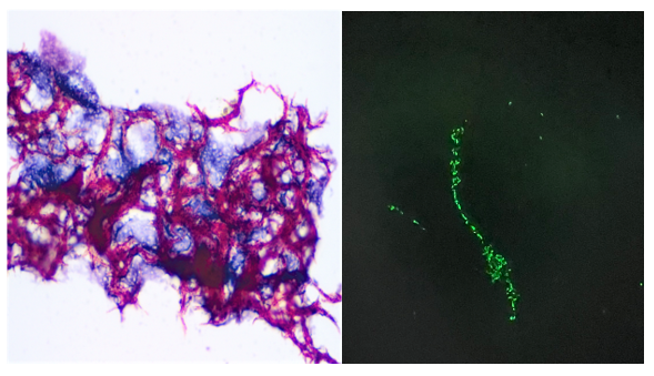
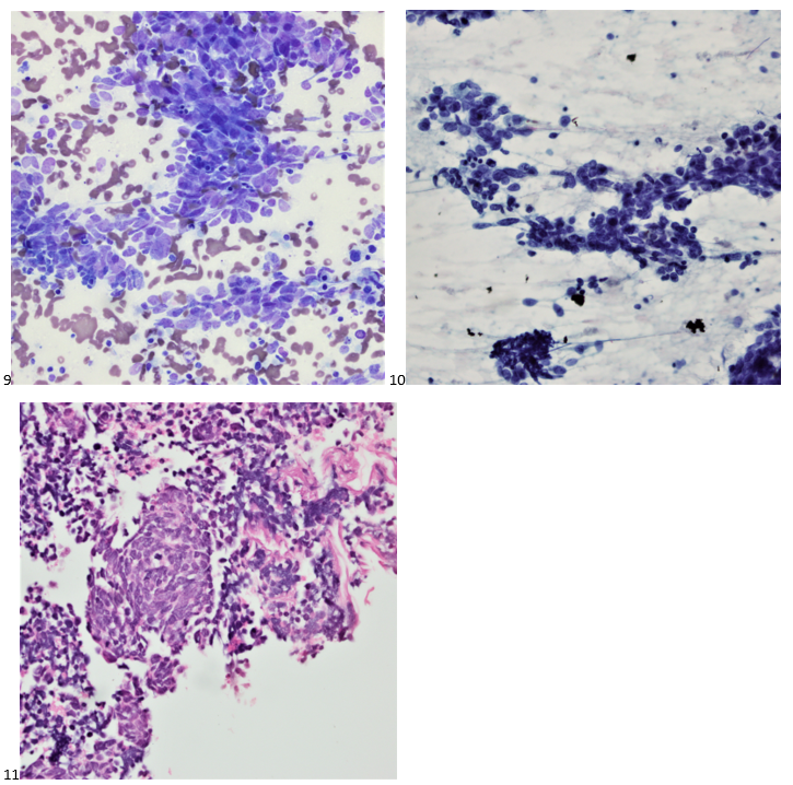
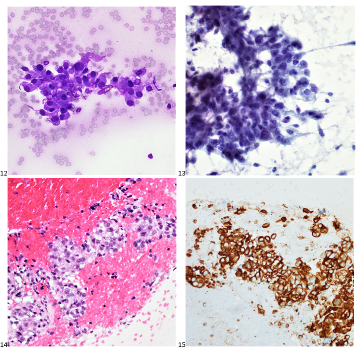
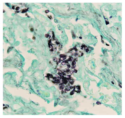
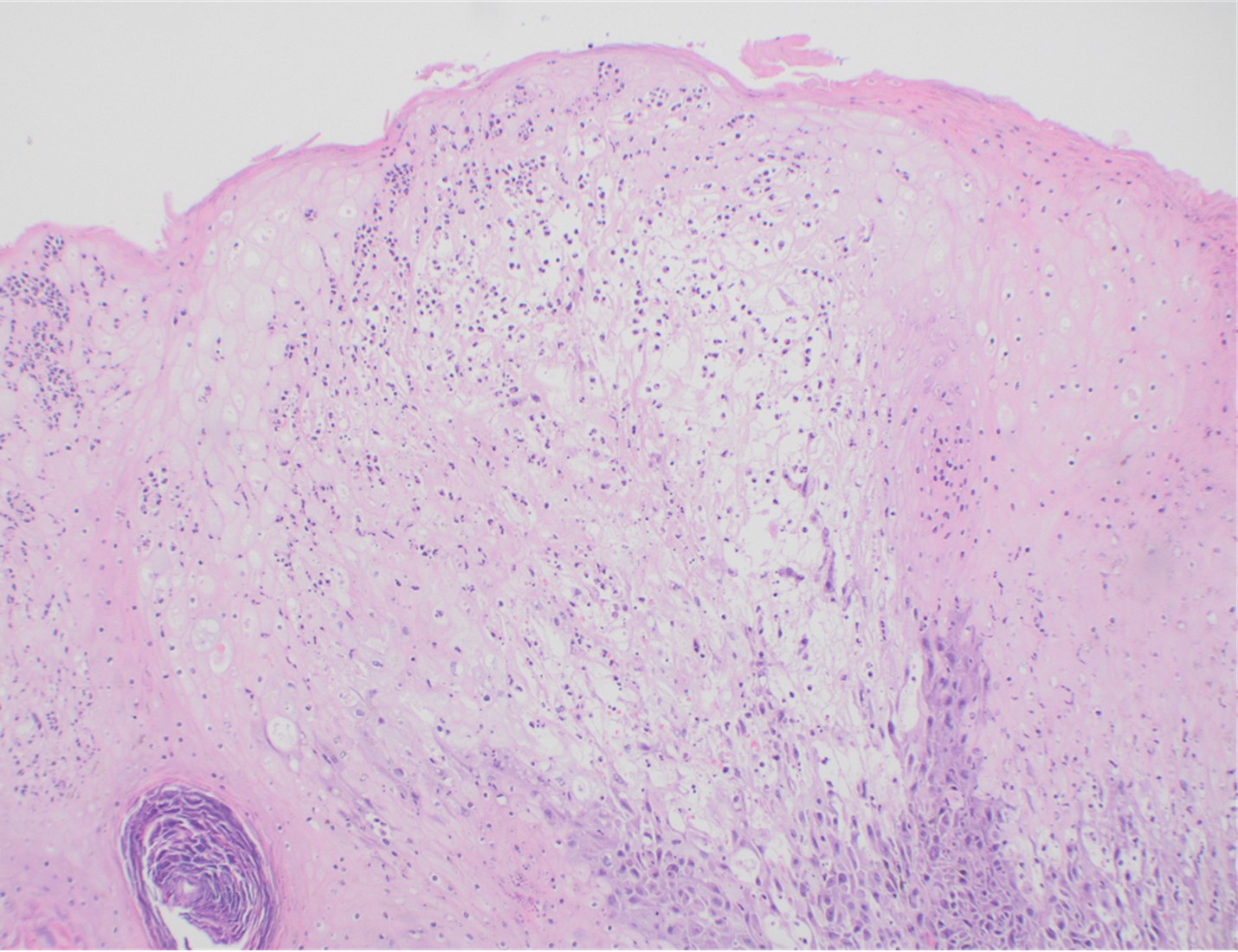
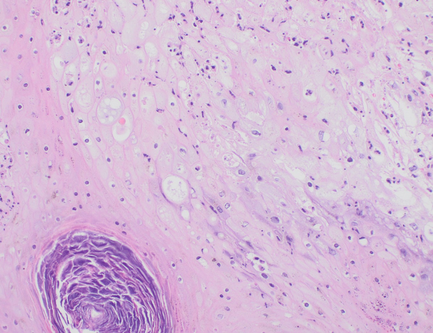
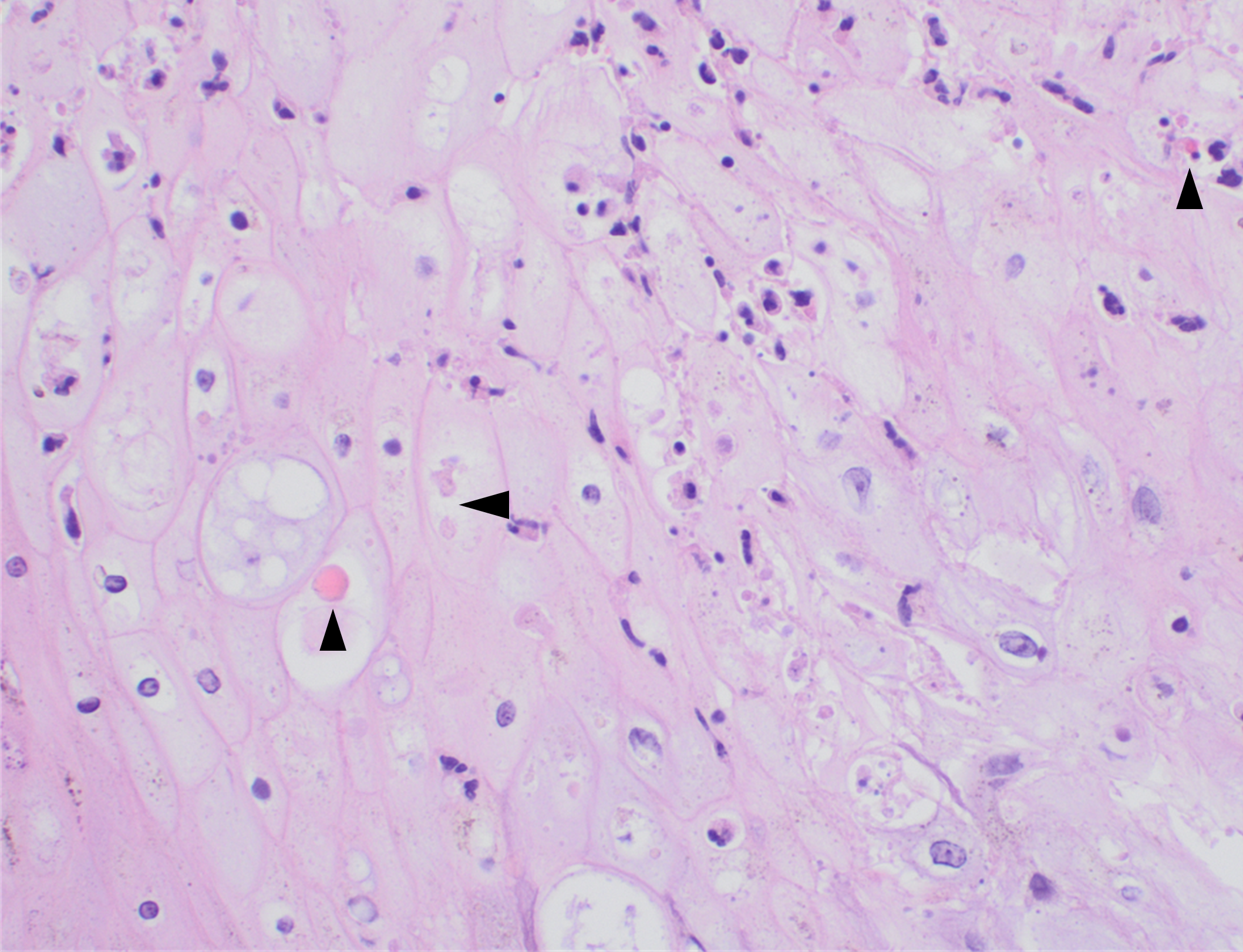
Histopathology slides (GMS and H&E stains in Fig A and B) show budding yeasts morphologically consistent with Cryptococcus. Mucicarmine stain was also positive. Lumber puncture was performed the next day and Cryptococcal antigen was positive, with a titer of 1:640. Interestingly, the CSF culture and Gram stain did not reveal any organisms.

Figure A. H&E shows encapsulated variably sized transparent/gray color yeasts with thin walls. Black arrows show organisms.
Figure B. GMS stains highlight very faint staining of capsule (black arrow). Yellow arrow highlights background inflammatory cells.

Discussion

Among several species of Cryptococcus, C. neoformans and Cryptococcus gattii are pathogenic, with C. neoformans causing meningitis in immunocompromised patients worldwide whereas C. gattii has a preference for immunocompetent individuals.1 Cryptococcal disease remains a major opportunistic infection and a leading cause of mortality in patients infected with HIV in much of the developing world. Most HIV-related meningitis cases are caused by Cryptococcus neoformans.2

Cryptococci are found in soil, due to contamination with pigeon droppings. The infection occurs through inhalation, with or without symptoms of pneumonia, with subsequent dissemination to the central nervous system (CNS) via blood. Imaging findings are often unspecific or negative. CT or MRI examination of the central nervous system is performed to rule out alternative diagnoses. The diagnosis of ‘meningitis’ is made with a lumbar puncture, which typically shows lymphocytosis, an increased protein and decreased glucose concentration. Few neutrophil granulocytes are often found in CSF. This is likely because neutrophil migration is inhibited by specific polysaccharides that are part of the cryptococcal capsule.3

Cryptococci can be seen directly in the sediment of centrifuged CSF stained with India ink. The sensitivity and specificity of India ink is poor; therefore, CSF Gram stain and culture, multiplex meningitis/encephalitis PCR, and lateral flow antigen (LFA) tests have replaced the use of India ink. The cryptococcal lateral flow antigen test should be performed in CSF and serum, in addition to Multiplex ME PCR panel, and is a preferred test because of high sensitivity (93-100%) and specificity (93-98%).4

High organism burden at baseline (indicated by quantitative CSF culture or CSF antigen titre) and abnormal mental status are the most important predictors of death, while high opening pressures and a poor inflammatory response in the CSF have also been associated with poor outcome.5

On H&E it has the characteristic appearance ofencapsulated variably sized yeasts (2-20 microns) with thin walls which can be highlighted with the GMS stain. Although the presence of a capsule differentiates Cryptococcus from Histoplasma capsulatum and Blastomyces dermatitidis with the H&E or mucicarmine stain, additional confirmation can be made with Fontana-Masson stainin the absence of capsules.6 Since both C.neoformans and C. gattii produce melanin, the pathology report by FM silver or H&E/GMS stain cannot further distinguish these two closely resembled species.

Occasionally, cryptococcal meningitis cases with sterile CSF culture and/or negative Cryptococcal CSF antigen are observed in HIV individuals, regardless of the CD4 counts.7,8 However, serum Cryptococcal antigen and blood culture may be positive in those individuals.7 In our case, the diagnosis of Cryptococcal meningitis was made by the pathology report and positive CSF Cryptococcal antigen.

-Fnu Sapna is a 2ndyear AP/CP pathology resident in the Department of Pathology at Montefiore Medical Center in Bronx, NY. She completed her Medical education at Chandka Medical College in Pakistan. Her interests are putting efforts to improve screening guidelines for diagnosis of preventable gynecological and breast cancers.

-Phyu Thwe, Ph.D, D(ABMM), MLS(ASCP)CM is Associate Director of Infectious Disease Testing Laboratory at Montefiore Medical Center, Bronx, NY. She completed her medical and public health microbiology fellowship in University of Texas Medical Branch (UTMB), Galveston, TX. Her interests includes appropriate test utilization, diagnostic stewardship, development of molecular infectious disease testing, and extrapulmonary tuberculosis.

One year of Chronic Cough after Past Resolution of Viral and Bacterial Respiratory Infections

Case History

A 74-year-old female from El Salvador with a medical history of hypertension, diabetes, osteoarthritis presented with persistent productive cough. The patient states that the symptoms started on November 2022 which was believed to be associated with ongoing COVID-19 infection. Despite recovery from COVID-19 infection, significant productive cough still remained. A visit to her primary care doctor revealed streptococcal throat infection and despite completing a course of Zithromax antibiotics, a subsequent chest x-ray revealed potential right upper lobe pneumonia and she reported productive cough with occasional blood streaks. Blood work up in the Emergency Department revealed leukocytosis (20.22 x10e3/mcL) with neutrophilia (14.56 x10e3/mcL neutrophils) and monocytosis (3.03 x10e3/mcL monocytes). A chest x-ray showed bilateral reticular and airspace opacities with an air-fluid level containing opacity overlying the left mid lung, likely representing a cavitary lesion. A follow up with a computerized tomography of the chest identified innumerable randomly distributed pulmonary nodules or cavities with upper lobe predominance. The largest cavity measures approximately 6.6 cm with air-fluid levels and debris. Additionally, a small pericardial effusion with thickened pericardium was also noted. A sputum sample was submitted for Acid-fast bacilli (AFB) culture and molecular testing. AFB stain was positive for acid-fast bacilli (Figure 1). The GeneXpert MTB/RIF assay detected M. tuberculosis with no rifampin resistance marker. Growth on the Lowenstein-Jensen agar after two weeks showed buff colored rough and dry colonies and was confirmed as M. tuberculosis on the MALDI-TOF.

Figure 1. Visualization of acid-fast bacilli directly from patient specimen. Using acid-fast stain, cording was observed (Figure 1A, left) and using fluorescent staining, fluorescent rods were observed (Figure 1B, right).

Discussion

Mycobacteria are aerobic, nonmotile, thin rod shape, non-spore forming bacilli that possess mycolic acid in its cell wall giving its acid-fast stain characteristics. Tuberculosis (TB) is an airborne disease caused by Mycobacterium tuberculosis (MTB). TB remains to be the leading cause of death from a single infectious agent worldwide. According to the CDC a total of 1.6 million people died from TB in 2021 (including 187, 000 people with HIV) 1,2. The M. tuberculosis complex (MTBC) includes M. tuberculosis, M. bovis, M. bovis, Bacille Calmette-Guérin strain (BCG), M. caprae, M. pinnipedii, M. mungi,M. africanum, M. microti, and M. canettii 3. M. tuberculosis produces cord factor, a glycolipid which is also known as trehalose dimycolate, that causes the bacteria to grow in parallel strands and that appeared like cord, or rope when cultured in liquid media (Figure 1A). The cord factor is present in the outer envelope and protects the bacteria from the host response 4.

The pathogenesis of human tuberculosis involves a complex interaction between host immune system and bacterial factors 4. M. tuberculosis is carried in airborne particles generated by infected individuals. The droplet nuclei traverse the mouth or nasal passages, upper respiratory tract, and bronchi to reach the alveoli of the lungs. The bacteria are then phagocytosed by alveolar macrophages and can inhibit maturation of phagosome and block formation of phagolysosome, allowing its unchecked replication in the macrophage which results in bacteria proliferation in the alveolar macrophage and air spaces. In immunocompetent hosts, the immune response (via TLR2, TLR9, Th-1 and IFN- ꝩ cascades) may contain the infection before significant tissue destruction or systemic illness 1-4. However, in the immunosuppressed hosts, the primary infection results in a broad clinical spectrum such as meningitis, miliary tuberculosis and extrapulmonary granulomas. Post-primary/secondary tuberculosis (reactivation TB) usually begins months to years after the establishment of systemic immunity in primary TB mostly in a period when the host immune response is weakened, following exogenous or a large inoculum of virulent bacilli overwhelming the host immunity system. Extrapulmonary manifestations will develop based on the organ system affected 5.HIV infection is the greatest risk factor for reactivation of TB as the virus causes functional abnormalities in CD4+ T cells and CD8+ T cells which confer protection against active TB. Other risk factors that promote the reactivation TB include aging, malnutrition, diabetes mellitus, renal failure, cancer and immunosuppressive therapy 5. The disease typically affects the lungs (pulmonary TB) but can affect other sites as regional lymph nodes, apex of the lung, larynx, kidneys, brain, bone, joints and pleura 1,2,5.

Diagnostic tests for TB detect either the bacteria or host immune response. Specimens recommended for diagnosis of mycobacterial infection are sputum, bronchial brushing/washings /biopsies, gastric aspirates (children) urine, blood, CSF, BM, body fluids, stool (only in HIV) 5,8. Specimens from sputum and other nonsterile sites should be liquefied with N-acetyl-L-cysteine and decontaminated with NaOH and for gastric aspirate neutralized with buffer 6-8. For diagnostic purposes, all persons suspected of having TB disease at any site should have at least three consecutive sputum specimens collected in 8 to 24 hours with at least one being an early morning one for AFB smear and culture 1,8.

The organisms can be visualized under a microscope using two principal methods: carbolfuchsin staining (e.g., Acid-fast stain), or using a fluorochrome (auramine-rhodamine and auramine-O stains) procedure (Figure 1B). Microscopy is the most rapid diagnostic method for the detection of tubercle bacilli but is less sensitive; it requires a minimum of 10,000 bacilli/mL of sputum to produce a positive result. Culture is the gold standard and more sensitive method for the detection of tubercle bacilli and is necessary for performing antimicrobial drug drug-susceptibility testing and genotyping. 6,9 However culture requires 3–6 weeks for growth which delays the initiation of anti-tuberculosis drug therapy. Two types of solid media are used for mycobacterial culture: egg based (Löwenstein-Jensen) and agar based (Middlebrook 7H10, 7H11, and selective 7H11). Colony morphology of Mycobacterium tuberculosis on solid media are dry, rough, raised, wrinkled, off white to buff colored. M. tuberculosis is commonly positive for niacin, nitrate reduction test, pyrazinamidase test, but negative for 68C catalase, tween 80 hydrolysis, and 5% NaCl tolerance. Molecular techniques such as nucleic acid amplification tests revolutionized tuberculosis diagnosis since M. tuberculosis nucleic acid material can be detected directly from specimen in less than 2 hours. Two commercial NAATs for the detection of M. tuberculosis complex are available in the United States: The Amplified MTD (Mycobacterium Tuberculosis Direct) test (Hologic, Marlborough, MA) and the Xpert MTB/RIF (Cepheid, Sunnyvale, CA).

Common serological approaches for detection of M. tuberculosis are the Tuberculin skin test (TST) or IFN- ꝩ release assays (IGRA’s) 1,9,10. The tuberculin skin test is performed by intracutaneous injection of purified protein derivative of M. tuberculosis, which induces a visible and palpable induration that peaks in 48 to 72 hours. A false-positive tuberculin skin test may result on individuals with prior vaccination with BCG (Bacillus Calmette-Guerin), an attenuated strain of M. bovis. BCG immunization does not affect the test result of IGRA assay. The IGRA are blood tests that measure a person’s immune reactivity to M. tuberculosis. Both T-Spot and QuantiFERON can aid in diagnosis M. tuberculosis but do not differentiate latent infection from tuberculosis disease. IGRAs are in vitro tests that measure the IFN- γ production by T cells responding to stimulation with specific TB antigens ESAT-6, TB7.7, and CFP-10, which are not present in the M. bovis strains. Results can be interpreted both qualitatively (positive, negative, or indeterminate) and quantitatively.

The regimen currently recommended for treatment of TB is isoniazid, rifampin, ethambutol, and pyrazinamide. The initial M. tuberculosis isolate should be tested for resistance to first-line medication. Second-line drug susceptibility testing should be limited to specimens from patients who have prior TB disease treatment, contact with a patient with known TB drug resistance or positive cultures after more than 3 months of treatment 10. Multidrug-resistant TB (MDR TB) disease is defined as resistance to isoniazid and rifampin, and Extensively drug-resistant TB (XDR TB) is characterized with resistance to isoniazid and rifampin, any fluoroquinolone, and at least one of three injectable second-line drugs (i.e., amikacin, kanamycin, or capreomycin). The duration of therapy depends on the drugs used, the drug susceptibility test results, and the patient’s response to therapy. Most patients are started with a 6-month regimen plan. A difficult challenge to M. tuberculosis treatment is patient compliance with lengthened therapy. Without treatment mortality rate for tuberculosis is more than 50% 11.

References

  1. World Health Organization, Global TB Programme. Global tuberculosis report 2022. 2022 November 19. https://www.who.int/publications/i/item/9789240061729
  2. Kamholz, S. L. 1996. Pleural tuberculosis, p. 483-491. In W. N. Rom and S. Garay (ed.), Tuberculosis. Little, Brown and Co., Boston, Mass.
  3. Yanti, B., et al. The role of Mycobacterium tuberculosis complex species on apoptosis and necroptosis state of macrophages derived from active pulmonary tuberculosis patients. BMC Res Notes. 2020; 13: 415. doi: 10.1186/s13104-020-05256-2
  4. Smith, Issar. Mycobacterium tuberculosis Pathogenesis and Molecular Determinants of Virulence Clin Microbiol Rev. 2003 Jul; 16(3): 463–496. doi: 10.1128/CMR.16.3.463-496.2003
  5. Wells, C.D, et al. HIV infection and multidrug-resistant tuberculosis: the perfect storm. J Infect Dis. 2007 Aug 15;196 Suppl 1:S86-107. doi: 10.1086/518665.
  6. Dunn, J.J., Starke, J.R., Revell, P.A. Laboratory Diagnosis of Mycobacterium tuberculosis Infection and Disease in Children. J Clin Microbiol 2016 Jun;54(6):1434-1441. doi: 10.1128/JCM.03043-15.
  7. Parashar D, Kabra S, Lodha R, Singh V, Mukherjee A, Arya T, Grewal H, Singh S. 2013. Does neutralization of gastric aspirates from children with suspected intrathoracic tuberculosis affect mycobacterial yields on MGIT culture? J Clin Microbiol 51:1753–1756.
  8. Clinical and Laboratory Standards Institute. 2008. Laboratory detection and identification of mycobacteria; approved guideline—1st edition. CLSI document M48-A. Clinical and Laboratory Standards Institute, Wayne, PA.
  1. Pai M, Nicol MP, Boehme CC. Tuberculosis Diagnostics: State of the Art and Future Directions. Microbiol Spectr 2016; 4.
  2. Dheda, K. et al. The epidemiology, pathogenesis, transmission, diagnosis, and management of multidrug-resistant, extensively drug-resistant, and incurable tuberculosis. Lancet Respir Med. 2017 Mar 15;S2213-2600(17)30079-6.
  3. World Health Organization. Tuberculosis Fact Sheet. 21 April 2023. https://www.who.int/news-room/fact-sheets/detail/tuberculosis

-Dr. Carla Ayala-Soriano was born and raised in Bayamon, Puerto Rico. She attended Universidad Autonoma de Guadalajara School of Medicine where she received her doctorate degree. She completed a Bachelor of Science in Biology at the University of Puerto Rico. She spent an additional year completing a Post Bachelor Certificate in Cytotechnology. Her academic interests include Cytopathology and Gynecologic Pathology. In her spare time, Dr. Ayala-Soriano enjoys cooking, traveling, listen to music, and outdoor activities. She is pursuing AP/CP training.

-Rebecca Yee, PhD, D(ABMM), M(ASCP)CM is the Chief of Microbiology, Director of Clinical Microbiology and Molecular Microbiology Laboratory at the George Washington University Hospital. Her interests include bacteriology, antimicrobial resistance, and development of infectious disease diagnostics.

Thyroid Tales, Part 2

Typically, our patients present to the endocrinology clinic after their thyroid nodules are incidentally found on staging or surveillance after being diagnosed with a primary cancer in another part of the body. Based on TI-RADS criteria, the clinician either monitors the nodule or refers the patient to radiology for a thyroid FNA. When we hear “thyroid nodule,” we rarely assume anything other than thyroid tissue. Whether the imaging favors benign or suggests a high risk of malignancy, we prepare ourselves to assess the FNA smears for follicular cells (and all the levels of atypia), colloid, macrophages, Hurthle cells, lymphocytes, etc. While we must keep an open mind, we are always caught off guard when we see anything other than thyroid-related cells. So as promised in the first edition of this post, here are four thyroid FNA cases with unsuspecting findings.

Case 1

A 47-year-old male presented with throat dysphagia and odynophagia. CT scan revealed a destructive mass within the thyroid gland with compression and invasion of the thyroid cartilage and seemed contiguous with a large pharyngeal mass, spanning approximately 8 centimeters. A follow-up PET scan noted multiple hypermetabolic thyroid masses within both lobes, direct invasion of the subglottic trachea and upper esophagus, and mediastinal lymphadenopathy.

FNA passes were obtained from the right lobe of the palpable thyroid mass.

Images 1-2. Thyroid, Right Lobe, FNA 1: DQ-stained smear; 2: Pap-stained smear.

Smears (Images 1 & 2) revealed poorly differentiated neoplastic cells, follicular cells, and colloid (not visualized). No features of papillary thyroid carcinoma, medullary carcinoma, or Hurthle cell neoplasm/carcinoma were identified.

Immunohistochemical stains performed on cell block sections showed the poorly differentiated neoplastic cells to be negative for thyroglobulin, TTF-1, and calcitonin; Follicular cells, which may probably be differentiated neoplasm, were positive for thyroglobulin, TTF-1, and negative for calcitonin. Unfortunately, the scant cellularity in the cell block specimen precluded additional stains. Giant cell and spindle cell features were not identified in this specimen. Morphological features are compatible with poorly differentiated carcinoma of the thyroid gland; however, metastasis from other sites cannot be excluded.

The patient then underwent a total laryngectomy and thyroidectomy (Images 3 & 4) and level IV neck dissection, bilateral modified radical neck dissection, and tracheostomy with reconstruction performed. The patient then underwent adjuvant radiation followed by palliative re-irradiation and chemotherapy after abnormal activity was noted throughout the neck. Treatment was discontinued due to severe disease progression.

Images 3-4. Thyroid, Thyroidectomy: 3: H&E section (200X); 4: H&E section (600X).

Final diagnosis: Poorly differentiated thyroid carcinoma with squamous differentiation arising in association with differentiated follicle derived carcinoma cells.


Case 2

A 68-year-old female presented with a 3.7 centimeter left lobe-filling thyroid nodule and a history of melanoma of the left anterior tibial region that was excised a decade prior. During that time, a sentinel lymph node biopsy identified microscopic metastasis. Seven years after her initial diagnosis, the patient underwent an excision of a right upper quadrant subcutaneous nodule, demonstrating metastatic melanoma. Three months after that excision, the patient had a low anterior resection of a rectosigmoid metastasis. A breast lesion was then identified five months later, and the patient underwent a mastectomy for melanoma involving the breast. Six months after her mastectomy, the patient had a segmental resection and excision of a left posterior thigh nodule, at which point she was enrolled in a clinical trial. The next month, four additional subcutaneous nodules were excised on the left thigh, calf, and arm. After 2 years of relatively stable disease, the patient underwent a partial gastrectomy, partial small bowel resection, and left lower extremity mass for recurrent melanoma. The last PET avid area to biopsy was the left-lobed thyroid nodule. Under ultrasound guidance, multiple FNA passes of the solid and hypervascular thyroid nodule. The smears (Images 5 & 6) and cell block (Image 7) featuring single cells with eccentric nuclei and prominent nuclei are presented below.

Images 5-8. Thyroid, Left Lobe, FNA 5: DQ-stained smear; 6: Pap-stained smear; 7: H&E Cell Block section (600X); 8: A103-positive.

Immunostains were performed on cell block sections, and the neoplastic cells are positive for A103 (Image 8), HMB45 (scattered cells), and SOX-10, while negative for CD45, TTF-1, thyroglobulin, and calcitonin.

The patient began treatment with temozolomide and completed 33 cycles of pembrolizumab. Her most recent metastasis demonstrated extensive tumor cell necrosis, and disease progression has slowed tremendously.

Final diagnosis: Melanoma.


Case 3

After developing sudden shortness of breath and chest tightness, a 40-year-old female patient presented to the emergency department. A large mediastinal mass compressing the heart and central structures in the chest was identified on CT scan. Two thyroid nodules were also noted during that time. The patient underwent a mediastinal biopsy, which demonstrated small cell lung cancer, and the patient underwent thoracic radiation and six cycles of chemotherapy, as well as whole brain radiation. Two years later, the patient established care with endocrinology for her 1.6 centimeter solid left lobe thyroid nodule and a 1.2 cm complex thyroid nodule in the right lobe. While the right nodule was consistent with a hyperplastic nodule, the smears and cell block of the left thyroid nodule are presented below (Images 9-11).

Images 9-11. Thyroid, Left Lobe, FNA 9: DQ-stained smear; 10: Pap-stained smear; 7: H&E Cell Block section (400X).

Immunohistochemical stains performed on cell block sections demonstrate the neoplastic cells were positive for TTF-1, AE1/AE3, synaptophysin, and CD56.

The patient then completed four subsequent cycles of chemotherapy with concurrent chemoradiation and is currently on active surveillance showing no evidence of disease for over 12 months.

Final diagnosis: Metastatic small cell carcinoma.


Case 4

A 59-year-old female presented to her primary care physician for gross hematuria and fatigue. Her thyroid workup demonstrated hypothyroidism on her thyroid function panel and a 2.3 centimeter solid and hypervascular thyroid nodule in the right lobe. Her urology workup revealed a 6.7 centimeter exophytic left kidney mass, and the follow-up CT scan identified a lytic lesion in the right iliac bone. The thyroid biopsy was performed in the endocrinology clinic while she was also establishing care with the urologic oncology team the same day. The smears and cell block specimen from multiple FNA passes are presented below (Images 12-14).

Images 12-15. Thyroid, Right Lobe, FNA 12: DQ-stained smear; 13: Pap-stained smear; 14: H&E Cell Block section (400X); 15: Vimentin-positive.

Immunocytochemical stains were performed on paraffin sections of the cell block. Tumor cells show positive staining for vimentin (Image 15), focal staining for e-cadherin, and negative staining for CK7, TTF-1, thyroglobulin, CD10, and RCC.

The patient was referred to radiology for a CT-guided biopsy of the lytic bone lesion, which demonstrated similar cells. The patient had a radical left nephrectomy, followed by sunitinib. The thyroid nodule was not responding to treatment, so they patient underwent a total thyroidectomy, which showed metastatic high-grade clear cell carcinoma with sarcomatoid progression, consistent with renal primary. In some areas, the thyroid follicles were proliferating and appear atypical, probably reactive to the metastatic carcinoma. A checkpoint inhibitor was added to the patient’s therapy, but the disease continued to progress, and the patient elected for palliative care.

Final diagnosis: Poorly differentiated carcinoma, consistent with metastatic renal cell carcinoma.

That’s a wrap! Stay tuned for the next series of cytology case studies!

-Taryn Waraksa-Deutsch, MS, SCT(ASCP)CM, CT(IAC), has worked as a cytotechnologist at Fox Chase Cancer Center, in Philadelphia, Pennsylvania, since earning her master’s degree from Thomas Jefferson University in 2014. She is an ASCP board-certified Specialist in Cytotechnology with an additional certification by the International Academy of Cytology (IAC). She is also a 2020 ASCP 40 Under Forty Honoree.

Microbiology Case Study: A 21 Year Old Male with Shortness of Breath

Case History

A 21-year-old male with no significant past medical history presented to the emergency department for acute hypoxemic respiratory failure. He also reported a productive cough with green sputum and unintentional 30 lb weight loss over the past 5 months. On presentation, his oxygen saturation was in the 80s on room air and initial labs were significant for lactic acidosis and a reactive HIV Ag/Ab. His HIV viral load was 1,359,029 copies/mL with a CD4 count of 37 cells/mm3 . Imaging included a chest X-ray and chest CT, which revealed left sided pneumothorax and diffuse ground glass opacities with scattered areas of consolidation. Based on his clinical presentation and CD4 counts <200 cells/mm3, the patient was started on trimethoprim/sulfamethoxazole (TMP/SMX) for PCP prophylaxis. A bronchoalveolar lavage was performed but had no growth on bacterial and fungal cultures. On hospital day 5, the left-sided pneumothorax was persistent despite multiple chest tube placements and a right-sided pneumothorax had formed, and the patient underwent video-assisted thoracoscopic surgery and pleurodesis. During the procedure, a ruptured bleb was found at the apex of the upper lobe of the left lung and a limited apical wedge resection was performed. Pathologic examination of the resection was significant for foamy exudate-filled alveoli on H&E staining.  GMS stain revealed cup-shaped organisms confirming the presence of Pneumocystis jirovecii. The patient was continued on TMP/SMX for 21 days total and weaned off supplemental oxygen with follow-up scheduled at an HIV clinic.

Figure 1. Hematoxylin and Eosin lung tissue sections show alveolar spaces filled with
 pink, foamy amorphous material (20x and 100x magnification)
Figure 2. GMS stain show alveolar spaces filled with Fungi 4 – 6 microns, cup / boat shaped cysts (100x magnification)

Discussion

Pneumocystis jirovecii pneumonia (PCP pneumonia) is a life-threatening infection found in immunocompromised patients, with approximately a third of patients affected being HIV-positive.1 Although the incidence of infection in HIV-positive patients is declining with modern therapies, it is still commonly seen in undiagnosed HIV-positive patients who present late in the course of the disease.2

Clinically, PCP pneumonia is characterized by dyspnea, tachypnea, cough, and fever in an immunocompromised patient. Chest imaging features include bilateral interstitial infiltrates and a “ground glass” appearance on CT. Because Pneumocystis is extremely challenging to culture, diagnosis relies on these clinical findings with confirmation by staining or PCR testing of bronchoalveolar lavage fluid or lung biopsy.3 Stains that can be used to identify PCP pneumonia include Gromori-methenamine silver (GMS) stain, calcofluor white (CW) stain, Toluidine Blue O (TBO) stain, with GMS and CW stains having the highest sensitivity.3,4 On GMS stain, the cyst wall of Pneumocystis will appear black with a “crushed ping-pong ball” or crescent shaped appearance.

Treatment with TMP/SMX should be started in patients with suspected PCP pneumonia while work-up is pending. Corticosteroids can also be added to the treatment regimen in patients with more severe respiratory symptoms (5). After completion of 21 days of therapy, a lower-dose of TMP/SMX should be continued in HIV-positive patients with CD4+ counts less than 200 for prophylaxis.5

References

  1. Roux, Antoine, et al. “Pneumocystis Jirovecii Pneumonia in Patients with or without AIDS, France.” Emerging Infectious Diseases, vol. 20, no. 9, 2014, pp. 1490–1497, https://doi.org/10.3201/eid2009.131668.
  2. White, P. Lewis, et al. “Pneumocystis Jirovecii Pneumonia: Epidemiology, Clinical Manifestation and Diagnosis.” Current Fungal Infection Reports, vol. 13, no. 4, 2019, pp. 260–273, https://doi.org/10.1007/s12281-019-00349-3.
  3. Bateman, Marjorie, et al. “Diagnosing Pneumocystis Jirovecii Pneumonia: A Review of Current Methods and Novel Approaches.” Medical Mycology, vol. 58, no. 8, 2020, pp. 1015–1028, https://doi.org/10.1093/mmy/myaa024.
  4. Procop, G. W., et al. “Detection of Pneumocystis Jiroveci in Respiratory Specimens by Four Staining Methods.” Journal of Clinical Microbiology, vol. 42, no. 7, 2004, pp. 3333-3335, https://doi.org/10.1128/jcm.42.7.3333-3335.2004.
  5. Vilar, et al. “The Management of Pneumocystis Carinii Pneumonia.” British Journal of Clinical Pharmacology, vol. 47, no. 6, 1999, pp. 605–609, https://doi.org/10.1046/j.1365-2125.1999.00966.x.

-Alice Ann Lever is a fourth-year medical student at the Medical College of Georgia. She is interested in hematopathology and surgical pathology.

-Hasan Samra, MD, is the Director of Clinical Microbiology at Augusta University and an Assistant Professor at the Medical College of Georgia.

Journey into Mystery: Unknown Source Exposures

In 1962, Marvel Comics introduced a new super-hero in their comic book titled “Journey into Mystery!” That character would become famous both in the book and eventually on the big screen. He was the mighty Thor. Through the years this Norse god of thunder would have many adventures and travel into many strange and unusual places all to protect his home of Asgard and to save the people of his adopted home planet, Earth. While the character of Thor willingly chose to journey into those many unknown places, those who work in the laboratory with bloodborne pathogens should not.

Evan popped the tops off of the serum separator tubes and placed them into the analyzer rack. He used a counter-mounted shield to protect himself from a splash. He picked up the rack containing five specimens and walked over to the chemistry analyzer to run them, but as he neared the analyzer his grip loosened, and he dropped the rack. It fell about an inch onto the analyzer and serum splashed up into Evan’s eyes. He did not know from which tube or tubes was the source of his exposure.

Rose was running late when she started her shift in the histology grossing lab. She did not notice that the small sharps container for scalpel blades was over full at the bench. When it was time to change her blade, Rose reached up without looking to eject the blade into the sharps container. She felt a sharp pain and saw that she had cut herself on several used blades that were sticking up out of the container access hole. Her injury had to be treated as an unknown source exposure.

If a bloodborne pathogen exposure occurs in the lab, there are several regulations that should be in place to help protect the exposed employee. OSHA’s Exposure Control Plan includes hepatitis vaccinations for employees, and follow up source testing instructions to discover the HIV and hepatitis status of the known source patient. Prophylaxis for an HIV exposure in the lab must be administered quickly to be effective, usually within 2 hours of the exposure, so rapid testing is key.

There are, unfortunately, accidents that occur for which the bloodborne pathogen source cannot be determined. The incidents described above could have been prevented, and they should have been, because treatment for an unknown source exposure is a journey no ne should want to make. In some cases, like with the sharps exposure, it is impossible to determine the source. In other cases, as with a rack of tubes, it is too costly and there is no time to test all possible exposure sources.

In some facilities, after an unknown source exposure, the policies call for complete serological testing of the exposed victim for HIV and hepatitis. This does not provide useful information, however, it only provides the serological status before the exposure, it does not alter the necessary treatment.

Treatment for an unknown source exposure usually consists of the immediate administration of prophylactic drugs. While these drugs are designed to help prevent the post-exposure development of HIV or hepatitis, they are known to be toxic to the body and can have many ill effects. Personal consequences can occur as well after such an exposure. As a precaution, the exposed victim may be told to avoid intimate relationships for six months. Clearly, this is not a journey anyone would willingly want to take.

All exposure incidents in the laboratory setting should be prevented, and the majority of them can be prevented easily. Pay attention to the surroundings and look for potential sources of exposure. Consistently use proper PPE including face protection whenever handling open specimens or performing maintenance on an analyzer where tubing or reservoirs are involved. Empty sharps containers when ¾ full, and never allow anyone to open them or dig through them, even for a lost specimen. The risk is too high.

In many ways, the work of a laboratorian should be a journey into mystery. There are test results to produce, diagnoses to be made, and new techniques to discover. With the work in the lab environment, all exposure risks should be assessed, and they should be mitigated using engineering controls, safe work practices, and PPE so that this work can be performed safely. Let the scientific mysteries be those that prevail and not the scary alien consequences of an unknown source exposure.  

Dan Scungio, MT(ASCP), SLS, CQA (ASQ) has over 25 years experience as a certified medical technologist. Today he is the Laboratory Safety Officer for Sentara Healthcare, a system of seven hospitals and over 20 laboratories and draw sites in the Tidewater area of Virginia. He is also known as Dan the Lab Safety Man, a lab safety consultant, educator, and trainer.

The Latest Pox, Up Close and Personal

Against the backdrop of COVID-19, the world experienced a multicounty outbreak of Mpox (formally monkeypox) beginning in May of 2022. Prior to that time, the virus was primarily known to circulate within central and west African nations causing zoonotic disease. Clinical presentations of Mpox comprise signs and symptoms including rash on the hands, feet, face or mucous membranes and patients may experience fever or an influenza-like illness.1 Historically, transmission was associated with travel to an endemic region and contact with an infected animal. Importantly, the outbreak in 2022 was associated with broad changes in Mpox epidemiology, as most infections were acquired via sexual transmission.

Pox viruses and Mpox

Pox viruses are members of the family Poxviridae, which are double stranded DNA viruses that replicate entirely in the cytoplasm of host cells. They have worldwide distribution and cause disease in humans and other animals. Infection typically manifests as the formation of lesions, skin nodules or rash. Mpox belongs to the genus Orthopoxvirus which also includes other clinically important viruses including variola virus (smallpox), vaccinia virus, and cowpox. In the context of diagnosis, differentiation between the members of the Orthopox family becomes important.

The duration of illness with Mpox is usually between 2-4 weeks, with a variable incubation time most often between 6-13 days. The Mpox rash has historically been more focused on the face and extremities,2 and will cycle through stages including encrustation, scabbing, and eventually resolution. During the 2022 outbreak, an increasing number of presentations involved the anogenital and oral regions, further highlighting the change in epidemiology. The window for transmission is currently an area of active research as new data suggests transmission can begin prior to the appearance of symptomology.3

Diagnosis – Molecular

Mpox is generally diagnosed using PCR testing from a swabbed lesion. At the onset of this emerging infectious disease, the CDC shared its algorithm and testing for Mpox with public health laboratories. The first-generation algorithm largely reflected its potential use as a tool for screening for bioterrorism agents, which included using two-tiered testing. The first test was designed to demonstrate that Orthopox DNA was present and rule out variola virus by targeting the Orthopox DNA polymerase gene found not present in Variola (E9L-NVAR). The second step was to target an Mpox-specific gene encoding the envelope protein (B6R).4 It soon was readily apparent that the only Orthopox virus in circulation was Mpox, so the CDC updated its guidance in late June 2022 to confirming diagnosis of Mpox with the single Orthopox DNA-polymerase PCR assay.

However, despite this modification to improve expediency and like the situation faced at the onset of the COVID-19 pandemic, the need for testing greatly exceeded what public health infrastructure could support. Thus, laboratories designed and validated laboratory developed tests (LDTs) to expand access to testing, thus enabling physicians to interrogate the causes of a patient’s rash more thoroughly. This flexibility was essential given rising cases numbers and relatively non-specific symptomology of Mpox. By May 2023, over 80 laboratories registered Mpox LDTs with the Food and Drug Administration,5 and commercial device manufacturers are now including it in new and forthcoming assays still in development.

Diagnosis – Histopathology

Although PCR testing is the mainstay of diagnosis, histopathologic evaluation of biopsy material from a lesion can also provide insight into the viral etiology. Mpox infected skin biopsies demonstrate similar histopathologic features of infections caused by other pox viruses. As the rash continues to evolve over time, representative histopathological changes can also be observed. Early lesions may demonstrate ballooning degeneration, acanthosis and spongiosis. More mature lesions progress to near total keratinocyte necrosis with exocytosis comprised of mixed cellular inflammatory infiltrate.6 Eosinophilic bodies may be identifiable in the cytoplasm of infected cells, commonly known as Guarnieri bodies, represent the mature virions produced in the cytoplasm of infected cells.

Recently, the histopathological description of 20 outbreak-associated clinical cases of Mpox from Spain was reported. Epidermal necrosis and keratinocytic ballooning were commonly encountered microscopic features associated with Mpox lesions.7 Figure 1 is a skin biopsy from a patient who presented with a vesicular eruption in September with a history of mpox, syphilis and herpes simplex infection whose lesions were worsening. It similarly shows ballooning degeneration, epidermal necrosis, exocytosis of neutrophils into the epidermis, and intracytoplasmic eosinophilic inclusions (Guarnieri bodies) (Figures 2-3).

Figure 1. Histopathology of MPOX from a biopsied skin lesion (4x magnification, H&E). Intact epidermis with evidence of ballooning keratinocyte degeneration and infiltration of neutrophils.
Figure 2. Histopathology of Mpox (10x magnification, H&E). Epidermis with a cross-section of follicular infundibulum (hair follicle) is in the bottom left. The keratinocytes to the right demonstrate marked vacuolar change and small eosinophilic bodies can be observed in a background of neutrophils and necrotic keratinocytes.
Figure 3. Histopathologic findings of MPOX in a biopsy of a skin lesion (40x magnification, H&E).
High power magnification of viral inclusions, guarnieri bodies, (arrowheads) in a background of necrotic keratinocytes and neutrophilic infiltrate.

Treatment

Mpox is much milder than smallpox despite similar rash manifestations. In cases of severe Mpox infection, therapies used for smallpox have been compassionately utilized, but supportive measures are the mainstay of management of uncomplicated cases. Vaccination is now available as both a pre-exposure prophylaxis and post-exposure prophylaxis. It is important to note that the clinical effectiveness of the currently used vaccine in the United States is not known; however, early data across 32 US jurisdictions showed that among males 18-49, those who were unvaccinated had an Mpox incidence 14 times higher than similarly aged males who received at least one dose of vaccine at least 2 weeks prior.8

Conclusion

The Mpox outbreak, declared a global health emergency in July of 2022, has reinforced the need for flexibility within laboratories and industry to respond to emerging infectious diseases. The global health emergency for Mpox was declared over on May 11, 2023, but cases are still going to sporadically occur and minor outbreaks will result. The rapid development of numerous LDTs was essential to support the overwhelmed public health infrastructure, and this continued flexibility is needed to appropriately respond to future public health emergencies.

References

  1. https://www.cdc.gov/poxvirus/mpox/symptoms/index.html. Accessed April 19th, 2023.
  2. Saxena et al. J. Med. Virol. 2022;95:e27902.  DOI: 10.1002/jmv.27902
  3. https://www.cdc.gov/poxvirus/mpox/about/science-behind-transmission.html Accessed May 19th, 2023
  4. Li Y, Olson VA, Laue T, Laker MT, Damon IK. Detection of monkeypox virus with real-time PCR assays. J Clin Virol. 2006 Jul;36(3):194-203. doi: 10.1016/j.jcv.2006.03.012. Epub 2006 May 30. PMID: 16731033; PMCID: PMC9628957.
  5. https://www.fda.gov/medical-devices/emergency-situations-medical-devices/monkeypox-mpox-and-medical-devices#Laboratories. Accessed May 3, 2023.
  6. Bayer-Garner IB. Monkeypox virus: histologic, immunohistochemical and electron-microscopic findings. J Cutan Pathol. 2005 Jan;32(1):28-34. doi: 10.1111/j.0303-6987.2005.00254.x. PMID: 15660652.
  7. Rodríguez-Cuadrado FJ, Nájera L, Suárez D, Silvestre G, García-Fresnadillo D, Roustan G, Sánchez-Vázquez L, Jo M, Santonja C, Garrido-Ruiz MC, Vicente-Montaña AM, Rodríguez-Peralto JL, Requena L. Clinical, histopathologic, immunohistochemical, and electron microscopic findings in cutaneous monkeypox: A multicenter retrospective case series in Spain. J Am Acad Dermatol. 2023 Apr;88(4):856-863. doi: 10.1016/j.jaad.2022.12.027. Epub 2022 Dec 26. PMID: 36581043; PMCID: PMC9794029.
  8. https://www.cdc.gov/poxvirus/mpox/clinicians/vaccines/vaccine-considerations.html. Accessed May 3, 2023.

-Clare McCormick-Baw, MD, PhD is an Assistant Professor of Clinical Microbiology at UT Southwestern in Dallas, Texas. She has a passion for teaching about laboratory medicine in general and the best uses of the microbiology lab in particular.

-Travis Vandergriff, MD is an Associate Professor and Board-Certified Dermatopathologist and practicing Dermatologist at UT Southwestern Medical Center.

-Andrew Clark, PhD, D(ABMM) is an Assistant Professor at UT Southwestern Medical Center in the Department of Pathology, and Associate Director of the Clements University Hospital microbiology laboratory. He completed a CPEP-accredited postdoctoral fellowship in Medical and Public Health Microbiology at National Institutes of Health, and is interested in antimicrobial susceptibility and anaerobe pathophysiology.

Toxicology and Forensic Pathology: More Than a Numbers Game

I was recently reviewing new toxicology reports from my pending autopsies, and came across a report with the following results: 

Looking at this in isolation, it would be easy to assume this person died from an overdose. Even low levels of fentanyl can be dangerous to an opioid-naive individual – a level this high is rare. Then there’s the added presence of fluoro fentanyl, a fentanyl analog, which would seem to support the notion of an overdose. The problem with this assumption? This person died from blunt force trauma as a pedestrian struck by a car. He was, according to witness accounts, walking and talking right until the moment of impact. Autopsy had shown multiple blunt force injuries incompatible with life.

This situation illustrates some of the complexity of postmortem forensic toxicology. Despite methodology being nearly the same, toxicology in a forensic setting differs in many important ways from that performed in a clinical setting.

The first major difference occurs in the pre-analytical phase. The results of clinical testing may be used to alter therapy or make a diagnosis. However, forensic toxicology results are meant to be used in a court of law, meaning the chain of custody needs to be maintained. If there is no documentation of who touched the sample and when, the integrity of the specimen can be called into question and results may be impermissible.

Not all forensic toxicology is performed on deceased patients. Specimens may be taken from the living during evaluation of an alleged assault, driving under the influence, or for workplace monitoring. In autopsy specimens though, postmortem redistribution (PMR) is another pre-analytical factor to consider. After death the stomach, intestines, and liver can serve as a drug reservoir and passively transfer the drug to surrounding vasculature. Other organs can also act as reservoirs, depending on where the drug is concentrated in life. Drugs which are highly lipid-soluble and/or have a high volume of distribution will diffuse down their gradient from adipose tissue into the bloodstream – antidepressants are notorious for this, and elevated postmortem levels need to be interpreted with caution.

Autopsy specimens are also more varied in type and quality than typical clinical specimens. Vitreous fluid, bile, and liver tissue are commonly collected at autopsy, in addition to central (heart) and peripheral (femoral or subclavian) blood. Femoral blood vessels, being relatively isolated from PMR-causing drug reservoirs, are a preferred source of specimens. Decomposition or trauma can limit the types or quantity of specimens and may even alter results. After death, bacteria from the GI tract proliferate and can produce measurable levels of ethanol in the blood. Decomposition also produces beta-phenethylamine, which can trigger a ‘positive’ result for methamphetamine on ELISA-based tests.

The post-analytical phase of autopsy toxicology also poses unique challenges. Lawyers and law enforcement will sometimes ask what the ‘lethal level’ of a drug is, and they’re invariably disappointed by my response. While there are published ranges of toxicity and lethality for most drugs, these are only general guidelines. There is no absolute lethal blood level for prescription or illicit drugs. Opioid users develop tolerance, making them relatively immune to a dose which would kill an opioid-naive person. In the example of the pedestrian described above, he had a long history of heroin abuse and could therefore tolerate much higher levels than most. For stimulants like cocaine and methamphetamine, there are no documented ‘safe’ levels as any amount could act as an arrhythmic agent. To add to the complexity, most overdose deaths involve multiple substances which may have synergistic effects and interactions that are difficult to parse.

Because of the reasons given above, the National Association of Medical Examiners still recommends full autopsy for possible overdoses. Deciding if a death was due to overdose is more complex than just reading a toxicology report – it requires interpretation and correlation with the autopsy findings and overall investigation.   

References

D’Anna T, et al. The chain of custody in the era of modern forensics: from the classic procedures for gathering evidence for the new challenges related to digital data. Healthcare. 2023 Mar;11(5):634.

Davis GG, et al. National Association of Medical Examiners Position Paper: Recommendations for the Investigation, Diagnosis, and Certification of Deaths Related to Opioid Drugs. Acad Forensic Pathol 2013 3(1):77-81.

Pelissier-Alicot AL, et al. Mechanisms underlying postmortem redistribution of drugs: A review. J Anal Toxicol. 2003 Nov-Dec;27(8):533-44.

You Can’t Hide Those Safety Eyes!

Jamie, the manager of a large metropolitan hospital lab, has many responsibilities. She must spend most of her time in the office, on the phone, or in meetings. She does find time to come out to speak with the employees, but only for a second to check on things or maybe make a request. During a recent safety audit, Jamie received feedback that several employees were seen working in the lab without using the proper PPE. One tech was working the bench without gloves, one individual had their lab coat on but not buttoned, and one auditor noticed that no one in the lab was wearing face or eye protection. This came as a shock to Jamie, she had never noticed this before. This doesn’t necessarily mean that Jamie is a bad manager, it could be that she was so focused on daily operation issues and she failed to notice other problems.

We have all heard the term “nose blind.” It’s when a person is around a bad smell so frequently that they become oblivious to its presence, and this can actually happen with vision as well. Have you ever heard the phrase, “you can’t see the forest for the trees,” or maybe the term “snow blind?” This phenomenon occurs when someone is concentrating so hard on one problem they may miss a more serious safety issue directly in front of them. Lucky for us, we have a tool to help those safety issues stand out. We have our “Safety Eyes!”

Ok, so what exactly are Safety Eyes? Are they some kind of new eye protection device that fit directly on your eyes? Are they indestructible eyes? Not exactly. Safety Eyes is a term used to describe the ability to spot current or potential safety issues more easily. It is the ability to walk into a room and immediately scan the environment for safety issues. This ability doesn’t just magically develop, it takes time and effort to master, and once you have it, you will begin to notice issues without even trying.

There are methods you can use to develop your safety eyes. Like any other sense, it is important to practice using it frequently so that its use becomes second nature to you. Think about this in terms of a wine sommelier. A sommelier may train for several years to acclimate their nose and palate in order to detect various nuances in different types of wine. It is through experience and exposure to many different types of wine that they are able to pick up on the slightest hint of a flavor or scent. This same repeated exposure works for sharpening your Safety Eyes as well. It is probably unlikely that you have a Safety Unicorn in your lab who can pick up on potential safety issues on their first day on the job. To become better at seeing safety issues, perform periodic rounding in the department and look for specific safety issues. Start by covering one specific safety area such as PPE use, waste management or fire safety. Your ability to quickly notice issues in these areas will sharpen, and you will be able to expand your newly honed power to other areas.

By developing your Safety Eyes, you will become more aware of various types of safety issues and where they are most likely to be encountered. It is easy to become “nose blind” to safety issues in a lab where you work every day. Start by simply using a checklist to focus specifically on one new safety area and soon the issues that may have been there all along will be more easily detected. Now that you can see the forest, you can make those important changes which will improve your overall lab safety culture!

-Jason P. Nagy, PhD, MLS(ASCP)CM is a Lab Safety Coordinator for Sentara Healthcare, a hospital system with laboratories throughout Virginia and North Carolina. He is an experienced Technical Specialist with a background in biotechnology, molecular biology, clinical labs, and most recently, a focus in laboratory safety.

Thoracic Aortic Disease at Autopsy: An Opportunity for Intervention

When I perform an autopsy, I know that anything I find will be discovered too late to save my patient’s life. I generally hope the autopsy report helps explain why and how someone died to family members, law enforcement, lawyers, and potential jurors.  It’s less common that an autopsy finding can have immediate impact on the healthcare of the decedent’s family, but this is the case with thoracic aortic disease.

Thoracic aortic aneurysms and/or dissections (TAAD) are relatively common mechanisms of sudden, unexpected, and natural death we encounter in forensic pathology. The classic teaching is that TAAD are caused by hypertension, bicuspid aortic valves, pregnancy, and cocaine use. Genetic disease1,2,3 is included late in the list, almost as an afterthought, with Marfan syndrome given as the prototype. However, the evidence for a genetic underpinning of TAAD has been steadily expanding. Family pedigrees in the late 1990s revealed up to 20% of patients with TAAD had an affected first-degree relative.4,5 If extended to 2nd degree relatives, up to 43% of patients with TAAD had at least one affected family member.6 There are also now more than 15 types of defined connective tissue diseases, and more than 29 genes identified which are mutated in heritable TAAD (H-TAAD). Clearly, the etiologies of hereditary TAAD (H-TAAD) expand far beyond Marfan syndrome.

H-TAAD can be categorized as “syndromic” or “non-syndromic” and has wide variability in clinical presentation. Syndromic forms show multisystem involvement, and often have externally apparent phenotypes. The most common forms of syndromic H-TAAD are Marfan syndrome, vascular Ehlers-Danlos, and Loeys-Dietz syndrome. However, the physical appearances associated with these syndromes can be subtle and aren’t always present.7 Non-syndromic H-TAAD affects only the aorta and aortic valve and includes both bicuspid aortic valve-associated TAAD and “familial” H-TAAD.

Thoracic aortic disease often remains subclinical until serious, life-threatening complications occur. Forensic pathologists can therefore play an important role in preventative health, proactively identifying patients at high risk for TAAD. Because a relatively large percentage of thoracic aortic disease is hereditary, it is prudent to ask whether forensic pathologists should initiate genetic testing in these situations.  Unfortunately, postmortem genetic testing is still out of reach for most Medical Examiner and Coroner systems. Insurance companies don’t reimburse for postmortem genetic tests, even when requested by healthcare providers of the surviving family. Selective testing of decedents with high-risk features may be more affordable, but there is no consensus on what those “high-risk” features are – and because of the variable penetrance and expressivity of H-TAAD, patients can present at nearly any age (even in their 80s).5,6

Despite the limitations of access to postmortem genetic testing, the most practical resource forensic pathologists have at our disposal is the telephone. According to 2022 guidelines from the American College of Cardiology and American Heart Association, aortic imaging is recommended in all first-degree relatives of patients with TAAD to screen for occult disease.8 Family members therefore need to be notified of this recommendation, regardless of the pathologist’s choice to pursue genetic testing. Developing a collaborative relationship with a local hospital or academic center may be beneficial; medical examiner offices then have a place to refer families for screening, and genetic counselors in these locations are better suited to evaluate the entire family and potentially order targeted genetic tests.9

Forensic pathologists are in a crucial position to recognize potential H-TAAD. Surgical outcomes for patients with TAAD are much better when performed prophylactically rather than emergently, so identifying aortic disease prior to rupture or dissection is crucial. Premature attribution to hypertension, or overreliance on the presence of Marfan-like features to identify hereditary disease, will result in missed opportunities to save lives. Forensic pathologists can therefore have a significant impact on public health, by recognizing the heritability of TAAD and contributing to improved screening of families at risk.

References

  1. Prahlow JA, Barnard JJ, Milewicz DM. Familial thoracic aortic aneurysms and dissections. J Forensic Sci. 1998 Nov;43(6):1244-1249.
  2. Gleason T. Heritable disorders predisposing to aortic dissection. Semin Thorac Cardiovasc Surg. 2005 17:274-281.
  3. Gago-Diaz M, Ramos-Luis E, Zoppis S. Postmortem genetic testing should be recommended in sudden cardiac death cases due to thoracic aortic dissection. Int J Legal Med. 2017 Sep;131(5):1211-1219.
  4. Biddinger A, Rocklin M, Coselli J, et al. Familial thoracic aortic dilatations and dissections: a case control study. J Vasc Surg. 1997;69:506-511.
  5. Coady MA, Davies RR, Roberts M, et al. Familial patterns of thoracic aortic aneurysms. Arch Surg. 1999;134:361-367.
  6. Chou AS, Ma WG, Mok SCM, et al. Do familial aortic dissections tend to occur at the same age? Ann Thorac Surg. 2017 Feb;103(2):546-550.
  7. Isselbacher EM, Cardenas CLL, Lindsay ME. Hereditary influence in thoracic aortic aneurysm and dissection. 2016. Circulation 133(24):2516-2528.
  8. Isselbacher EM, Preventza O, Hamilton Black 3rd J, et al. 2022 ACC/AHA Guideline for the diagnosis and management of aortic disease: A report of the American Heart Association/American College of Cardiology Joint Committee on Clinical Practice Guidelines. Circulation. 2022 Nov 2. Online ahead of print.
  9. Krywanczyk A, Rodriguez ER, Tan CD, Gilson T. Thoracic aortic aneurysm and dissection: Review and recommendations for evaluation. Am J Forensic Med Pathol. 2023 Mar 6.

-Alison Krywanczyk, MD, FASCP, is currently a Deputy Medical Examiner at the Cuyahoga County Medical Examiner’s Office.

Microbiology Case Study: New Presentation of Chest Pain and Cough

Case History

A 35 year old male presented to the emergency department with left-sided chest pain, a new cough and pain that gets worse with inspiration. The past medical history was significant for HIV, recurrent syphilis, rectal high-grade dysplasia, proctitis with possible rectal abscess with concomitant Herpes Simplex Viral infection and Cytomegalovirus colitis. This patient was not compliant with antiretroviral therapy (ART) and his CD4 count was <100 at admission. In the ED, a chest X-ray was performed which revealed bilateral patchy, peripheral mass-like opacities with ground glass halos (Figure 1). Routine respiratory cultures of sputum and bronchoalveolar lavage (BAL) revealed no significant observations; mainly respiratory flora was reported. However, hematology staining of BAL revealed multiple fungal elements consistent with Pneumocystis jirovecii organisms (Figure 2).

Bilateral patchy airspace opacities with a loculated left pleural effusion (Figure 1, top). Wright Giemsa staining of Pneumocystis jirovecii cysts (with intracystic bodies, dots in the cysts) in a bronchoalveolar lavage specimen (Figure 2, bottom).

Discussion

Previously classified as a protozoa, Pneumocystis jirovecii (formerly known as Pneumocystis carinii) is currently considered a fungus based on nucleic acid and biochemical analysis. Stringer et al. proposed the change in nomenclature in early 2002, in honor of the Otto Jirovec, who was credited by some, as the original descriptor of the organism in human hosts.1 A few Pneumocystis species have been described in a wide variety of mammalian hosts but P. jirovecii is only capable of infecting humans and is not capable of infecting other animals.4 P. jirovecii infection mostly affects the immunocompromised patients and can lead to severe, life-threatening disease. HIV is one of the most commonly encountered underlying diseases, but individuals with cancer, transplant recipients and hosts receiving immunosuppressive medication can be at risk. The route of transmission is thought to be person to person through air transmission. Immunocompetent hosts can act as reservoirs of the organisms and spread it to the immunocompromised. The use of antiretroviral therapy and prophylactic medication on HIV patients has substantially decreased the incidence of PJ infection in this population.2, 3 Defects in cellular immunity, specifically CD4+ T-cell-mediated immunity is a predisposing factor for the development of severe Pneumocystis infection. The disease is usually a pneumonia that can have a slow progression or progress rapidly. Fever, nonproductive cough, tachypnea, and severe dyspnea with hypoxia are the most common symptoms.

Induced sputum, bronchoalveolar lavage fluid, or lung tissue are the commonly accepted specimens received in clinical laboratories for direct diagnosis of P. jirovecii. Microscopically, the life cycle of Pneumocystis consists of at least two different life cycle forms of Pneumocystis organisms: the trophic form and the cyst form. The trophic form generally measures ∼2 µm in greatest diameter and in contrast, the cyst is significantly larger, measuring ∼8–10 µm in greatest diameter. The rigid Pneumocystis cyst wall is formed of β-glucan, which warrants detection of systemic Pneumocystis infections using Fungitell testing.4 Trophic and cyst forms can be detected with Papanicolaou, Gram-Weigert, or Wright Giemsa, Gomori methenamine silver (GMS), or calcofluor white. The sensitivity of the interpretation of these stains depends upon the expertise of the observed to differentiate Pneumocystis from artifacts and other fungi such asand Histoplasma capsulatum. The use of monoclonal antibodies with Immunofluorescent antibody stains directed against human Pneumocystis epitopes, can enhance direct detection of this organism in clinical specimens.5

Because the sensitivity of special stains and the microscopy depends, in part at least, on the experience and skill of the microscopist, polymerase chain reaction (PCR) has become a newer and promising testing method for P.jirovecii DNA detection. It is recommended that PCR should be done in cases with only mild to moderate immunosuppression because these individuals may have a lower burden of the fungus and clinical and radiologic findings are less developed compared to severely immunosuppressed patients.6 Another advantage of PCR is the ability to quantify the amount of P. jirovecii in the specimen, which has been suggested to help distinguish individuals who may be colonized versus those with true P. jirovecii associated pneumonia.7

The drug of choice for prophylaxis against or treatment of P. jirovecii  is trimethoprim-sulfamethoxazole. Prophylaxis may generally be started on HIV positive patients once the CD4+ is <200cells/microL, CD4% is <14%, and/or patients have a detectable viral load.8

Ilianne Vega Prado is a PGY-3 pathology resident at George Washington University. She has worked as a Microbiology technologist for a few years before completing her medical degree. Her academic and research interest include antibiotic stewardship, cytopathology, and breast pathology.

-Rebecca Yee, PhD, D(ABMM), M(ASCP)CM is the Chief of Microbiology, Director of Clinical Microbiology and Molecular Microbiology Laboratory at the George Washington University Hospital. Her interests include bacteriology, antimicrobial resistance, and development of infectious disease diagnostics.