Microbiology Case Study: 24 Year Old with Loss of Consciousness

Case History

A 24 year old African American male presents to the emergency department after he lost consciousness and fell at home.  Currently, he complains of a significant headache, double vision and confusion. He also states he has a cough and shortness of breath with exertion. Over the past month, he has generally felt unwell and reports recurrent subjective fevers, night sweats, a 20 pound unintentional weight loss and nausea with a loss of appetite.  On physical exam he is found to be febrile (103.1°F), with coarse breath sounds over the right chest. His neurological exam is normal. Chest x-ray shows right lobe infiltrates and bilateral perihilar opacities. A lumbar puncture is performed and showed an elevated opening pressure (28 cm H2O) with an increased white cell count (58% neutrophils, 32% lymphocytes). Blood, sputum and CSF specimens are sent to the microbiology laboratory for Gram stain and culture.

Laboratory Identification

crypto1
Figure 1. Cerebral spinal fluid with narrow based budding yeast forms surrounded by a thick capsule and background neutrophils and lymphocytes (Gram stain, 1000x).
crypto2
Figure 2. Growth of a white, mucoid yeast on Sabouraud’s agar with chloramphenicol after 4 days incubation at 30°C.


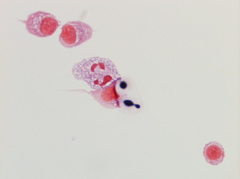
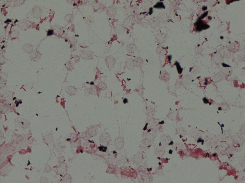
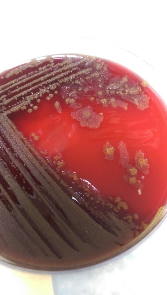
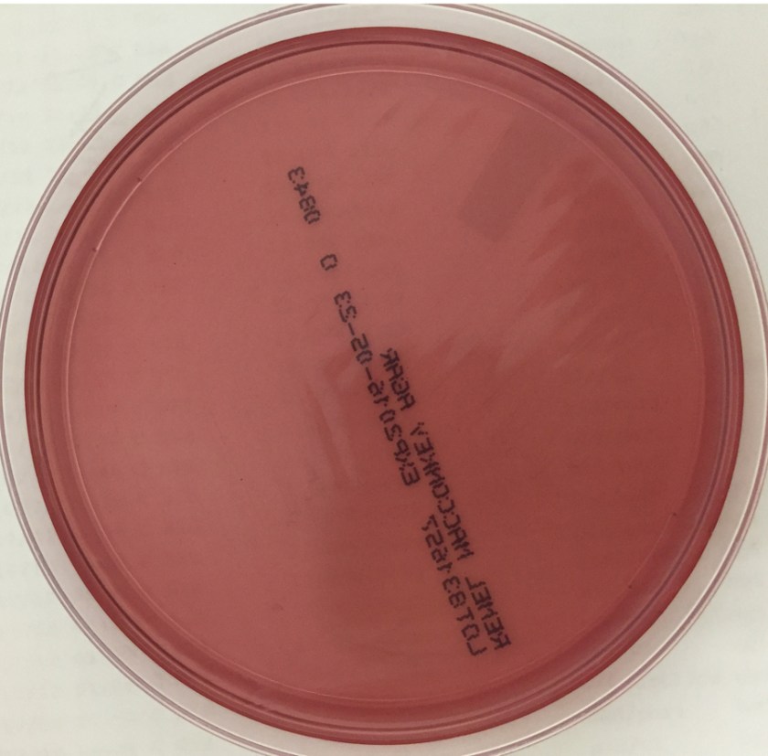
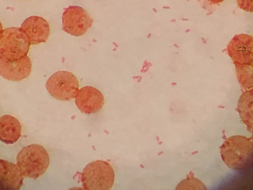
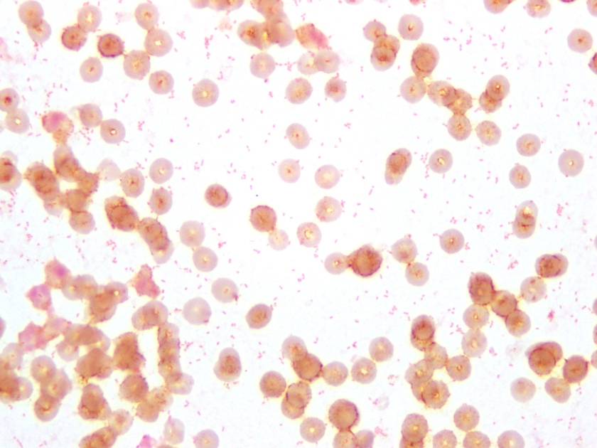
The centrifuged Gram stain of the CSF showed few yeast forms that exhibited narrow based buds and were surrounded by outline of a thick capsule (Figure 1).  Bacterial and fungal culture of the CSF revealed a white, creamy yeast that grew after 2-3 days incubation (Figure 2). The organism was identified by MALDI-TOF as Cryptococcus neoformans. A cryptococcal antigen test was performed on the CSF and showed a titer of 1:1024. In addition, C. neoformans grew from 4/4 of the patient’s blood cultures. While waiting for the culture results, the patient was found to be HIV positive for a viral load of 975,882 vc/ml and an absolute CD4 count of 17 cells/cm2. No other significant pathogens grew from any of the other cultures obtained.

Discussion

Cryptococcus neoformans is an encapsulated yeast found widely in nature.  It is typically found in soil that is contaminated with pigeon droppings or bat guano that has a high nitrogen content allowing the organisms to proliferate. C. neoformans is acquired via the inhalational route particularly when dust is generated. Individuals with a higher risk of infection include those who work in poultry farms, excavators and spelunkers. In addition, immunosuppressed people, especially with cellular immunodeficiencies such as HIV, those with hematopoietic malignancies and those taking immunosupressants have an increased risk of acquiring infection with C. neoformans. While the organism commonly causes pulmonary infections, fungemia and disseminated disease with cutaneous involvement have been reported. C. neoformans has a particular tropism for the central nervous system and often initially presents as meningoencephalitis without evidence of disease elsewhere.

Historically, the first step in diagnosis of suspected meningitis due to Cryptococcus in the HIV era was to perform an India ink preparation on a CSF specimen. This test served to identify the variably sized (2-20 µm), narrow based budding yeast forms due to the prominent capsule not allowing the ink to reach the cell wall of the organism and creating a halo like appearance around the yeast. While this test provided a rapid diagnosis and was inexpensive to perform, its lack of sensitivity has paved the way for detection of the cryptoccocal polysaccharide antigen via a latex agglutination method. This test can be performed on serum & CSF specimens and provides both diagnostic (qualitative) and prognostic (quantitative) information. By following titers by this method throughout the disease course and therapy, clinicians can monitor response to treatment (declining titers) and relapsed infections (increasing titers).

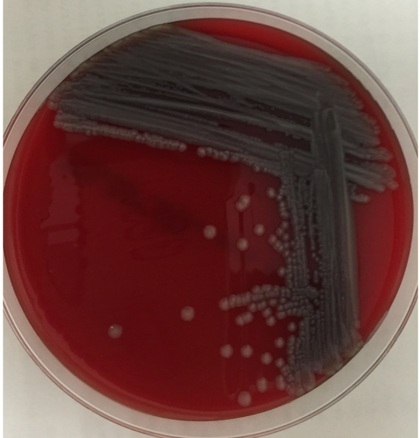
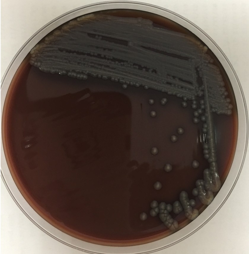
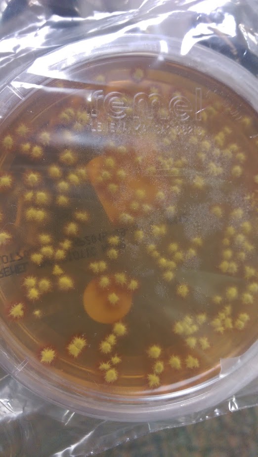
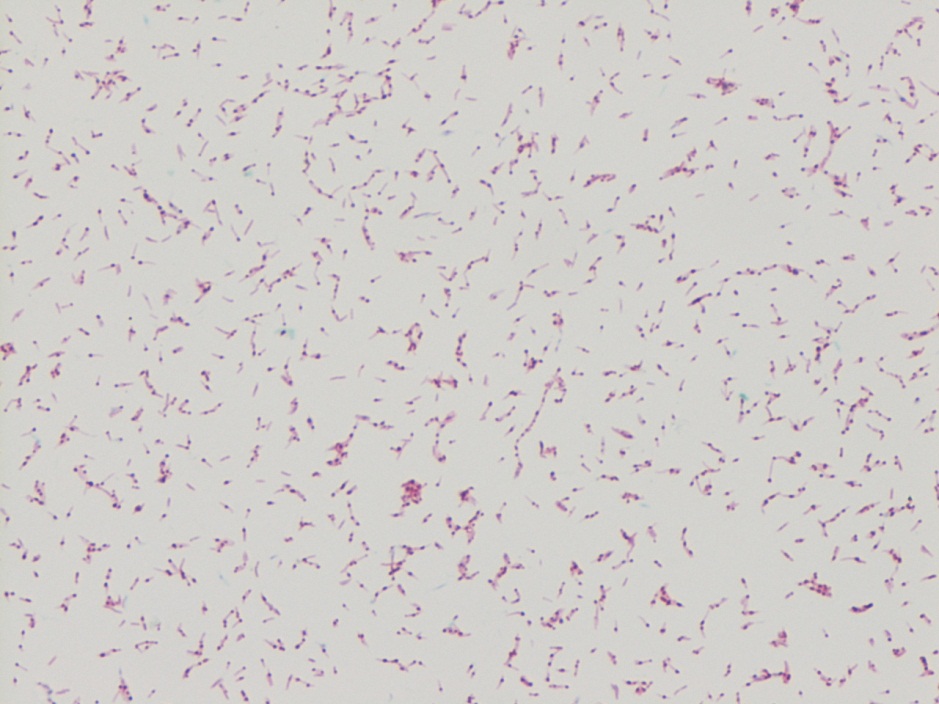
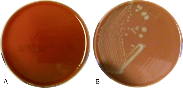
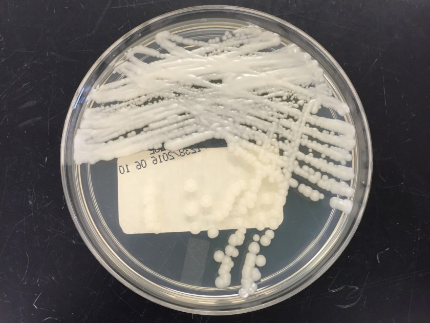
In culture, C. neoformans appears as white, creamy to mucoid colonies which grow well on Sabouraud dextrose agar within 3 days.  It is positive for both urea and phenoloxidase, which is exemplified by the colony’s reddish-brown pigmentation on bird seed agar. The presence of pigment on this agar helps to differentiate C. neoformans and C. gatti from other cryptococcal species, a distinction that may be important therapeutically as the latter are often more resistant to standard treatments. Microscopically on cornmeal agar, C. neoformans shows variability in size and is uniformly spaced among each yeast form due to the presence of the thick capsule. This characteristic is described as resembling glass beads. No pseudohyphae are present. Other commonly employed laboratory methods currently used to identify C. neoformans include automated instruments, such as the Vitek, and MALDI-TOF mass spectrometry.

In the case of our patient, he received two weeks of induction with amphotericin B and flucytosine. He was discharged home on oral fluconazole maintenance therapy as well as bactrim and azithromycin for prophylaxis from other infectious organisms. It is important to note that C. neoformans is resistant to echinocandins and this group of antifungals should not be used for treatment.


TV

-Tudor Vladislav, MD, is a 2nd year Anatomic and Clinical Pathology resident at the University of Mississippi Medical Center.

Stempak

-Lisa Stempak, MD, is an Assistant Professor of Pathology at the University of Mississippi Medical Center in Jackson, MS. She is certified by the American Board of Pathology in Anatomic and Clinical Pathology as well as Medical Microbiology. Currently, she oversees testing performed in both the Chemistry and Microbiology Laboratories.  Her interests include infectious disease histology, process and quality improvement and resident education. 


Microbiology Case Study: A 62 Year Old Male with Coronary Artery Disease

A 62 year old male with a past medical history of CAD, CABG x 4, HTN, DMII, OSA on CPAP, and GERD was admitted for acute onset of chest pressure that radiated to his back. He also complained of nausea, vomiting. He had a similar episode of pain two weeks ago which resolved with nitroglycerin. The patient was found to have Type 1A aortic dissection on CTA. Decision was made to proceed to the OR emergently. Status post the operation, he continued to have hemodynamic instability and evidence of pneumonia. He had been intermittently febrile with leukocytosis (WBC=12.66). Blood cultures were drawn and were positive for gram negative bacilli in one bottle.

Gram stain demonstrating Gram-negative rods.
Blood agar plate with dry, yellow colonies.

Identification:

Pseudomonas luteola was identified on the MALDI-TOF.

P. luteola was originally identified as Chryseomonas, but later changed to be a part of the Pseudomonas family. It is an opportunistic pathogen found in damp environments. It is a gram negative rod of 0.8 μm to 2.5 μm and is a motileaerobe. Its motility is created by multitrichous flagella. Colonies produce a yellow-orange pigment. P. luteola can be differentiated from most other motile yellow-pigmented nonfermenters by a negative oxidase reaction and from the Enterobacteriaceae by its strict aerobic growth. Optimal temperature for growth is 30°C, although it can grow at 42°C and not at 5°C. It grows best on heart infusion agar supplemented with 5% horse blood, but is also able to grow on TSA, Nutrient Agar, MacConkey or CASA Agar. The pathogenic form of P. luteola is a saprophyte and it can cause septicemia, peritonitis, endocarditis in patients with health disorders or with indwelling devices, and meningitis. Most strains are susceptible to broad-spectrum antibiotics, such as cephalosporins and ciprofloxacin.

Based on the history, the clinical team was unsure if it was a false positive/contaminant or truly a pathogen. The patient did have grafts and bioprosthetic material and due to the virulence of Pseudomonas, they decided to treat with cefepime and remove the central line. The patient clinically improved after removal of the line, which favored a line infection.

-Mustafa Mohammed, MD is a 2nd year anatomic and clinical pathology resident at the University of Vermont Medical Center.

Wojewoda-small

-Christi Wojewoda, MD, is the Director of Clinical Microbiology at the University of Vermont Medical Center and an Assistant Professor at the University of Vermont.

Microbiology Case Study: 8 Day Old with Meningitis

Case:

An 8-day-old baby was brought by his family to the Emergency Department. The baby had become irritable in the past 12 hours and was refusing to eat. He was born via vaginal birth at 38 weeks gestation and the pregnancy was uncomplicated besides an intrapartum fever for which mom received antibiotics during labor and baby received 48 hours of antibiotics after birth. In the ED, a lumbar puncture was performed and the resulting cerebral spinal fluid (CSF) had a protein of 353 mg/dL and glucose of <1 mg/dL. The CSF contained 2,935 nucleated cells with a differential of 76% polymorphonuclear cells, 14% macrophages, and 10% lymphocytes. Cytospin Gram stain of the CSF showed many white blood cells but no microorganisms. After 24 hours, an organism was growing on blood and chocolate agar, but not on MacConkey or Colistin Naladixic Acid (CNA) agar (Figure 1A, 1B, and 1C). Before this organism could be identified, the patient’s blood culture signaled positive with the same organism (Figure 2).

eliz1
Figure 1A. Growth on blood agar.
eliz2
Figure 1B. Growth on chocolate agar.
eliz3
Figure 1C. Growth (or lack thereof) on MacConkey agar.
eliz4
Figure 2. Gram stain of blood culture.

Discussion:

The organism was identified by MALDI-TOF MS (Matrix-assisted laser desorption/ionization time-of-flight mass spectrometry) as Elizabethkingia meningoseptica. This organism was previously named Chyrsiobacterium meningosepticum and before that Flavobacterium meningosepticum.

E. meningoseptica is an oxidase-positive, indole-positive, nonmotile, glucose nonfermenting, Gram-negative rod. It grows well on blood and chocolate agars after 24-48 hours of incubation, but does not grow on MacConkey agar. Colonies appear smooth and can be non-pigmented, like our isolate, or contain a slightly yellow or salmon pigment. Elizabethkingia spp. are environmental organisms which are ubiquitous in water and soil. They can also become nosocomial pathogens due to colonization of hospital sinks and other medical devices associated with water (humidifiers, ice chests, respirators, ect.).

E. meningoseptica is a rare cause of neonatal meningitis with prematurity being the greatest risk factor for infection. E. meningoseptica meningitis has a mortality rate of up to 57% and causes severe sequelae such as brain abscesses and developmental delay in those that survive the infection. Besides neonates, Elizabethkingia spp. have been associated with a variety of infections in immunocompromised patients and currently Wisconsin is in the middle of an outbreak of > 50 patients with Elizabethkingia anopheles bloodstream infections.

E. meningoseptica have a very unusual susceptibility pattern for Gram-negatives rods, which is shared by the closely related organisms Sphingomonas, Chryseobacterium, and Empedobacter spp. They produce β-lactamases making them resistant to most β-lactam antibiotics including carbapanems and aztreonam. E. meningoseptica is generally resistant to colistin and anaminoglycosides as well. They have variable resistance to vancomycin, rifampin, and fluroquinolones, making treatment options scant.

Our case patient cleared his blood and CSF of E. meningoseptica and is clinically improving. Only time will tell the extent of long term sequelae caused by this infection.


References:

  • Manual of Clinical Microbiology, 11th edition
  • Mehmet Ceyhan and Melda Celik, “Elizabethkingia meningosepticum (Chryseobacterium meningosepticum) Infections in Children,” International Journal of Pediatrics, vol. 2011, Article ID 215237, 7 pages, 2011. doi:10.1155/2011/215237
  • 2016 Wisconsin Outbreak of Elizabethkingia anopheles https://www.dhs.wisconsin.gov/disease/elizabethkingia.htm


-Erin McElvania TeKippe, Ph.D., D(ABMM), is the Director of Clinical Microbiology at Children’s Medical Center in Dallas Texas and an Assistant Professor of Pathology and Pediatrics at University of Texas Southwestern Medical Center.


Microbiology Case Study: 7 Year Old Male with Rash on his Scalp

A 7 year old Congolese male presented with pruritic, erythematous, non-flaky rash on top of his scalp for the past 3 weeks. The rash in non-painful, but continues to spread. His mother has been applying hydrocortisone cream nightly, with no improvements.

Colony morphology on fungal media.
Organism morphology on lactophenol analine blue scotch tape prep.


Laboratory Identification

A hair sample was obtained for fungal culture. Colonies were yellow and waxy with feet-like projections. Microscopic morphology on lactophenol analine blue scotch tape prep revealed broad hyphae with tortuous branches. The hyphae lacked obvious micro and macro conidia, raising the suspicion for Trichophyton violaceum.


Discussion

Trichophyton violaceum is an anthropophilic fungus seen predominantly in North Africa, East Asia and parts of the Middle East. It forms slow growing with glabrous colonies. Microscopically, broad tortuous hyphae are seen. Microconidia and Macroconidia are notably absent. T. violaceum causes Tinea Capitis, which can be acquired through scalp contact with the dermatophyte, either with direct contact with an infected individual or an object. It can also affect skin, nails and beards. It manifests clinically as pruritic scaly patches with alopecia, often producing black dots. Affected hairs demonstrate an endothrix infection.


-Mustafa Mohammed, MD is a 2nd year anatomic and clinical pathology resident at the University of Vermont Medical Center.

Wojewoda-small

-Christi Wojewoda, MD, is the Director of Clinical Microbiology at the University of Vermont Medical Center and an Assistant Professor at the University of Vermont.


Microbiology Case Study: A 39 Year Old Male with Headaches and Fatigue

Case History:

A 39 year old African American male presents to the Emergency Department with a three day history of headaches and fatigue. The patient describes his headache as a sharp, constant pain that is exacerbated by movement. He also has been experiencing blurred vision and feeling unstable on his feet. His past medical history is significant for HIV diagnosed 8 years ago. On admission, his absolute CD4 count is 104 with a viral load of 150 vc/ml. He is compliant with his anti-retroviral therapy and Bactrim prophylaxis. Physical exam is unremarkable except for oral thrush. Imaging reveals multifocal areas of enhancement throughout the brain and cerebellum with associated vasogenic edema, which was thought to most likely represent an infectious process. The patient was taken to the operating room for a brain biopsy. Tissue was sent to surgical pathology and the microbiology laboratory for evaluation.

Laboratory identification:

blasto1
Figure 1. Brain tissue with pyogranulomatous inflammation and a budding yeast form (H&E 400x).
blasto2
Figure 2. Brain tissue with many yeast forms present exhibiting broad based budding (GMS 400x).
blasto3
Figure 3. Growth of a white-tan mold on Sabouraud dextrose, SAB with chloramphenicol and Mycosel agar slants (left to right).
blasto4
Figure 4. Narrow septate hyphae with fine conidiophores giving rise to round shaped conidia (Lactophenol cotton blue, 400x)


The histology results were reviewed first and the H&E stain showed a pyogranulomatous inflammatory response with budding yeast forms. These findings were better visualized on the GMS stain which illustrated numerous yeast forms ranging in size with frequent broad based budding. In the microbiology laboratory, a white-tan mold grew on Sabouraud dextrose, SAB with chloramphenicol and Mycosel agar slants after 18 days of incubation at 30°C. On microscopic examination, the lactophenol cotton blue prep revealed narrow septate hyphae with round conidia at the end of fine conidiophores (characterized as a lollipop appearance). These finding are consistent with a diagnosis of Blastomyces dermatiditis and were confirmed with a DNA probe. Other pertinent laboratory results included urinary antigens positive for both Blastomyces and Histoplasma and a negative cryptococcal antigen in the serum.

Discussion:

Blastomyces dermatiditis is a thermally dimorphic fungus found in the Midwest, Ohio and Mississippi River valleys and the south central portion of the United States. The infection is obtained by inhalation of spores from decaying wood along rivers and patients typically show symptoms consistent with pneumonia. In some cases, particularly in immunocompromised patients, dissemination to the skin, bone and central nervous system can occur.

Morphologic diagnosis can be made from surgical pathology specimens which show broad based, budding yeast forms with a double contoured cell wall, ranging in size from 8-15 µm. In the microbiology laboratory, the mold form grows slowly (2-3 weeks) and is characterized by narrow septate hyphae with delicate conidiophores bearing round to oval conidia which are described as lollipop-like. Due to that fact that Blastomyces is not able to be differentiated from the mold form of Paracoccidioides brasiliensis and Chrysosporium spp., confirmation of the identification is necessary. Traditionally, this was done by temperature induced culture conversion of the mold form to the yeast form but due to the availability of rapid, commercially available DNA probes specific for the exoantigen of Blastomyces dermatiditis, this test is more commonly utilized currently.

Other supplemental tests used in the diagnosis include a Blastomyces urinary antigen which shows good sensitivity in both pulmonary and disseminated disease. In our case, the positive urinary antigen for Histoplasma was considered to be a false positive result due to known cross reactivity with Blastomyces as a result of shared polysaccharides.

Cryptococcus neoformans was also included in the differential diagnosis due to the patient’s HIV status, presenting symptoms and the fact it is a common cause of fungal meningitis in individuals with CD4 counts below 200. This infection was ruled out due to a negative serum cryptococcal antigen. In addition, in tissue Cryptococcus would show variably sized (2-20 µm), narrow based yeast forms with a prominent polysaccharide capsule and grow quickly as a yeast in fungal culture.

In the case of our patient, he was started on Amphotericin B based on the histology results. He experienced resolution of his headaches and recovered well following surgery. His was discharged to an extended care facility for 6 weeks of continued IV antifungal therapy before being placed on oral voriconazole.


SC

-Srinivasa Chekuri, MD, is a 4th year Anatomic and Clinical Pathology resident at the University of Mississippi Medical Center.

Stempak

-Lisa Stempak, MD, is an Assistant Professor of Pathology at the University of Mississippi Medical Center in Jackson, MS. Currently, she oversees testing performed in both the Chemistry and Microbiology Laboratories.


Microbiology Case Study: 77 Year Old Male with Asthma

A 77 year old male with history of asthma, atrial fibrillation, and recurrent respiratory distress when visiting Vermont presented to the ED with progressive dyspnea and wheezing for the past 4 days. Two days prior, he required a “breathing treatment” at his PCP. One day ago, he saw his PCP and was prescribed prednisone and azithromycin. He denies cough, fevers, or chills. He used his albuterol and Advair inhalers which barely helped. He was found to be in Afib with RVR to the 160s, a respiratory rate in the 40s, and an oxygen saturation of 70%.

Kinyoun stain revealing broad rods with cross-barring.
Colony growth on Lowenstein-Jensen medium.


Lab Identification

The organism was auramine fluorescent stain positive from the broth. The AFB culture bottle was sub-cultured to agar based medium and Lowenstein-Jensen medium, which yielded small yellow colonies. Kinyoun stain revealed broad rods with cross-barring. The organisms produced a yellow pigment when exposed to light, and a nucleic acid probe for Mycobacterium kansasii was positive.


Discussion

Mycobacterium kansasii was discovered in 1953 by Buhler and Pollack. It is an acid fast bacillus that produces yellow pigment when exposed to light (photochromogen). The bacilli are thick, long and cross-barred and have been described as ladder-like. It is prevalent in the Midwest and Southeast, and is the second most common cause of nontuberculous mycobacteria disease in patients with AIDS. Mycobacterium kansasii manifests as lung disease that clinically appears similar to tuberculosis. It can also cause local disease of the skin and subcutaneous tissue, as well as lymphadenitis and disseminated disease. Symptoms are more severe in immunocompromised hosts. Mycobacterium kansasii is generally acquired via either aspiration or local inoculation from the environment, with little evidence to support person to person transmission.


-Mustafa Mohammed, MD is a 2nd year anatomic and clinical pathology resident at the University of Vermont Medical Center.

Wojewoda-small

-Christi Wojewoda, MD, is the Director of Clinical Microbiology at the University of Vermont Medical Center and an Assistant Professor at the University of Vermont.

Microbiology Case Study: 6 Year Old Girl with Headache

A 6 year-old girl with a history of posterior fossa ependymoma presented with a one month history of fever, headaches, vomiting and more recently, neck stiffness. Additional history includes remote tumor resection followed by radiation and chemotherapy resulting in remission, with a residual ventriculoperitoneal shunt (VPS). Her parents reported she was in good health until approximately 1 month prior to presentation and is up to date on her immunizations. She was previously seen by her primary care physician for her symptoms and treated her with amoxicillin for suspected strep throat. Upon admission, she received supportive therapy for her symptoms after she was found to have tumor recurrence on imaging. The patient was scheduled for resection approximately two weeks after discharge and on post-operative day two she developed fever, vomiting and neck stiffness again. At this time, blood cultures were drawn and a lumbar puncture (LP) was performed. Cerebrospinal fluid (CSF) from both the LP and VPS submitted for fluid analysis (Table 1) and culture.

Table 1: Cerebrospinal Fluid Analysis

Spinal Fluid LP VPS
Appearance Clear Clear
Nucleated cells 1075 cells/μL 628 cells/μL
RBC 150 cells/μL 35 cells/μL
Polys 94% 87%
Lymphs 2% 6%
Mono/Macrophage 4% 7%
Glucose 68 mg/dL 13 mg/dL
Protein 69 mg/dL 164 mg/dL
haem1
Figure 1. Gram stain of the pathogen isolated from aerobic blood culture, showing gram-negative coccobacilli, sometimes in pairs. The same organism was seen on the patient’s CSF Gram stain.
haem2
Figure 2. Aerobic blood culture on (A) 5% sheep blood agar plate (BAP), showing no growth and on (B) chocolate agar plate (CAP), showing round, smooth, opaque grey-yellow colonies.


Culture results:

The CSF Gram stain showed rare, paired, Gram-negative diplococci, which could raise suspicion for Neisseria meningitidis, however the typical flattened sides of adjacent bacteria were not observed. Rather, the morphology was more consistent with Gram-negative coccobaccilli, which is better demonstrated on Gram stain of the blood culture (Figure 1). Culture of both the CSF and blood specimens grew fairly large, smooth, round, opaque grey-yellow colonies on CAP, however showed no growth on BAP (Figure 2), suggesting a fastidious organism requiring growth factors. The colonies were both catalase and oxidase positive. The organism was identified as Haemophilus influenzae by MALDI-TOF MS (matrix-assisted laser desorption/ionizations time-of-flight mass spectrometry). This H. influenzae isolate was non-typeable by slide agglutination serotyping performed at the state public health laboratory.

Discussion:

H. influenzae are small, pleomorphic, gram-negative rods or coccobacilli that are non-motile. They are facultative anaerobes that grow best between 35-37°C with 5% CO2. H. influenzae is a fastidious species, requiring hemin (X factor) and nicotinamide-adenine-dinucleotide (NAD/V factor) for growth, which are both available in chocolate agar, but not blood agar. On chocolate agar, the colonies are non-hemolytic, typically large, smooth, round and convex with an opaque, colorless or grey hue. Encapsulated strains, including H. influenzae serotype b (Hib), appear mucoid and are typically small, grey colonies on CAP. Isolates are catalase and oxidase positive. H. influenzae displays the “satellite phenomenon” when grown near Staphylococcus aureus. This occurs when colonies of S. aureus lyse nearby red blood cells releasing hemin and NAD in the media. The presence of extracellular hemin and NAD allow colonies of H. influenzae to grow in the immediate vicinity of S. aureus.

H. influenzae is widely distributed in humans, colonizing the nose and throat and is spread from person-to-person via direct contact or respiratory droplets. Severe infections, including pneumonia, bacteremia and meningitis, affect predominantly infants and children. The American Academy of Pediatrics recommends routine vaccination with the Hib conjugate vaccine for infants aged 2 through 6 months (2 or 3 doses, depending on vaccine product) followed by a booster dose at age 12 through 15 months. Hib is the only serotype preventable by vaccine. Prior to routine vaccination in the US, approximately 20,000 children under the age of 5 were infected with H. influenzae and 3-6% died each year.

References:

1. Ledeboer N, Doern G. 2015. Haemophilus, p 667-684. In Jorgensen J, Pfaller M, Carroll K, Funke G, Landry M, Richter S, Warnock D (ed), Manual of Clinical Microbiology, 11th Edition. ASM Press, Washington, DC.
2. http://www.cdc.gov/meningitis/lab-manual/chpt09-id-characterization-hi.html
3. http://www.cdc.gov/hi-disease/index.html


-Petra Rahaman, MD is a 4th year Anatomic and Clinical Pathology resident at UT Southwestern Medical Center.

-Erin McElvania TeKippe, Ph.D., D(ABMM), is the Director of Clinical Microbiology at Children’s Medical Center in Dallas Texas and an Assistant Professor of Pathology and Pediatrics at University of Texas Southwestern Medical Center.

The Gloves Are Off … Or Are They?

The manager of the microbiology laboratory walked into the monthly staff meeting to discuss safety. Her first announcement was that the one clean hand washing sink in the department was going to be removed. The techs were shocked, and some were angry. Didn’t the manager care about infection prevention and control? Didn’t she know that hand hygiene should always occur after PPE is removed and before leaving the lab? The manager waited for the reactions to subside, then she explained that since the staff treated the lab as a clean area in many instances, that there should be no need for hand washing. The staff went on to argue that they were working with microbiological pathogens, and that they did wear lab coats and gloves, especially when handling specimens and setting them up for cultures. Some of those specimen containers were pretty disgusting, in fact.

That was when the manager dropped the charade. She had no real intention of removing the sink, but she wanted to make a point. She was tired of watching her staff reading culture plates with no gloves. She had spoken about it before, but no one agreed- they had been handling incubated plates for years.

One of the most common issues lab managers and safety professionals face is maintaining Personal Protective Equipment (PPE) compliance in the work area. An effective weapon in this battle is telling stories of lab incidents with bad outcomes, or explaining the consequences of this unsafe behavior. That is a valuable piece of lab safety education. It is unfortunate that we sometimes have to learn from others’ mistakes, but when it comes to safety, that’s better than learning from your own. Some lab accidents and exposures can be career-altering or career-ending.

OSHA’s Bloodborne Pathogen Standard states that PPE (specifically gloves) must be worn when there is a risk of exposure. That is as specific as they get on the topic. Anytime patient specimens are handled or opened, it follows that gloves should be worn. That means that in the microbiology area, staff is handling specimens and agar plates with gloves while they streak plates and set up gram stain slides. These contaminated gloves are handling plate after plate, and then those plates are placed into the incubators. Like any other contaminated item in the lab, those plates should be treated and handled with gloves until properly discarded. That means that gloves are necessary when removing plates from the incubator, and when reading those cultures. Not only is staff handling contaminated plates, but they are working with bacterial and fungal colonies. There is a high risk of exposure in those processes.

OSHA also requires PPE under its Chemical Hygiene Standard (or Lab Standard). Gloves are required when handling chemicals, so they would be needed when performing simple chemical tests (oxidase, catalase, etc.) and when performing gram stains. Make sure you use chemical-resistant gloves when selecting the appropriate PPE for these tasks.

In 2010, OSHA responded to an inquiry specifically about the use of gloves while handling culture plates in the microbiology laboratory. The letter “strongly suggests” the use of gloves for the task, but OSHA’s own standards already address the issue and clearly require the need for PPE in that situation.

The story at the beginning of this entry is true- there was a lab manager who was fed up with her staff not wearing gloves, so she told them she was removing the sink. She was kidding, but she made her point. In that microbiology lab they all wear gloves to read cultures today.

Laboratory-acquired infections occur every year, and some of the easiest ones to investigate are the cases in which techs are infected with pathogenic bacteria. It is fairly easy to trace the sources of those exposures. What is the staff doing in your microbiology laboratory? Are they doing everything they can to prevent exposure to pathogens? As a manager or safety professional, are you enforcing the use of PPE when exposure is possible? Keep your staff from becoming a safety statistic- provide PPE, teach consequences of unsafe behaviors, and monitor the continual use of those safe work practices in your lab.


Scungio 1

-Dan Scungio, MT(ASCP), SLS, CQA (ASQ) has over 25 years experience as a certified medical technologist. Today he is the Laboratory Safety Officer for Sentara Healthcare, a system of seven hospitals and over 20 laboratories and draw sites in the Tidewater area of Virginia. He is also known as Dan the Lab Safety Man, a lab safety consultant, educator, and trainer.

Microbiology Case Study: Asymptomatic 12 Year Old Girl

A 12 year old girl who recently emigrated from Nepal was seen in clinic to establish care. She was entirely asymptomatic. Stool ova and parasite exam was performed and on the permanent trichrome-stained section, the following parasites were identified.


Image (A)
Image (A)
Image (B)
Image (B)
Image (C)
Image (C)
Image (D)
Image (D)
Image (E)
Image (E)

Laboratory Identification:

The first image (A) is morphologically diagnostic for Dientamoeba fragilis trophozoites. They are approximately 10 microns in diameter and have 1-2 nuclei, which appear fractured. The next two images are diagnostic for Endolimax nana cysts (B) and trophozoites (C). The cysts are approximately 7 microns in diameter and most have 4 nuclei with blot-like karyosomes that are red on trichrome stain with clearing around the nuclei. The trophozoites are approximately 10 microns in diameter with a single large blot-like karyosome that is red on trichrome stain. The last two images are diagnostic for Entamoeba coli cysts (D) and trophozoites (E). The cysts are approximately 20 microns in diameter and have five to eight nuclei with karyosomes that are red on trichrome stain. The trophozoites are approximately 22 microns in diameter and have a single nucleus with a large kayosome that is darkly staining on trichrome stain. There is peripheral chromatin that is ring-like or clumped.

Discussion:

Dientamoeba fragilis, an ameboflagellate, is a potential pathogen that can be associated with diarrhea, vomiting, abdominal pain, and anorexia, particularly in children. Transmission is via ingestion of contaminated food and water. Some studies postulate co-transmission via helminth eggs, particularly with Enterobius vermicularis. Historically, this intestinal parasite is only known to have a trophozoite form. However, there are now case reports describing the presence of cysts and precysts in humans.1 Treatment is with metronidazole or paromomycin in patients who are symptomatic.

Endolimax nana and Entamoeba coli are protozoa that are considered non-pathogenic and therefore no treatment is necessary. However, when identified, they should be reported since their presence indicates exposure to contaminated food and water. Transmission is via ingestion of cysts. Once in the small bowel, they ex-cyst and migrate to the large bowel where they divide by binary fission and produce cysts. Both cysts and trophozoites are passed in stool.

Reference

  1. Stark D, Garcia LS, Barratt JLN, Phillips O, Roberts T, Marriot D, Harkness J, Ellis JT. Description ofDientamoeba fragilis cyst and precystic forms from human samples. Journ Clin Micro. 2014; 52: 2680-2683.


-Joanna Conant, MD is a 4th year anatomic and clinical pathology resident at the University of Vermont Medical Center.

Wojewoda-small

-Christi Wojewoda, MD, is the Director of Clinical Microbiology at the University of Vermont Medical Center and an Assistant Professor at the University of Vermont.

Microbiology Case Study: A 33 Year Old Female with Abdominal Pain

Case History:

A 33 year old African American female presents to the hospital complaining of mild abdominal pain for the past couple of days. She is 17 weeks pregnant and has a history of two prior spontaneous abortions at 15 and 16 weeks due to a shortened cervix. She is afebrile and denies any vaginal bleeding or leakage of amniotic fluid. A complete blood count reveals mild leukocytosis and anemia. On physical examination, her cervix is 2 cm dilated with bulging membranes. She is admitted for a possible cerclage placement, and an amniocentesis is performed to rule out infection prior to the procedure. The microbiology lab received 20 ml of clear, amber fluid for Gram stain and bacterial culture.

fuso1
Figure 1. Direct Gram stain from the amniotic fluid showing many neutrophils and fusiform Gram negative bacilli (1000x oil immersion).
fuso2
Figure 2. Wright-Geimsa stain of the amniotic fluid specimen containing many acute inflammatory cells which have engulfed the fusiform bacteria (1000x oil immersion).
fuso3
Figure 3. Small, greyish-white colonies growing on Brucella blood agar after 48 hours of incubation under anaerobic conditions at 35°C.

Laboratory Identification:

The Gram stain showed moderate fusiform Gram negative bacilli in a background of many acute inflammatory white blood cells. Bacterial cultures grew small, greyish-white colonies as Brucella blood agar and routine blood agar after 48 hours of incubation under anaerobic conditions at 35°C. No growth was observed on kanamycin-vancomycin laked blood (KVLB) agar. The organism was identified by MALDI-TOF as Fusobacterium nucleatum and confirmed using the Vitek anaerobic identification card.

Discussion:

Fusobacterium nucleatum is an anaerobic, Gram-negative rod that is non-spore forming. It is considered normal flora of the oral cavity and gastrointestinal & genitourinary tracts of healthy adults. F. nucleatum has been implicated in the pathogenesis of oropharyngeal infections, especially in neutropenic patients with mucositis after receiving chemotherapy or bone marrow transplant. It is an important etiologic agent in a wide spectrum of extraoral infections including bacteremia, brain abscess, osteomyelitis and infections of the genitorurinary tract, including the fetal membranes. There have been many documented cases linking infections with F. nucleatum to chorioamnionitis, preterm birth, and neonatal sepsis. The mode of transmission of F. nucleatum to the amniotic fluid can be as a result of direct extension from the vaginal tract, hematogenous spread or as recently implicated, orogenital transmission.

Given that F. nucleatum is the most common of Fusobacterium species found in clinical specimens and it’s potential to cause significant disease, early identification of the pathogen is important. It grows well on a non-selective anaerobic agar and its growth is inhibited on Bacteroides bile esculin (BBE) and kanamycin-vancomycin laked blood (KVLB) agars. After 48 hours of incubation under anaerobic conditions, the colonies measure 1-2 mm in diameter and have been noted to have a characteristic internal flecking quality that is referred to as “speckled opalescence”. On Gram stain, the fusiform cells of F. nucleatum are long (usually 5-10 µm in length), slender filaments with tapered ends and may contain spherical swellings. In regards to biochemical testing, it is indole positive and lipase negative. Disk testing for Fusobacterium spp. shows the bacteria are resistant to vancomycin and susceptible to kanamycin and colistin.

While susceptibility testing is not routinely performed for all anaerobes, testing is indicated for organisms in pure culture isolated from normally sterile sites or for those more virulent organisms for which susceptibilities cannot be predicted. In the case of Fusobacterium spp., penicillin and ampicillin resistance among isolates of has been reported due to beta-lactamase production and it is recommended that all Gram negative anaerobes have a beta-lactamase screen performed. F. nucleatum is routinely susceptible to metronidazole, clindamycin and beta-lactam beta-lactamase inhibitor combination antibiotics.

In the case of our patient, her diagnosis of F. nucleatum in the amniotic fluid specimen precluded her from obtaining a rescue cerclage procedure. She was transferred to labor and delivery for a uterine evacuation secondary to the intra-amniotic infection and delivered a non-viable fetus. She received ampicillin and gentamicin as intravenous antibiotics.


sims

-Brooke Sims, MD, is a third year Anatomic and Clinical Pathology resident at the University of Mississippi Medical Center.

Stempak

-Lisa Stempak, MD, is an Assistant Professor of Pathology at the University of Mississippi Medical Center in Jackson, MS. Currently, she oversees testing performed in both the Chemistry and Microbiology Laboratories.