A 7 year old Congolese male presented with pruritic, erythematous, non-flaky rash on top of his scalp for the past 3 weeks. The rash in non-painful, but continues to spread. His mother has been applying hydrocortisone cream nightly, with no improvements.

Laboratory Identification
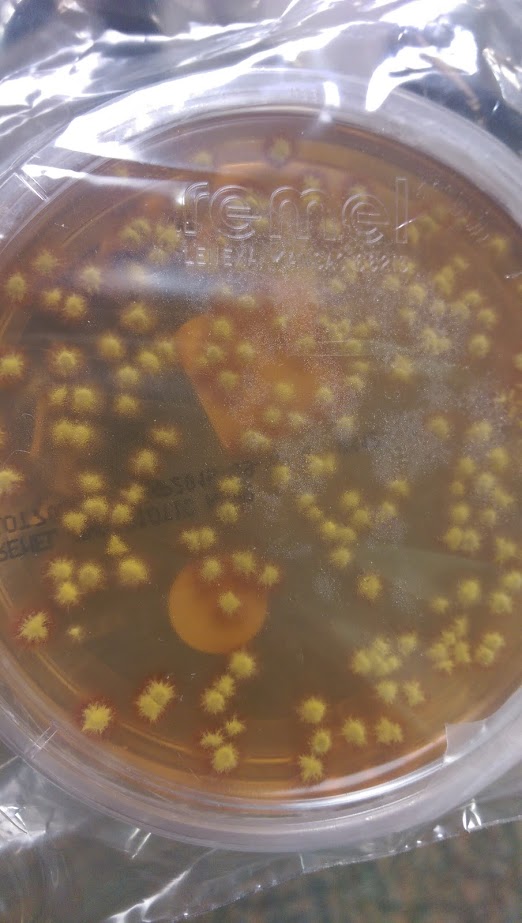
A hair sample was obtained for fungal culture. Colonies were yellow and waxy with feet-like projections. Microscopic morphology on lactophenol analine blue scotch tape prep revealed broad hyphae with tortuous branches. The hyphae lacked obvious micro and macro conidia, raising the suspicion for Trichophyton violaceum.
Discussion
Trichophyton violaceum is an anthropophilic fungus seen predominantly in North Africa, East Asia and parts of the Middle East. It forms slow growing with glabrous colonies. Microscopically, broad tortuous hyphae are seen. Microconidia and Macroconidia are notably absent. T. violaceum causes Tinea Capitis, which can be acquired through scalp contact with the dermatophyte, either with direct contact with an infected individual or an object. It can also affect skin, nails and beards. It manifests clinically as pruritic scaly patches with alopecia, often producing black dots. Affected hairs demonstrate an endothrix infection.
-Mustafa Mohammed, MD is a 2nd year anatomic and clinical pathology resident at the University of Vermont Medical Center.

-Christi Wojewoda, MD, is the Director of Clinical Microbiology at the University of Vermont Medical Center and an Assistant Professor at the University of Vermont.

Great mycology case, thanks 🙂