Analysis of a DNA sequence can be accomplished via a method called electrophoresis. Electrophoresis is a term that basically describes the movement of molecules by way of an electric current and separation of those molecules based on size. This process occurs through agarose or polyacrylamide gels, which serve as a way to limit migration of molecules as they move from the negative anode to positive anode. Small molecules move through the gel matrix faster than larger molecules.
How does it work?
Each phosphate group from a DNA molecule is ionized
↓
DNA becomes negatively charged
↓
DNA migrates towards the positive pole (anode)
Factors Affecting Electrophoretic Separation
- Strength of the electric current (voltage)
- Concentration and type of the buffer
- Gel density
- Size of the DNA
| AGAROSE CONCENTRATION AND SEPARATION RANGES | |
| Agarose Concentration (%) | Separation Range (base pair size) |
| 0.3 | 5,000 – 60,000 |
| 0.6 | 1,000 – 20,000 |
| 0.8 | 800 – 10,000 |
| 1.0 | 400 – 8,000 |
| 1.2 | 300 – 7,000 |
| 1.5 | 200 – 4,000 |
| 2.0 | 100 – 3,000 |
As agarose concentration increases, the separation range decreases
| TYPES OF ELECTROPHORESIS SYSTEMS | |
| Pulsed Field Electrophoresis |
|
| Field Inversion Gel Electrophoresis
(FIGE) |
|
| Polyacrylamide Gel Electrophoresis
(PAGE) |
|
| Capillary Electrophoresis |
|
Understanding Buffer Systems
In order to change the pH of a buffered solution by one point, either the acidic or basic form of the buffer must be brought to a concentration 1/10th that of the other form.
Test Your Knowledge
- Based on the diagram, determine the sizes (to the best approximation) of the DNA fragments for each of the samples:
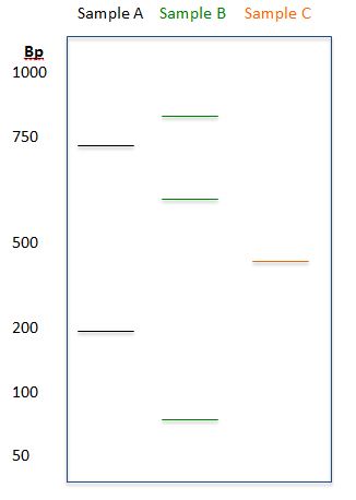
Answer:
- Sample A: 200bp, 700bp
- Sample B: 90bp, 600bp, 875bp
- Sample C: 400bp
-LeAnne Noll, BS, MB(ASCP)CM is a molecular technologist in Wisconsin and was recognized as one of ASCP’s Top Five from the 40 Under Forty Program in 2015.

One thought on “Separation and Detection of Nucleic Acids via Electrophoresis Methods”