Microbiology Case Study: A 58 Year Old Female with Lung Nodules

Case History

A 58 year old female with past medical history significant for Type II diabetes, hypertension, hyperlipidemia, chronic diastolic heart failure, and hypothyroidism was hospitalized for following a fall and was found to have compression fractures. The hospitalization was complicated by flash pulmonary edema requiring intubation. CT chest obtained during this hospitalization demonstrated lung nodules, which were biopsied and cultured.

Laboratory Identification

The bacterial and mycobacterial cultures grew gram positive bacilli which were positive on Modified Kinyoun stain. They were negative for Auramine/Rhodamine. The organism grew on several media, including 7H11, Chocolate, and Buffered Charcoal Yeast Extract (BCYE). They formed chalky, white-pink colonies. The organism was confirmed as Nocardia nova by a reference laboratory.

nocard1

Image 1. Modified acid fast bacilli on Modified Kinyoun stain.

nocard2

Image 2. Chalky white-pink colonies on BCYE agar.

Discussion

Nocardia nova is a weakly acid fast, aerobic filamentous, beaded, gram positive bacilli with right-angled branching. It is identified by a Modified Kinyoun stain. Nocardia grows best on BCYE agar; however it also can grow within 3-5 days on blood and chocolate agar. It forms chalky white-pink colonies. Molecular testing is performed to speciate Nocardia, primarily 16S ribosomal RNA gene sequencing, as well as mass spectrometry. Most infections can be treated with sulfonamides for 6-12 months, however, the CDC recommends performing speciation and susceptibility testing on every isolate due to specific susceptibility profiles and drug resistant strains. Our patient was treated with high dose sulfamethoxazole/trimethoprim and meropenem.

Nocardia nova is commonly found in soil and is one of several pathogenic Nocardia species. Nocardia is often inhaled and presents as a chronic pulmonary infection with cough, shortness of breath, and fever. Nocardia can also cause pleural effusions, empyema, pericarditis, abscesses, or dissemination to deep organs, especially the brain. Nocardia can also be contracted though trauma, causing cutaneous diseases such as a mycetoma or cellulitis. Because of its low virulence, Nocardia generally affects immunocompromised patients, however those with preexisting pulmonary disease can also be susceptible to infection.


-Mustafa Mohammad, MD is a 3rd year anatomic and clinical pathology resident at the University of Vermont Medical Center.

Wojewoda-small

-Christi Wojewoda, MD, is the Director of Clinical Microbiology at the University of Vermont Medical Center and an Assistant Professor at the University of Vermont.

Hematopathology Case Study: A 69 Year Old Female with Persistent Monocytosis

Case History

A 69 year old female with a past medical history significant for endometrial adenocarcinoma, traumatic brain injury, atrial fibrillation, hypertension, hyperlipidemia, and persistent monocytosis (absolute monocyte count ranging from 1.6-3.7 K/uL) who had an indeterminate lesion identified in the T5 vertebra, read as “hemangioma, although surrounding edema is worrisome for malignancy” upon staging imaging for history of endometrial carcinoma.

CBC at the time was: WBC 9.8; HGB 12.9; HCT 37.6; PLT 154; MCV 92 fL; MCH 31.7.

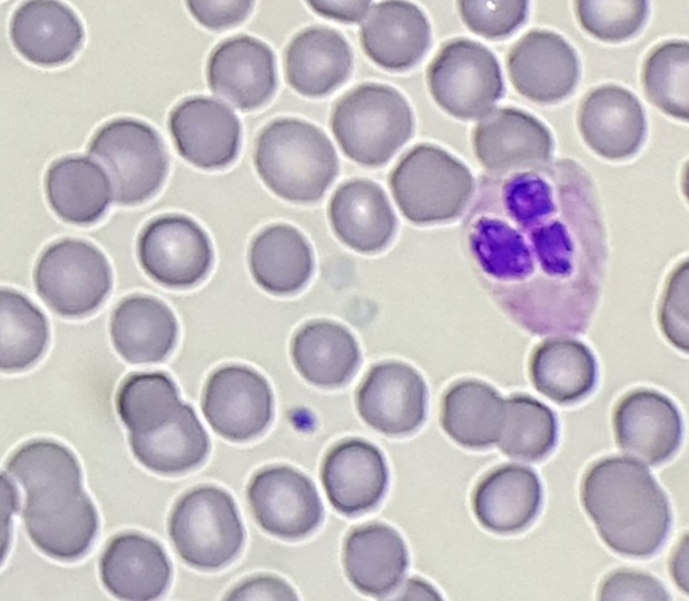
Automated differential showed: 43.0 Neutrophils; 34.8 Lymphocytes; 20.0 Monocytes; 1.4 Eosinophils; 0.2 Basophils; 0.6 Immature granulocytes.

Absolute monocyte count was 1.95 K/uL.

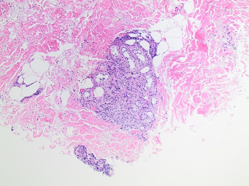
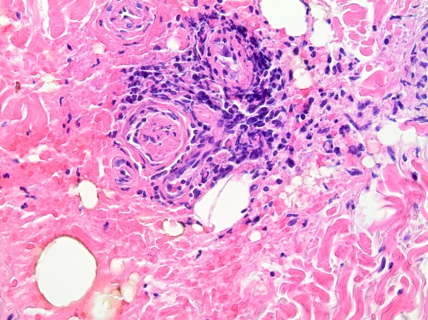
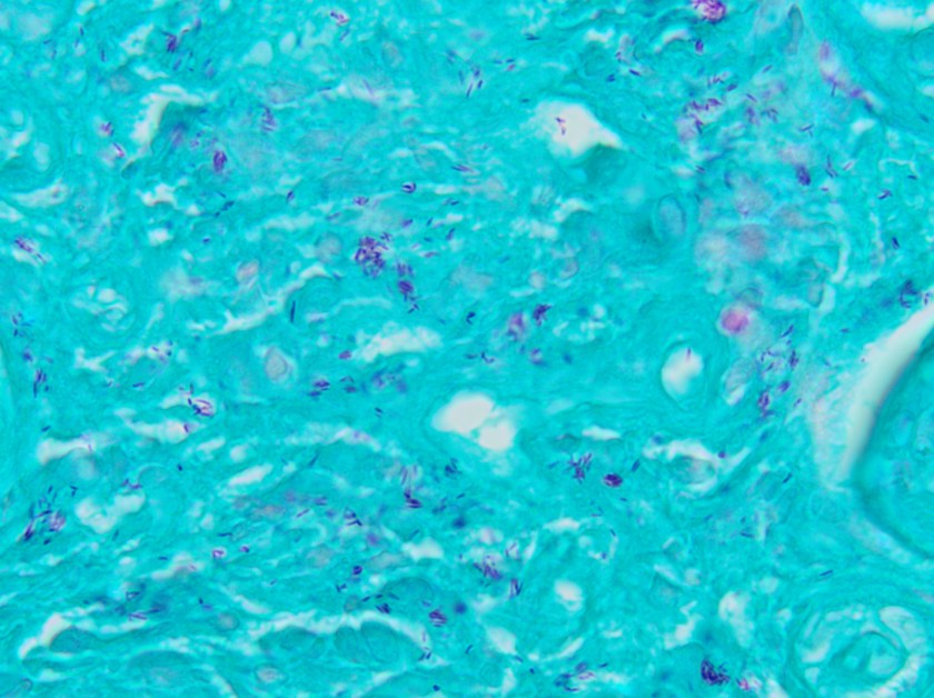
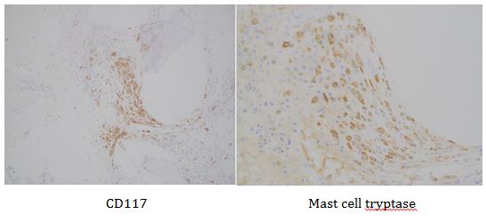
This lesion was biopsied and given the findings, a subsequent bone marrow biopsy was performed on 5/4/2017. The bone marrow core biopsy contained multiple compact aggregates of spindle shaped cells with hypogranular cytoplasm, morphologically compatible with atypical mast cells. Within these aggregates, numerous eosinophils are present. By immunohistochemistry, the mast cells are brightly positive for CD117 and mast cell tryptase. Concurrent bone marrow aspirate flow cytometry demonstrated a small population of mast cells that co-expressed CD2 and CD25.

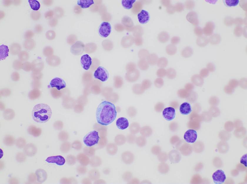
Of note, the patient was found to have a persistent absolute monocytosis. Flow cytometry revealed an abnormal population of monocytes that displayed aberrant phenotypic expression of CD2 and CD56 (subset).

Next-generation sequencing revealed two truncation mutations in the TET2 gene (K988* in 34.6% of the reads and Q1138* in 36.4% of the reads). Cytogenetic analysis revealed a normal female karyotype (46,XX).

myelomono1

myelomono2

myelomono3

Overall, an immunophenotypically abnormal population of mast cells and monocytes are present in the context of a long-standing absolute monocytosis and the presence of two TET2 truncating mutations, supporting a diagnosis of systemic mastocytosis with an associated hematologic non-mast cell lineage disorder (best classified as chronic myelomonocytic leukemia).

Discussion

A diagnosis of systemic mastocytosis is a combination of clinical, morphologic, immunophenotypic, and molecular analyses, as required by the World Health Organization (WHO 2008). By current consensus guidelines, SM variants are partly distinguished by clinicopathologic criteria referred collectively as B and C findings. B findings include: >30% of bone marrow mast cells (MC) on biopsy and/or serum tryptase levels >200 ng/mL; increased marrow cellularity/dysplasia without meeting diagnostic criteria for another myeloid neoplasm; or enlargement of liver, spleen, or lymph nodes without evidence of organ damage. C findings include: evidence of organ damage caused by a local MC infiltrate, such as abnormal liver function and/or ascites, hypersplenism, cytopenias, large osteolytic lesions/fractures, and malabsorption with weight loss caused by MC infiltrate in the gastrointestinal tract.

Systemic mastocytosis commonly occurs in two types with different clinical courses based upon the aforementioned findings. Indolent SM (ISM) is defined by the absence of C findings. Smoldering SM is a subtype of ISM that displays 2 or more B findings. ISM may become more aggressive and a descriptive term of advanced SM refers to a category including aggressive SM (ASM), mast cell leukemia (MCL), and “SM with an associated myeloid neoplasm.” The latter entity comprises more than 90% of cases that have previously been referred to as SM with an associated hematologic non-mast cell lineage disorder (SH-AHNMD).

ASM and MCL are characterized by organ damage and histologic characteristics. ASM often exhibits multifocal bone marrow infiltration of atypical mast cells that are often spindled in shape with hypogranular or immature morphology. Marked fibrosis often accompanies the infiltrate as well as a KIT D816V mutation. MCL is codified by more than 20% of the marrow aspirate nucleated cells represent by mast cells and on core biopsy, a compact infiltrate is often identified with usually low level fibrosis. In MCL, circulating mast cells are greater than 10% of nucleated cells but according to Gotlib et al., the aleukemic MCL (less than 10% circulating mast cells) is more common.

In the context of our patient, myeloid neoplasms associated with SM are often represented by MDS, MPN, or MDS/MPN overlap disorders, and occasionally AML.

Associated lymphoid or plasma cell neoplasms have been described, but in a much lower frequency.

In accordance with the diagnostic implications, KIT D816V mutational analysis is important therapeutically. Most patients with SM harbor the KIT D816V mutation (>80% in one clinical series; 90-100% in research studies using purified MCs), which is a considered imatinib-resistant mutation. Midostaurin (a second generation TKI) may provide some disease response while nilotinib or dasatinib are usually less likely to lead to a durable response. The rare patients who have a juxtamembrane domain KIT mutation are much more likely to respond to imatinib or masitinib.

For disease response, criteria were first published in 2003 by Valent, et al. In a reiterated version published in 2007, the evaluation of clinical evidence of organ damage (C findings), was the foundation for determining appropriate response. Another facet to determining response was in relation to BM MC burden, serum tryptase level, and organomegaly, which further subcategorized the levels of major response (MR). MR was defined as normalization of 1 or more C findings. In turn, MR was divided into 3 categories:

  1. Complete remission (resolution of MC infiltrates in organs, serum tryptase less than 20 ng/mL, and disappearance of SM-associated organomegaly)
  2. Incomplete remission (decrease in MC infiltrates in organs and/or serum tryptase levels and/or visible regression of organomegaly by >50%)
  3. Pure clinical response (without decrease in MC infiltrates, serum tryptase levels, or organomegaly)

Partial response (PR) is defined as incomplete regression of 1 or more C findings and include good partial response (GPR; >50% regression of 1 or more C findings) and minor response (<50% regression).

Lastly, the Mayo Clinic published revised response criteria in 2010 which established minimal baseline laboratory abnormalities for organ damage to be evaluated in order to allow for more accurate assessment of response to therapy that is clinically more relevant.

Overall, systemic mastocytosis is a rare entity that displays a range of presentations that can be described as indolent up to an aggressive (advanced) phenotype. The hallmarks for diagnosis include histologic, immunophenotypic, molecular, and clinical findings.


References

  1. Gotlib, J et al. “International Working Group-Myeloproliferative Neoplasms Research and Treatment (IWG-MRT) & European Competence Network on Mastocytosis (ECNM) consensus response criteria in advanced systemic mastocytosis,” Blood, 2012.
  2. Horny HP et al. “Mastocytosis,” In: Swerdlow S et al. WHO Classification of Tumours of Haematopoietic and Lymphoid Tissues. Lyon: IARC Press; 2008:53-63
  3. Valent P et al. “Aggressive systemic mastocytosis and related mast cell disorders: current treatment options and proposed response criteria.” Leuk Res. 2003;27(7):635-641.
  4. Pardanani A, et al. “A critical reappraisal of treatment response criteria in systemic mastocytosis and a proposal for revisions. Eur J Haematol. 2010;84(5):371-378.


PhillipBlogPic-small

-Phillip Michaels, MD is a board certified anatomic and clinical pathologist who is a current hematopathology fellow at Beth Israel Deaconess Medical Center, Harvard Medical School, Boston, MA. His research interests include molecular profiling of diffuse large B-cell lymphoma as well as pathology resident education, especially in hematopathology and molecular genetic pathology.

Microbiology Case Study: A 60 Year Old Male with Longstanding Skin Lesions

Case History

A 60 year old male from Louisiana presents to his family doctor with a chief complaint of longstanding skin lesions for approximately the last two years. On physical exam, there are several sharply defined reddish-brown plaques on his upper back and extremities. He reports sensory loss involving his chest, back and upper extremities. The lesions have not responded to conventional topical anti-fungal treatments. Punch biopsies along the margin of the most active lesion were obtained and sent to the Microbiology laboratory for bacterial, fungal and mycobacterial cultures and to the Pathology Department for histologic diagnosis.

Tissue sections

mycolep1
Image 1. Section from the right upper extremity skin punch biopsy demonstrates a normal basket-weave stratum corneum and normal epidermis with nodular superficial and deep granulomatous inflammatory infiltrate. A Grenz zone, a narrow layer beneath the epidermis that is not infiltrated or involved in the same way as are the lower layers of the dermis, is noted (H&E, 40x).
mycolep2
Image 2. Inflammation engulfing eccrine glands in the deep portion of the dermis (H&E, 100x).
mycolep3
Image 3. Portion of punch biopsy demonstrating perineural inflammation consisting predominantly of mononuclear cells (H&E, 400x).
mycolep4
Image 4. Fite stain highlighting numerous acid fast bacilli within macrophages surrounding the eccrine glands (1000x oil immersion).


On histologic examination of the skin biopsy, nodular, superficial and deep granulomatous inflammation was noted surrounding eccrine glands and engulfing nerves (Images 1-3). Fite staining illustrated numerous acid fast bacilli (Image 4) and, given the geographic location of the patient and clinical symptoms, was felt to be highly suggestive of Mycobacterium leprae. The case was sent for confirmatory testing by polymerase chain reaction (PCR). All cultures collected were negative.

Discussion

Mycobacterium leprae is a chronic, granulomatous disease which presents as anesthetic skin lesions and peripheral neuropathy with nerve thickening. While rare in the United States (US) today, historically it was one of most prominent pathogens in Mycobacterium genus apart from M. tuberculosis. In the past, leprosy (also known as Hansen’s disease) was prevalent throughout Europe, but due to systematic control programs aimed at underserved and rural locations, the number of cases drastically decreased and countries with the majority of recent cases include India, Brazil and Indonesia. According to National Hansen’s Disease Registry, a total of 178 cases were reported in the US in 2015. Of these, 72% (129) of cases were reported in Arkansas, California, Florida, Hawaii, Louisiana, New York and Texas. Transmission to those who are in prolonged and close contact with an infected person is thought to occur via shedding from the nose. While humans are the only known reservoir of leprosy, infections with organisms indistinguishable from M. leprae have been detected among wild armadillos in parts of the southern US.

The diagnosis of M. leprae is largely a clinical one as the organism is not able to be grown on artificial media, but histology and confirmatory PCR are useful adjuncts. Skin biopsies should be full thickness and include the deep dermis. Ideally, the most active edge of the most active lesion should be biopsied. There is a spectrum of M. leprae which ranges from few lesions and a paucity of bacilli (tuberculoid leprosy) to widespread skin involvement with numerous bacilli (lepromatous leprosy).  Histologically, there are granulomatous aggregates of epithelioid cells, multinucleate giant cells and lymphocytes and inflammation often engulfs sweat glands and nerves. Small lesions that have poorly defined borders and are found on the elbows, knees or ears are where bacilli tend to be located. A Fite stain is useful to highlight the acid fast bacilli located in the macrophages within the inflammatory nodules. M. leprae PCR can also be performed on blood, urine, nasal cavity specimens and skin biopsies as a sensitive diagnostic technique. PCR can also be used to detect certain genes that confer resistance to common treatment drugs such as rifampin, ofloxacin and dapsone.

As with other mycobacterial diseases, the treatment for M. leprae infections consists of a long term multidrug regimen. The six most commonly used medications include rifampin, dapsone, clofazimine, minocycline, ofloxacin, and clarithromycin.


kt

-Katie Tumminello, MD, is a fourth year Anatomic and Clinical Pathology resident at the University of Mississippi Medical Center. 

Stempak

-Lisa Stempak, MD, is an Assistant Professor of Pathology at the University of Mississippi Medical Center in Jackson, MS. She is certified by the American Board of Pathology in Anatomic and Clinical Pathology as well as Medical Microbiology. She is the director of the Microbiology and Serology Laboratories.  Her interests include infectious disease histology, process and quality improvement and resident education. 

Hematology Case Study: An 80 Year Old Male with History of CLL

Case History

80 year old male patient with history of CLL presented to the emergency room with cough and not feeling well. He was diagnosed with CLL 4 years ago; had been asymptomatic and hence had not received any treatment. CBC done in the emergency room revealed a markedly elevated WBC count of 136 K/uL, decreased hemoglobin of 6.4 g/dl and mildly decreased platelet count at 131 K/uL.

Examination of peripheral blood smear revealed marked lymphocytosis (114.91 K/uL). Majority of the lymphocytes were small with round to oval nuclei. Few larger cells with morphology consistent with prolymphocytes were also noted (overall <5%). Further there was increased polychromasia and spherocytes were easily identified. The patient’s blood type was A positive and the antibody screen was positive. Direct antiglobulin test was positive (IgG) and the antibody identification panel was consistent with the presence of a warm autoantibody. His bilirubin and LDH were both elevated at 3.1 g/dl and 574 U/L, respectively.

The findings were consistent with warm immune mediated hemolysis.

aiha-cll
Image 1. Prolymphocyte, smudge cell, and abundant lymphocytes.

Discussion

Autoimmune hemolytic anemia (AIHA) due to the presence of warm agglutinins is mostly always due to the presence of IgG antibodies that react with protein antigens on the red blood cell (RBC) surface at body temperature.

Underlying causes or conditions that may be associated with AIHA include the following:

  • Preceding viral infections (usually in children).
  • Typical AIHA due to the presence of warm agglutinins has been described in patients with HIV infection.
  • Autoimmune and connective tissue diseases (eg, systemic lupus erythematosus, autoimmune lymphoproliferative syndrome).
  • Immune deficiency diseases, such as common variable immunodeficiency.
  • Malignancies of the immune system (eg, non-Hodgkin lymphoma, chronic lymphocytic leukemia [CLL], with a higher incidence in those treated with purine analogs).
  • Prior allogeneic blood transfusion, hematopoietic cell transplantation, or solid organ transplantation

The incidence of autoimmune hemolytic anemia (AIHA) in patients with CLL is difficult to determine with certainty. As many as one-third of patients with CLL may develop AIHA over the course of their illness unrelated to treatment modality. The prevalence increases with disease stage, from a rate of approximately 4 percent in Binet stage A to 10 percent in stages B and C. The incidence of AIHA may be higher following purine analog treatment.

Vajpayee,Neerja2014_small

-Neerja Vajpayee, MD, is the director of Clinical Pathology at Oneida Health Center in Oneida, New York and is actively involved in signing out surgical pathology and cytology cases in a community setting. Previously, she was on the faculty at SUNY Upstate for several years ( 2002-2016) where she was involved in diagnostic work and medical student/resident teaching.

Microbiology Case Study: A 51-Year-Old Woman with a Tick Bite

Case History

A 51-year-old female with a past medical history of chronic lower back pain, depression, and anxiety presented to an outpatient dermatology clinic for a lesion on her left cheek that was present for years but has recently grown and become irritated.  Additionally, she reported a “skin taggy thing” that she first noticed on her posterior neck about two days ago.  Upon physical examination, a tick was observed latched onto the right posterior neck.  After being alerted to the presence of a tick, the patient did disclose that she was in the woods three days prior.  The bite site was locally anaesthetized and the tick was removed and sent to the laboratory for arthropod identification.  Furthermore, the patient received a single prophylactic dose of doxycycline 200 mg.

deertick1
Image 1. Ixodes scapularis, adult female, dorsal surface: an anterior capitulum (A) that contains mouth parts (paired palpi (B) and a median (partially intact) hypostome (C)) and the basis capituli (D) with two porose areas (E). The scutum (F) is inornate. 
deertick2
Image 2. Ixodes scapularis, adult female, ventral surface: eight coxa (G) of paired legs I-IV, a genital aperture (H), two spiracular plates (I), and an anal groove (J) that is anterior to the anus (K).


Discussion

Ixodes scapularis (black-legged ticks), also known as deer ticks (their preferred hosts are white-tailed deer), are small arachnids.  As obligate ectoparasites of vertebral hosts, I. scapularis are also willing to feed on humans.  Importantly, infected arthropods can be vectors of multiple pathogens including: the spirochete, Borrelia burgdorferi, that causes Lyme disease; the intracellular gram-negative bacterium, Anaplasma phagocytophilum, that causes human granulocytic anaplasmosis; the Apicomplexan parasites, Babesia spp, that cause babesiosis; and the flavivirus, Powassan virus, that causes encephalitis.

The I. scapularis life cycle, ranging from one to two years in length, is composed of four developmental stages: egg, larva, nymph, and sexually dimorphic adult.  Compared to nymphs and adults that have eight legs, larvae are smaller and have six legs.  The term “three-host cycle” implies that during each of the three motile stages, I. scapularis takes a blood meal from a different host animal, at which time the tick’s saliva is injected and transmission of pathogens can occur.

Hard ticks possess an anterior capitulum (Image 1, A) whereas soft ticks lack a capitulum.  The capitulum is made up of mouth parts that are attached to the basis capituli.  The mouth parts refer to paired appendages called palpi (Image 1, B) that are parallel to a median hypostome (Image 1, C).  The hypostome holds teeth-like structures, called denticles, arranged in a specific formula useful for identification.  The mouth parts (palpi and hypostome) are longer than the width of the basis capituli (Image 1, D) and this ratio is also useful in identification.  The dorsal surface of the basis capituli has two porose areas (Image 1, E) in adult females that secrete wax to waterproof eggs.  The dorsal shield, called a scutum (Image 1, F), is absent in soft ticks and inornate compared to other hard ticks.  In adult males, the scutum covers nearly the entire dorsum.  Other hard ticks have eyes (lateral markings on the scutum) and festoons (grooved bulges on the posterior margin) that are both absent in I. scapularis.  The ventral surface demonstrates coxa (Image 2, G) that are the basal segments of paired legs, numbered I-IV from anterior to posterior.  Posterior to coxa IV are paired spiracular plates (Image 2, I), external openings of the respiratory system.  A median genital aperture (Image 2, H) is present in adults.  The distinct anal groove (Image 2, J) is an inverted U-shaped curve located anterior to the anus (Image 2, K) in all Ixodes species, as opposed to posterior or indistinct anal grooves of other genera of hard ticks.

Following arthropod identification in parasitology, microbiology laboratory reports include: the genus (Ixodes), species (scapularis), developmental stage (adult female), level of engorgement (unengorged), and status of mouth parts (partially intact).  The genus and species are of medical importance because of their characteristic associations with various human pathogens.  Of the developmental stages, nymphs and adults are most frequently associated with human transmission of A. phagocytophilum.  Also, larvae are unable to transmit B. burgdorferi because the spirochete is not transmitted vertically to eggs; as such, a blood meal from a reservoir host is required.  Nymphs, being smaller in size than adults, are more likely to complete feeding undetected and thereby transmit B. burgdorferi.  Feeding is necessary for adult females to achieve fertility; therefore males are less likely to be discovered on hosts.  The level of engorgement, an estimate of feeding time, is relevant because approximately 36 hours are required for B. burgdorferi to multiply in the tick’s midgut and migrate to salivary glands for transmission to a host, by which time ticks are visibly engorged.  Lastly, if the mouth parts (palpi and hypostome) are intact, this suggests that there was only brief host contact and it is less likely that pathogen transmission occurred.

Reference

-Patterson FC and Winn WC. Practical identification of hard ticks in the parasitology laboratory. Pathology Case Reviews 2003; 8(4):187-198.


-Adina Bodolan, MD is a 1st year anatomic and clinical pathology resident at the University of Vermont Medical Center.

Wojewoda-small

-Christi Wojewoda, MD, is the Director of Clinical Microbiology at the University of Vermont Medical Center and an Assistant Professor at the University of Vermont.

Hematology Case Study: A 12 Year Old Female with Thrombocytopenia.

Case History

A 12 year old female presented with thrombocytopenia. Previous platelet count performed at a different facility showed a platelet count of <100K.  Patient signs show history of bruising, no history of trauma, intermittent epistaxis.

Family history shows no history of anemia or hypothyroidism from either parent. Incidental finding of hypothyroidism was revealed for this patient when laboratory testing was performed.

gray-platelet-small
Light staining, “gray” platelets.

Laboratory results

DAT: Negative

PT 11.7/INR 1.1

PTT 38.3

Platelet aggregation studies: Decreased response to ADP-Collagen-Epinephrine and Arachidonic Acid. Results of which are consistent with platelet dysfunction due to storage pool defect.

vonWillberand panel shows within range results for Factor 8, vW antigen and vW Ristocetin.

Peripheral blood smear shows light staining (gray) appearance of platelets.

Diagnosis: Gray Platelet Syndrome


Discussion

Gray platelet syndrome (GPS) is an inherited platelet disorder that presents with thrombocytopenia and characteristic pale/gray appearance of platelets under light microscopy. This gray appearance of platelets is due to the absence of alpha granules and their constituents.

According to Gunay-Aygun et al., the diagnosis of GPS requires demonstration of the absence or marked reduction of α-granules in platelets observed by electron microscopy (EM). Megakaryocytes also show decreased α-granules. Platelet dense bodies and lysosomes are unaffected. Alpha granules, the most abundant vesicles in platelets, store proteins that promote platelet adhesiveness and wound healing when secreted during platelet activation. Some α-granule proteins (eg, platelet factor 4 and β-thromboglobulin) are synthesized in megakaryocytes and packed into the vesicles, whereas others are either passively (eg, immunoglobulins and albumin) or actively (eg, fibrinogen) taken up from the plasma by receptor-mediated endocytosis. Proteins synthesized in megakaryocytes are markedly reduced in GPS, whereas other α-granule constituents are less affected. Studies of granule membrane-specific proteins have shown that platelets and megakaryocytes of GPS patients have rudimentary α-granule precursors. Therefore, the basic defect in GPS is thought to be the inability of megakaryocytes to pack endogeneously synthesized secretory proteins into developing α-granules. (Gunay-Aygun et al, 2010).

Most patients who present with GPS are characteristically macrothrombocytopenic and the number of megakaryocytes in the bone marrow appears normal. However platelet survival is reduced. This inability of megakaryocytes to survive is due to the alpha granule deficiency of this disorder therefore leading to thrombocytopenia. Myelofibrosis and splenomegaly is also apparent on patients with GPS but severe hemorrhage is unlikely, bleeding tendencies tend to be mild to moderate for GPS.

Most patients had bleeding symptoms from infancy with the average onset of 2 years of age. Average age of diagnosis is 10-14 years of age; some patients who have Gray Platelet Syndrome have presented with initial diagnosis of ITP (idiopathic thrombocytopenic purpura).

Reference

Gunay-Aygun, M., Zivony-Elboum, Y., Gumruk, F., Geiger, D., Cetin, M., Khayat, M., . . . Falik-Zaccai, T. (2010). Gray platelet syndrome: natural history of a large patient cohort and locus assignment to chromosome 3p. Blood, 116(23), 4990-5001. doi:10.1182/blood-2010-05-286534


-Written in collaboration with Stephanie Foster, BS MLS

ledesma_small

-Carlo Ledesma, MS, SH(ASCP)CM MT(ASCPi) MT(AMT) is the program director for the Medical Laboratory Technology and Phlebotomy at Rose State College in Midwest City, Oklahoma as well as a technical consultant for Royal Laboratory Services. Carlo has worked in several areas of the laboratory including microbiology and hematology before becoming a laboratory manager and program director.

Microbiology Case Study: A 65 year Old Male with Decubitus Ulcer

Case History

A 65 year old male with a history of T7-8 paraplegia and lengthy history of a decubitus ulcer presented for surgical debridement of the wound. An ischial bone biopsy for culture was performed.

Lab Identification

The primary gram stain demonstrated mixed gram positive and gram negative organisms, and the tissue sample from the wound grew Corynebacterium striatum and Bacteroides fragilis. The bone sample grew Enterococcus faecalis and anaerobic gram positive cocci. He was initially treated with ertapenem alone but daptomycin was added to cover enterococcus and he continued this dual regimen. Daptomycin was discontinued due to elevated creatinine kinase levels and he was transitioned to intravenous vancomycin. He completed 42 days of ertapenem and vancomycin. He was transitioned to oral amoxicillin/clavulanic acid and he has not developed new fever, chills, sweats, fatigue or increased drainage from his wound.

corstri1

Image 1. Blood agar plate with gray-white, moist, smooth, non-hemolytic bacterial colonies.

corstri2

Image 2. Chocolate agar plate with gray-white, moist, smooth, bacterial colonies.

The blood and chocolate agar plates grew bacterial colonies while the MacConkey agar had no growth. The gram stain was consistent with Corynebacterium and mass spectrometry identified the organism as Corynebacterium striatum.

Discussion

Corynebacterium striatum is a gram positive bacilli that is part of normal skin and mucosal flora. In immunocompromised patients or through direct inoculation of a sterile site, C. striatum can cause infectious endocarditis, bacteremia, pneumonia, lung abscess, arthritis and chorioamnionitis. Studies have shown that C. striatum also can cause wound infections in patients with underlying disease and previous antibiotic use. Foreign medical devices can also be infected by C. striatum, and removal of the device may be necessary. Vancomycin is used to treat C. striatum due to the variable susceptibility to other antibiotics.


-Mustafa Mohammad, MD is a 3rd year anatomic and clinical pathology resident at the University of Vermont Medical Center.

Wojewoda-small

-Christi Wojewoda, MD, is the Director of Clinical Microbiology at the University of Vermont Medical Center and an Assistant Professor at the University of Vermont.

Microbiology Case Study: A 14 Year Old Female with Neck Swelling

Case History

A previously healthy 14-year-old female presents to the emergency department with three days of progressive facial and neck swelling. The swelling started on the left side. Two days ago she visited her primary care physician where she had negative monospot and mumps IgM testing.  She is fully vaccinated, but was exposed to a mumps outbreak at school.

Discussion

Our patient was diagnosed with mumps by positive RT-PCR from a buccal swab. The mumps virus is a member of the Paramyxoviridae family which includes notable human pathogens parainfluenza, Hendra, and Nipah viruses. Members of this family are enveloped, helical viruses with single-stranded, non-segmented RNA genomes with negative polarity. Mumps is an obligate human pathogen that replicates in the epithelial cells of the upper respiratory tract and subsequently moves to regional lymph nodes. It is spread from person to person via direct contact with respiratory secretions or contact with contaminated fomites. Mumps is a highly contagious disease with as high as 85% of naïve individuals becoming infected after contact with a mumps infected individual. It spreads most efficiently in areas where there is close contact among individuals for prolonged periods of time such as college campuses and close-knit religious communities.

Prior to vaccination for mumps in the 1960s, greater than 150,000 cases of mumps occurred each year in the US. The incubation period for infection is 16-18 days, with the majority of infected persons being asymptomatic or having mild respiratory symptoms. Orchitis causing sterility in post-pubescent males is the main concern of mumps infection but other rare but serious complications include mastitis and oophoritis in females, meningoencephalitis, pancreatitis, and deafness.

Due to sporadic outbreaks of measles since the introduction of the vaccine, the vaccine schedule has been revised from one dose of the MMR (measles, mumps, and rubella) vaccine at age 12-15 months to include another MMR booster at age 4-6 years. We are currently in the middle of yet another outbreak with nearly 6,000 cases of mumps reported to the CDC in 2016 and a high rate of infections reported thus far in 2017 (Figures 1 and 2).

mumps1
Figure 1. Number of cases identified by the CDC in 2017 by state. (Figure courtesy of the CDC Mumps website at https://www.cdc.gov/mumps/outbreaks.html. Content source: National Center for Immunization and Respiratory Diseases [NCIRD], Division of Viral Diseases)
mumps2
Figure 2. Number of cases of mumps per year identified by the CDC.
(Figure courtesy of the CDC Mumps website at https://www.cdc.gov/mumps/outbreaks.html. Content source: National Center for Immunization and Respiratory Diseases [NCIRD], Division of Viral Diseases)


Diagnostic Testing for Mumps

Serological testing for IgM and RT-PCR from a buccal swabs are the mainstay of mumps diagnosis. IgM becomes positive in the first 3-4 days after symptom onset and will remain positive for 8-12 weeks. IgG becomes positive 7-10 days following symptom onset and will remain at high levels for many years and detectable for life. In a vaccinated individuals, IgM testing has less utility as it may be non-reactive or weakly positive following a secondary immune response.

RT-PCR from a buccal swab specimen is the most sensitive test for diagnosis of mumps. It should be performed as soon as a patient is symptomatic, as testing by this method is the most sensitive in the first few days following symptom onset and becomes less sensitive as time goes on.

Urine specimens can be used to isolate mumps in viral culture. Urine is not positive for mumps until greater than 4 days post symptom onset and is less sensitive than PCR performed on the bucal swab. For these reasons, viral isolation from urine is no longer a commonly used test for diagnosis of mumps, although viral culture is still considered the gold standard for mumps conformation.

Resolution

The patient and her family were counseled on the infectious nature of mumps. She was instructed to remain in isolation at home for 6 days after resolution of swelling.


References

  1. Manual of Clinical Microbiology, 11th edition
  2. CDC Mumps Website (www.cdc.gov/mumps/index.html)


Erin McElvania TeKippe, PhD, D(ABMM), is the Director of Clinical Microbiology at Children’s Medical Center in Dallas Texas and an Assistant Professor of Pathology and Pediatrics at University of Texas Southwestern Medical Center.

Microbiology Case Study: A 74 Year Old Man’s BAL Specimen

Case History

A laboratory received a bronchioalveolar lavage specimen on a 74 year old male, who is now deceased. The potato flake agar grew white, mucoid colonies, while the Mycosel plate had no growth. The colony was identified as Cryptococcus neoformans by mass spectrometry.

crypto1-1

Image 1. White mucoid colonies on potato flake agar.

Discussion

C. neoformans and C. gattii produce white, mucoid colonies on a variety of agars that usually become visible within 48 hours. Urea and phenoloxidase are positive. L-Canavanine Glycine Bromothymol Blue (CGB) agar helps differentiate C. neoformans colonies from C. gattii, with C. neoformans producing a light green-blue color and C. gattii producing a cobalt blue color. C. neoformans is also described as resembling glass beads on cornmeal agar due the presence of its thick capsule. C. neoformans is generally 5-10 µm in size, however size is variable and they can be increased. Historically, India ink preparation was described to identify the organism due the capsule extruding the ink. Current identification methods include a rapid latex agglutination test for antigen, and mass spectrometry can also be used to identify C. neoformans.

C. neoformans and C. gattii are basidiomycetous, encapsulated yeasts found all over the world. They are commonly found in areas frequented by birds and bats. Patients with recent travel to caves or work in environments that expose them to chickens are at higher risk of infection due to inhalation of C. neoformans. While C. neoformans generally causes pulmonary infections, patients who are immunosuppressed can have disseminated cryptococcosis with CNS involvement. The clinical presentation of cryptococcosis due to the two species is generally indistinguishable. Cryptococcosis can be treated with amphotericin B and flucytosine or fluconazole.


-Mustafa Mohammad, MD is a 3rd year anatomic and clinical pathology resident at the University of Vermont Medical Center.

Wojewoda-small

-Christi Wojewoda, MD, is the Director of Clinical Microbiology at the University of Vermont Medical Center and an Assistant Professor at the University of Vermont.

Microbiology Case Study: A 58 Year Old Female with Fever, Headache, and Vomiting

Case History

A 58 year old female presented to the emergency department with complaints of a fever (reaching 102.9°F) and headache with associated nausea and vomiting for the past 24 hours. Her past medical history was significant for a resection of a recurrent hemangiopericytoma by the neurosurgery service three weeks prior. The patient also noted clear drainage from this surgical site which had begun 5 days ago. Other symptoms noted at presentation included decreased appetite and dehydration. She denied back & neck pain, photophobia or stroke and seizure-like symptoms. Her vital signs were all within normal limits. On physical exam, a healing surgical wound was noted in the posterior auricular area with clear drainage, but no blood or exudates were visualized.  She had no tenderness when her spine was palpated and neurologic exam showed a left sided facial droop and tongue deviation which were noted previously and attributed to her multiple central nervous system surgeries. Complete blood count (CBC) showed a mild increase in white blood cells and anemia. An external ventricular drain was placed and cerebral spinal fluid (CSF) was sent to the microbiology lab for culture. Blood cultures and a swab from the surgical wound were also collected.

Laboratory Identification

sermar1.png

Image 1. Gram stain of the cytospin CSF showed many acute inflammatory cells and numerous Gram negative bacilli (1000x).

sermar2

Image 2. Growth of large, glossy, reddish-orange colonies on sheep blood agar (image taken after 72 hours of incubation).

sermar3.png

Image 3. Growth of large, deep red colonies on MacConkey agar (image taken after 72 hours of incubation).


Gram stain of the CSF showed numerous acute inflammatory cells and many Gram negative bacilli (Image 1). Culture of the CSF and wound swab showed large, glossy red colonies on sheep blood and MacConkey agars (Images 2 & 3). Analysis of the colony by matrix assisted light desorption ionization time of flight mass spectrometry (MALDI-TOF MS) identified the organism as Serratia marscescens.

Discussion:

Serratia marscescens is a facultative Gram negative bacillus that is a member of the Enterobacteriaceae family. S. marscesens is ubiquitous in the environment and the most frequent and clinically important species in the genus. Although S. marscesens usually doesn’t cause infection in healthy individuals, it is notorious for colonizing and causing infections in hospitalized patients, particularly those who are immunocompromised, in intensive care units (especially intubated patients) and those with indwelling catheters.  While respiratory infection are most common, S. marscesens has also been implicated in numerous other opportunistic infections such as urinary tract infections, wound infections and septicemia. Brain abscesses and meningitis are less common. S. marscesens has been implicated as the cause of outbreaks in hospitals and can often be traced back to pieces of medical equipment including nebulizers, bronchoscopes, laryngoscopes and contaminated solutions. Person to person transmission is also recognized and thought to be predominantly transmitted via direct contact.

In the laboratory, S. marscesens can be identified by its characteristic non-diffusible red pigment, prodigiosin. Care should be taken when interpreting the lactose reaction on MacConkey agar, as the red pigment may be confused with a positive reaction, while S. marscesens is known to be lactose negative.  As a member of the Enterobacteriaceae family, S. marscesens is able to ferment glucose, reduce nitrate to nitrite and has a negative oxidase reaction. A unique feature of this genus is that all Serratia spp. produce three proteolytic enzymes: lipase, gelatinase, and DNase. Commercial systems, including MALDI-TOF MS, are helpful in the identification of S. marscesens as well.

Treatment of Serratia marscescens infections can be difficult due to various antimicrobial resistance mechanisms, such as expression of extended spectrum beta lactamases (ESBLs), AmpC cephalosporinases and carbapenemases, exhibited by the organism. In the case of our patient, she was empirically started on vancomycin and piperacillin-tazobactam and taken to surgery for wound wash out, removal of hardware and repair of CSF leak. Her antibiotics were changed to meropenem and gentamicin. She was discharged to a rehabilitation facility and received meropenem for a total of 6 weeks.


DM

-David Marbury, MD, is a 3rd year Anatomic and Clinical Pathology resident at the University of Mississippi Medical Center.

Stempak

-Lisa Stempak, MD, is an Assistant Professor of Pathology at the University of Mississippi Medical Center in Jackson, MS. She is certified by the American Board of Pathology in Anatomic and Clinical Pathology as well as Medical Microbiology. Currently, she oversees testing performed in both the Chemistry and Microbiology Laboratories. Her interests include infectious disease histology, process and quality improvement and resident education.