Microbiology Case Study: A 52 Year Old Man with End Stage Renal Disease

Case History

A 52-year-old man with multiple medical issues including a history of end stage renal disease on hemodialysis, chronic pancreatitis status post distal pancreatectomy, intravenous drug use through dialysis catheter, and multiple types of bacteremia presented with chills and abdominal pain. Labs on admission included a white blood cell count of 28.64 k/cmm, hemoglobin 8.8 g/dL, and platelets 581 K. He was diagnosed with a pancreatitis flare and admitted for pain management, with further labs drawn. After one day, he felt much better and was discharged with a pending blood culture to follow up on. At 61 hours, one bottle flagged positive with yeast seen on gram stain.
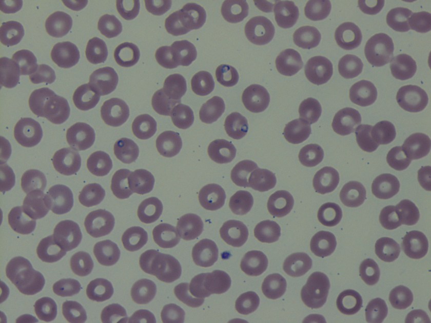
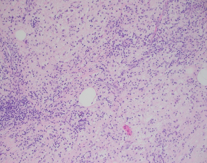
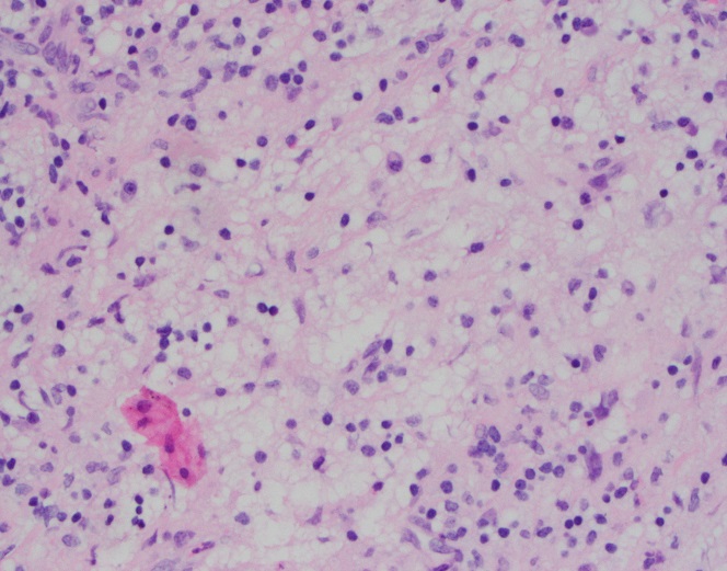
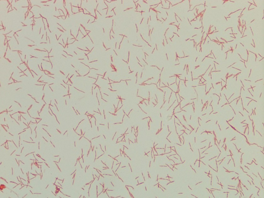
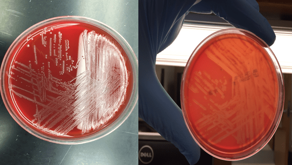
Laboratory findings

cryptlaur1
Image 1: potato flake agar with creamy tan-white colonies.
cryptlaur2.png
Image 2: calcofluor white fluorescent stain showing round yeast forms.

The organism was identified as Cryptococcus laurentii via MALDI-ToF MS. A follow-up fungal culture was negative, however, repeat blood culture grew Stenotrophomonas maltophilia. His tunneled catheter was removed, and two days later the patient required urgent interventional radiology access for dialysis. He completed a two-week course of ceftazidime and was discharged. 

Discussion 

Cryptococcus laurentii is a very rare fungal pathogen. It is a psychrophilic organism, growing ideally at 15 °C, and is the most common yeast found in tundra.1 Major reservoirs include soil, food, and pigeon excrement.2 C. laurentii usually causes infection in immunocompromised hosts, although rare incidents of infection in immunocompetent patients have been reported. Reported manifestations have included fungemia, meningitis, peritonitis, pneumonia, pyelonephritis, keratitis, and skin infection.3

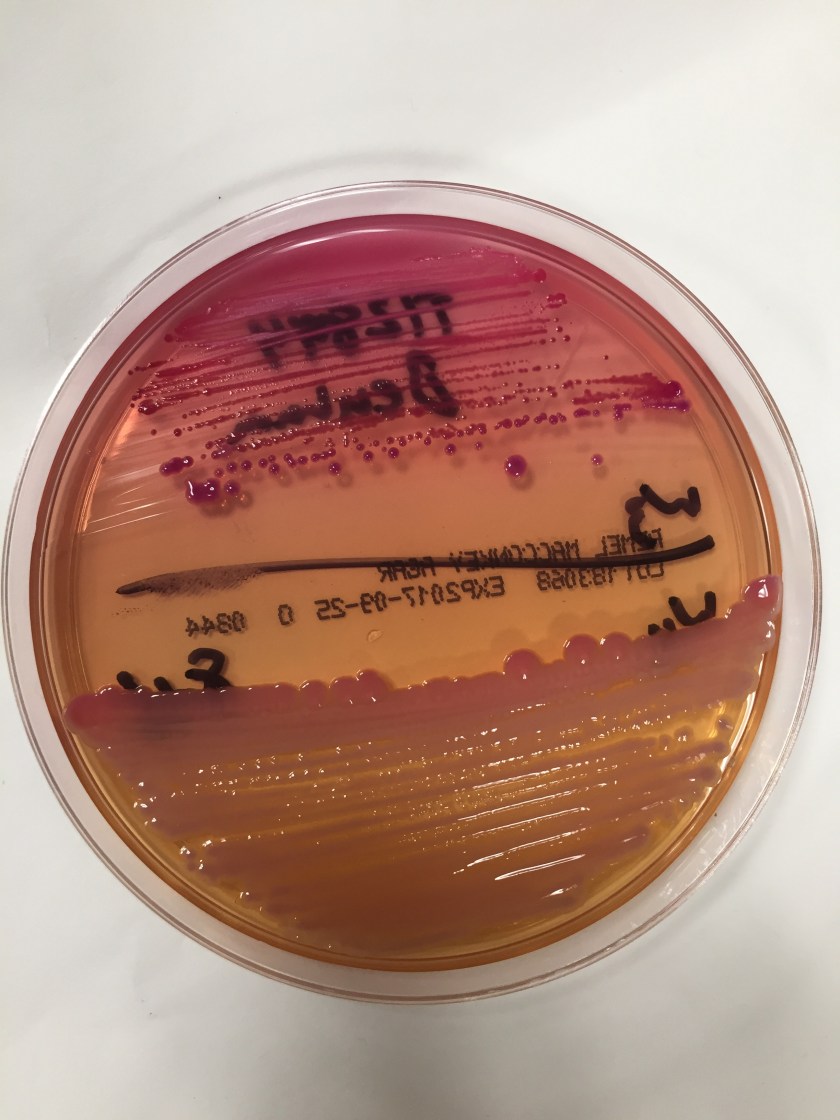
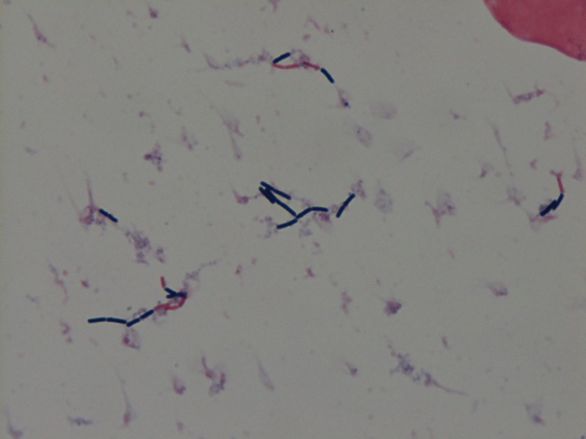
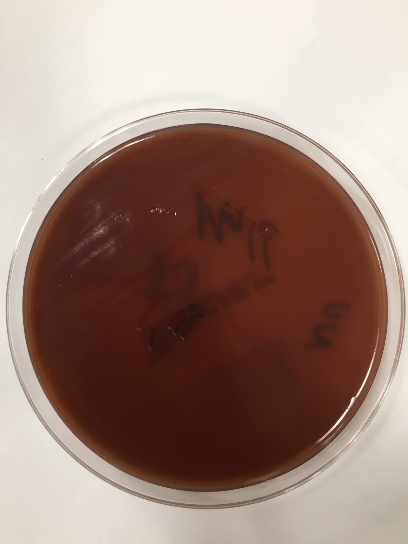
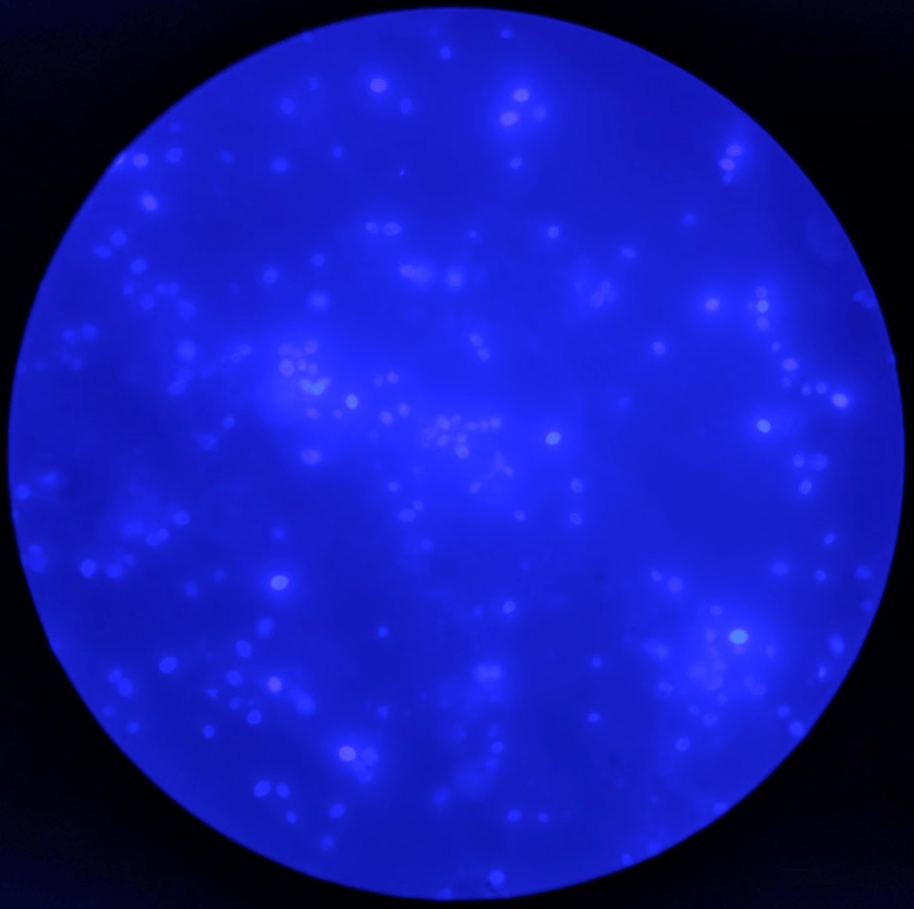
Cryptococcus laurentii is a urease-positive organism. Gram stain shows large budding yeasts without hyphae. The yeast grows on routine agar as whitish-yellow creamy colonies and on birdseed agar as whitish or greenish colonies. Staining with calcofluor highlights encapsulated yeast forms. Molecular diagnosis can be accomplished by ribosomal RNA sequencing of the internal transcribed spacer and D1/D2 regions. Treatment in most cases has been with fluconazole, although in one case of peritoneal dialysis catheter-related peritonitis, voriconazole was used due to low fluconazole susceptibility.4

References

  1. Molina-Leyva A, Ruiz-Carrascosa JC, Leyva-Garcia A, Husein-Elahmed H. Cutaneous Cryptococcus laurentii infection in an immunocompetent child. International Journal of Infectious Diseases. 2013;17(12). doi:10.1016/j.ijid.2013.04.017.
  2. Johnson, L. B., Bradley, S. F. and Kauffman, C. A. Fungaemia due to Cryptococcus laurentii and a review of non-neoformans cryptococcaemia. Mycoses. 1998;41: 277–280. doi:10.1111/j.1439-0507.1998.tb00338.x
  3. Furman-Kuklińska K, Naumnik B, Myśliwiec M. Fungaemia due to Cryptococcus laurentii as a complication of immunosuppressive therapy – a case report. Advances in Medical Sciences. 2009;54(1). doi:10.2478/v10039-009-0014-7.
  4. Asano M, Mizutani M, Nagahara Y, et al. Successful Treatment of Cryptococcus laurentii Peritonitis in a Patient on Peritoneal Dialysis. Internal Medicine. 2015;54(8):941-944. doi:10.2169/internalmedicine.54.3586.


-Prajesh Adhikari, MD is a 3rd year anatomic and clinical pathology resident at the University of Vermont Medical Center.

Wojewoda-small

-Christi Wojewoda, MD, is the Director of Clinical Microbiology at the University of Vermont Medical Center and an Associate Professor at the University of Vermont.

Microbiology Case Study: A 21 Year Old Female with a Sore Throat

A 21 year-old female presented to the emergency department with a sore throat. Her symptoms started two weeks prior to presentation. She was seen at student health last week and given Tamiflu, but her sore throat has grown progressively worse. She now has pain with swallowing and cannot swallow liquids. Upon examination the patient has no rash, no fever, and is not in respiratory distress. She does have left tonsillar fullness causing her uvula to be slightly deviated to the right along with an enlarged left cervical lymph node. Her complete blood count (CBC) was elevated at 19.7 x10³/ul (reference range 4-10 x103/ul) with 12% lymphocytes, 2% monocytes, and 83% neutrophils. A rapid antigen test for S. pyogenes or Group A Streptococcus was negative. A CT exam of the neck was performed and a peritonsillar abscess of 1 x 1.3 x 1.6 cm was identified. The abscess was drained resulting in 1 ml of yellow purulent fluid which was sent to the microbiology lab for culture. The following was Gram stain was prepared from the abscess material.

Fusobacterium necrophorum Gram stain

Discussion

The Gram stain of this abscess showed 4+ PMNs and 4+ small, pleomorphic gram negative bacilli. Anaerobic culture grew Fusobacterium necrophorum, identified by MALDI-TOF MS with a confidence score of 2.2. F. necrophorum is a non-motile, non-pigment forming, pleomorphic gram negative bacilli. It is a strict anaerobe that tests catalase negative, indole positive, and lipase positive on egg yolk agar. Anaerobic antibiotic disk testing for this organism shows susceptibility to kanamycin and colistin with resistance to vancomycin.

The two most clinically relevant species of Fusobacterium are F. nucleatum and F. necrophorum. Because they are strict anaerobes which are often not recovered in culture, Fusobacterium spp. are an under-recognized cause of disease. F. necrophorum colonizes the oral cavity, and like other colonizing anaerobes, it tends to cause infections near the mucosal surface where it resides. F. necrophorum most commonly causes pharyngitis, recurrent tonsillitis, and other odontogenic infections. In adolescents, 10% of tonsillitis that is not caused by S. pyogenes can be attributed to F. necrophorum. These infections can progress to septic thrombophlebitis of the internal jugular vein (Lemierre’s syndrome), bacteremia, and rarely F. necrophorum can cause abscesses throughout the body. Because it is an anaerobic bacterium, susceptibility testing is rarely performed on isolates of F. necrophorum. They are highly susceptible to β-lactam–β-lactamase inhibitor combinations, carbapenems, and metronidazole.

Lemierre’s syndrome was of great concern in our patient since it is most commonly observed in adolescents and young adults that were previously healthy, like our patient. Fortunately, CT scan of the neck showed no indication of thrombophlebitis in our patient. After drainage of the abscess, she felt much better and was able to tolerate liquids. The patient was discharged from the ED with a course of amoxicillin/clavulanate (augmentin). Upon follow up in ENT clinic she gave a more through history of 4-5 episodes of sore throat over the past year.

References 

  1. Manual of Clinical Microbiology, 11th edition
  2. Principles and Practices of Infectious Disease, 7th edition


-Erin McElvania, PhD, D(ABMM), is the Director of Clinical Microbiology NorthShore University Health System in Evanston, Illinois.

Microbiology Case Study: Generalized Weakness after a Trip to the United Kingdom

Case History
A 68 year old man presents with complaints of generalized weakness that started 3 weeks after his trip to the United Kingdom. The patient endorses night sweats, myalgia, fever, headaches, decreased appetite, mild nausea but no vomiting or diarrhea. He denied any history of recent rashes or arthralgia. The patient lives in a tick endemic area in the Northeastern part of the United States. Approximately 2 months ago he noticed an engorged tick while in the shower for which he completed a prophylactic course of doxycycline. He has sufficient titers for Lyme antibody; however no antibodies were present for Anaplasma. Blood smears were ordered for review.

Lab Identification
On the thin blood smears, there were multiple vacuolated, pleomorphic, ring-form like organisms seen in multiple infected red blood cells. No extracellular organisms were identified.

babesia
Image 1. Protozoa species on thin blood smears stained with Giemsa (100x oil immersion).

The organism was identified as a species of Babesia with 0.8% parasitemia and confirmed by PCR as Babesia microti.

Discussion
Babesia species are infectious protozoa which infect and cause lysis of red blood cells. Symptoms develop over the course of weeks to months and vary in severity. The most common symptoms are nonspecific flu-like symptoms (e.g., fever, chills, body aches, weakness, fatigue). If left untreated patients can develop hemolytic anemia, thrombocytopenia, disseminated intravascular coagulation, hemodynamic instability, and possibly death.1

The main agents of human babesiosis are B. microti in the northeastern and upper midwestern regions of the United States, B. duncani in the western regions of the United States, and B. divergens in Europe. Occasionally on blood smears Babesia parasites can be difficult to differentiate from Plasmodium falciparum; Babesia species however are pleomorphic, vacuolated, and can appear inside red blood cells or outside red blood cells whereas Plasmodium falciparum are typically only seen inside red blood cells.1 In most cases the diagnosis for babesiosis can be made on the basis of morphological features on thick and thin smears. However, for patients with subclinical symptoms, very low parasitemia with undetectable organisms on blood smears and a high clinical suspicion for babesiosis serologic and molecular testing can be offered. Serologic testing has 88-96% sensitivity and 100% specificity in patients with no concurrent history of malarial infections. There is some possibility of cross-reactions in serum specimens in patients with malarial infections.2 Molecular techniques such as PCR are excellent tools for the purposes of screening and in addition can help to differentiate amongst the different variants of Babesia species.3 As in this case PCR was used to rule out possible infection with B. divergens due to the patient’s recent travel history to the United Kingdom. The two major antimicrobial regimens for babesiosis are atovaquone plus azithromycin  for mild infection or quinine plus clindamycin for more severe infections.1

References

  1. Resources for Health Professionals. (2013, July 19). Retrieved August 21, 2017, from https://www.cdc.gov/parasites/babesiosis/health_professionals/index.html#dx
  2. Krause PJ, Telford S RI, Ryan R, et al. Diagnosis of babesiosis: Evaluation of a serologic test for the detection of Babesia microti J Infect Dis 1994;169:923-926.
  3. Hojgaard A, Lukacik G, Piesman J. Detection of Borrelia burgdorferiAnaplasma phagocytophilumand Babesia microti, with two different multiplex PCR assays. Ticks and Tick-borne Diseases 2014 (5):349–351.


-Noman Javed, MD is a 1st year anatomic and clinical pathology resident at the University of Vermont Medical Center.

Wojewoda-small

-Christi Wojewoda, MD, is the Director of Clinical Microbiology at the University of Vermont Medical Center and an Associate Professor at the University of Vermont.


Hematopathology Case Study: A 42 Year Old Female with Right Breast Mass

Case History

A 42-year-old female presented with a right breast mass at an outside hospital that was concerning for carcinoma. A core needle biopsy was performed of right breast mass and the case was sent for expert consultation.

Diagnosis

SHML5x
H&E, 5x
SHML10x
H&E, 10x
SHML20x
H&E, 20x
SHML50x
H&E, 50x

Sections of core needle biopsy material are composed primarily of adipose tissue shows a dense lymphohistiocytic infiltrate with histiocytes being the dominant cell type. Admixed plasma cells are present within the infiltrate. The histiocytes have abundant granular cytoplasm with irregular nuclear contours and some nuclei containing inconspicuous nucleoli. Frequent lymphocytic emperipolesis is identified. Immunohistochemistry performed at the outside facility show positivity for S100 and CD163 within the histiocytes, further highlighting the lymphocytic emperipolesis. Cytokeratin immunostains are negative.

Overall, the morphologic and immunophenotypic findings are consistent with a diagnosis of extranodal sinus histiocytosis with massive lymphadenopathy (Rosai-Dorfman disease).

Discussion

Sinus histiocytosis with massive lymphadenopathy (SHML) was first described by Rosai and Dorfman in 1969, however, similar findings may be present in extranodal sites thus earning the designation of Rosai-Dorfman disease (RDD). Although primarily present in lymph nodes, RDD may involve extranodal sites with sinuses and skin being the most frequently affected tissue types. Clinically, RDD often maintains a benign and self-limited course but may undergo exacerbations and recur, requiring surgical management. On histologic examination, RDD involves a rich inflammatory infiltrate with histiocytes, plasma cells, and lymphocytes. The histiocytes usually display a unique phenotype in which lymphocytes are phagocytosed, a process termed emperipolesis. By immunohistochemistry, these histiocytes are positive for S-100 and histiocytic markers (CD68 and CD163) and are negative for CD1a1.

The largest cohort studied involved 423 cases with 182 having extranodal manifestations2. Chest involvement was first reported by Govender et al. in 1997 in a 34-year-old female3. Overall, RDD is considered rare with a slight male predilection and young African-Americans being the most commonly affected. Sites involved ranging from most common to least common include lymph nodes, skin, upper respiratory tract, and bone4.

Extranodal sinus histiocytosis with massive lymphadenopathy, also known as Rosai-Dorfman disease, is a rare pathologic entity that histologically shows a dense lymphohistiocytic infiltrate and emperipolesis, a hallmark of the disease. Although lymph nodes are the most common site of involvement, extranodal sites may be affected and RDD should remain in the differential for lesions that contain abundant histiocytes, plasma cells, and lymphocytes as well as the classic feature of emperipolesis.

References

  1. Komaragiri et al.: Extranodal Rosai–Dorfman disease: a rare soft tissue neoplasm masquerading as a sarcoma. World Journal of Surgical Oncology 2013 11:63.
  2. Penna Costa AL, Oliveira e Silva N, Motta MP, Athanazio RA, Athanazio DA, Athanazio PRF: Soft tissue Rosai–Dorfman disease of the posterior J Bras Pneumol 2009, 35:717–720.
  3. Govender D, Chetty R: Inflammatory pseudotumour and Rosai–Dorfman disease of soft tissue: a histological continuum? J Clin Pathol 1997, 50:79–
  4. Montgomery EA, Meis JM: Rosai–Dorfman disease of soft tissue. Am J Surg Pathol 1992, 16:122–129.


PhillipBlogPic-small

-Phillip Michaels, MD is a board certified anatomic and clinical pathologist who is a current hematopathology fellow at Beth Israel Deaconess Medical Center, Harvard Medical School, Boston, MA. His research interests include molecular profiling of diffuse large B-cell lymphoma as well as pathology resident education, especially in hematopathology and molecular genetic pathology.


Microbiology Case Study: A 53 Year Old Female with Extreme Dysphagia

Case History 

A 53 year old Caucasian female presented to the emergency department with extreme dysphagia and pain in her mouth and throat. Her past medical history was significant for gastric adenocarcinoma for which she underwent a resection and received chemotherapy & radiation treatment. She had been hospitalized previously due to radiation esophagitis. On physical examination, she was cachectic but her vital signs were normal. Numerous ulcers were observed on her tongue and buccal mucosa. Her count blood count revealed she was pancytopenic with a white blood cell count of 0.19. An infectious disease work up was initiated and included blood and throat cultures as well as viral cultures of the oral ulcers for herpes simplex virus.

Laboratory Identification

capno1
Image 1. Gram stain of a bacterial isolate that grew from blood culture showed long fusiform gram negative rods, which slightly tapered at both ends (100x oil immersion).

The blood culture was positive for long gram negative fusiform rods that tapered at both ends (Image 1). The organism grew as very small, whitish-yellow colonies on both blood and chocolate agars after 24 hours incubation in 5% CO2 at 37°C. Biochemical reactions for both catalase and oxidase were negative. The isolate was identified by addition biochemical reactions by the reference bench as Capnocytophaga sputigena. Her throat and viral cultures were negative for additional pathogens.

Discussion 

The Capnocytophaga genus is comprised of nine species that grow as facultative anaerobes and have a characteristic fusiform appearance on Gram stain. Organisms from this genus make up the normal flora of the oral cavity of humans as well as the oral microbiota of dogs and cats. Capnocytophaga spp. contribute to periodontal disease in adolescents and adults and the majority of disseminated infections arise from this endogenous source. The individuals at most at risk for septicemia include those that are immunocompromised (mainly neutropenic patients), alcoholics, intravenous drug users or those that lack a spleen.

In the laboratory, Capnocytophaga spp. is often first recognized by its characteristic Gram stain which shows long, fusiform gram negative rods that taper at both ends. Organisms with similar appearing Gram stain morphology include Fusobacterium spp. and Leptotrichia buccalis, but both of these bacteria exhibit anaerobic growth in contrast to Capnocytophaga spp which grows aerobically. Capnocytophaga isolates tend to grow slowly and require enriched media and increased CO2 concentrations. The Capnocytophaga genus can further be broken down into a catalase- and oxidase-negative group and a catalase- and oxidase-positive group. Species in the first group include C. sputigena, C. gingivalis and C. granulosa. A notable species in the latter group includes C. canimorsus, which when it causes infection in humans it is most likely due to bites or contact with healthy dogs (25% colonization rate) or cats (15% colonization rate). Species differentiation can be challenging as some automated identification instruments can only identify to the genus level and many labs may not offer extensive biochemical work ups. However, the databases for the Bruker and Vitek MALDI-TOF MS currently include many of the species listed above.

In general, Capnocytophaga spp. are susceptible to broad spectrum cephalosporins, carbapenems, tetracyclines and fluoroquinolones. Resistance has been documented for aminoglycosides and colistin. In the case of our patient, her systemic infection was thought to be due to severe mucositis and the endogenous Capnocytophaga sputigena gained access to her blood stream via the numerous ulcers present. She responded well to antibiotic therapy and was discharged home.


Stempak

-Lisa Stempak, MD, is an Assistant Professor of Pathology at the University of Mississippi Medical Center in Jackson, MS. She is certified by the American Board of Pathology in Anatomic and Clinical Pathology as well as Medical Microbiology. She is the director of the Microbiology and Serology Laboratories. Her interests include infectious disease histology, process and quality improvement, and resident education.

Microbiology Case: An Unusual Case of Cholangitis

Case History
A 64 years old male with a past medical history of atrial fibrillation, obstructive sleep apnea, and hypertension presented to the emergency room due to fevers and chills status post stent removal by endoscopic retrograde cholangiopancreatography (ERCP) 1 day earlier. The patient was admitted 6 weeks prior with Klebsiella bacteremia secondary to cholangitis due to an obstructive stone requiring ERCP with sphincterotomy and stent placement. In the emergency room the patient was febrile to 102.7F. Workup included an abdominal x-ray, right upper quadrant ultrasound, and CT abdomen and pelvis all of which were consistent with expected pneumobilia of the biliary tree due to his recent ERCP. On labs his lipase and liver function tests were within normal limits. Blood cultures were drawn and the patient was empirically started on piperacillin/tazobactam. Blood cultures flagged positive after 12 hours.

Laboratory Identification
Gram smear revealed gram negative bacilli. On the blood agar plates there were two different colony morphologies identified. Colony (A) was beta-hemolytic, oxidase positive, and white appearing on blood agar. Colony (B) was gamma-hemolytic, oxidase negative, and greyish appearing on blood agar. Both colonies were lactose fermenters on the MacConkey agar.

Kleb-Aero1
Image 1. Gram stain from a positive blood bottle showing gram negative bacilli (100x oil immersion).
Kleb-Aero2.jpg
Image 2. Aerobic growth on blood agar showing two different colony morphologies. Colony (A) appears white with beta hemolysis and colony (B) appears grey with gamma hemolysis.
Kleb-Aero3.jpg
Image 3. Aerobic growth on chocolate agar showing two different colony morphologies. Colony (A) appears white and raised while colony (B) appears grey and flat.
Kleb-Aero4
Image 4. Comparison of the two morphologically different colonies sub-cultured from blood agar to MacConkey agar. Both colonies (A) and (B) are lactose fermenters.

Using mass spectrometry, the MALDI-TOF positively identified the two organisms as Aeromonas species (colony A) and Klebsiella pneumoniae (colony B). The MALDI-TOF was unable to differentiate between A. hydrophilia and A. caviae species.

Discussion
Klebsiella pneumoniae is a known opportunistic pathogen implicated in nosocomial bacterial gastrointestinal infections. There are several proposed mechanisms by which this organism causes cholangitis which include ascension from the small bowel, contamination of the portal blood, or via translocation of the bowel wall following hematogenous seeding.1

On the contrary, Aeromonas species are not native to the human gastrointestinal tract. These organisms are commonly found in freshwater and marine environments. They are gram negative, oxidase positive, facultative anaerobes. Most gastrointestinal infections caused by Aeromonas species are thought to be due to transient colonization of the GI tract and present asymptomatically or with mild diarrheal disease.6 Extra-intestinal wound infections are possible in the setting of a traumatic aquatic injury and cases of bacteremia have been reported; however these occur in the setting of malignancy or severe hepatobiliary disease.3

In the literature, there are 41 reported cases of hepatobiliary or pancreatic Aeromonas species infection. In almost all of these cases there are no documented aquatic environmental exposures. In one case series, 8/17 (47%) cases were due to nosocomial infections.3 One possible source for these infections can be the hospital water. Despite chlorination, Aeromonas species can be cultured from hospital water supply.4 Since many patients can be asymptomatic while transiently being colonized with Aeromonas species, it is possible that following an ERCP procedure, some organisms can be translocated from the GI tract to the biliary tree causing cholangitis.

To diagnose Aeromonas species a gram smear and biochemical testing should identify gram negative, rod shaped, non-spore forming, oxidase positive, glucose fermenting, facultative anaerobe organisms that are resistant to the vibriostatic agent O/129 and are unable to grow in 6.5% NaCl.2 Their pattern of hemolysis on blood agar can be variable, although most species are beta-hemolytic. Mass spectrometry can further be used to identify at the level of the species. Most Aeromonas strains are resistant to penicillin and ampicillin and some automated MIC systems such as BioMeriuex Vitek may not be able detect the beta-lactam resistance.2 Susceptibility studies should therefore be performed using standard agar dilution, broth microdilution, or using the Kirby-Bauerdisk diffusion method.7

Most Aeromonas species are susceptible to trimethoprim-sulfamethoxazole (TMP-SMX) and fluoroquinolones.5 There are some reported cases of fluoroquinolone resistance in patients that have a history of leech therapy. Aeromonas species can be isolated from the gut of the Hirudo medicinalis leech. These patients often receive systemic chemoprophylaxis to ciprofloxacin before undergoing leech therapy.5

References:

  1. Kochar R, Banerjee S. Infections of the biliary tract. Gastrointest Endosc Clin N Am. 2013 Apr;23(2):199-218.
  2. Morris, G.B., Horneman, A. (2017). Aeromonas Infections. UpToDate. Waltham, Mass.: UpToDate. Retrieved from uptodate.com.
  1. Clark NM, Chenoweth CE. Aeromonas infection of the hepatobiliary system: report of 15 cases and review of the literature. Clin Infect Dis. 2003 Aug 15;37(4):506-13.
  1. Picard B, Goullet P. Seasonal prevalence of nosocomial Aeromonas hydrophila infection related to Aeromonas in hospital water. J Hosp Infect 1987; 10:152–5.
  1. Patel KM, Svestka M, Sinkin J, Ruff P 4th. Ciprofloxacin-resistant Aeromonas hydrophila infection following leech therapy: a case report and review of the literature. J Plast Reconstr Aesthet Surg. 2013 Jan;66(1):e20-2.
  1. Gracey M, Burke V, Robinson J. Aeromonas-associated gastroenteritis. Lancet 1982; 2:1304–6.
  1. Methods for Antimicrobial Dilution and Disk Susceptibility Testing of Infrequently Isolated Fastidious Bacteria. 3rd ed. CLSI guideline M45. Wayne, PA: Clinical and Laboratory Standards Institute; 2016.


-Noman Javed, MD is a 1st year anatomic and clinical pathology resident at the University of Vermont Medical Center.

Wojewoda-small

-Christi Wojewoda, MD, is the Director of Clinical Microbiology at the University of Vermont Medical Center and an Associate Professor at the University of Vermont.

Hematopathology Case Study: A 64 Year Old Man with Widespread Lymphadenopathy

Case history

A 64-year-old, previously healthy man presented with a history of cervical and axillary lymphadenopathy of unknown duration. He did not endorse night sweats, weight loss, or fever. Radiologic examination (CT chest and MRI abdomen) revealed numerous enlarged mediastinal, peritracheal, periaortic, periportal and retroperitoneal lymph nodes. He underwent excisional biopsy of a 3.5 cm axillary lymph node.

foll-lymph

Microscopic Description

Histologic examination of the node revealed distortion of nodal architecture by a proliferation of neoplastic-appearing follicles. Follicles were distinct from one another, and closely packed. In areas the follicles were present back-to-back. Follicular centers were comprised of mostly small, cleaved centrocytes and showed no obvious zonation. There was loss of tingible body macrophages.

Immunophenotyping

Immunohistochemical analysis revealed CD20-positive B cells in a follicular pattern. The germinal centers revealed an underlying follicular dendritic meshwork highlighted by staining for CD21. Interestingly, while the germinal centers demonstrated immunopositivity for BCL-6, there was minimal to absent CD10 staining on follicular B cells. Analysis of BCL-2 staining revealed only few cells to be positive within the follicular centers, consistent with resident follicular helper T cells (Th cells). Equivalent numbers of CD3 and CD5 positive T cells were noted in the interfollicular zones. The Ki-67 proliferation index was estimated at 15-20% within follicular centers. Flow cytometric phenotyping demonstrated a lambda light chain restricted clonal B-cell population expressing CD20, CD19 and, FMC7. These neoplastic B-cells were negative for CD5 and CD10 expression.

Diagnosis

The morphologic features were consistent with Follicular Lymphoma; however the phenotype (BCL-2 negativity in follicular centers) was unusual for this diagnosis. Fluorescence in situ hybridization (FISH) was negative for an IgH/BCL-2 fusion; however, a BCL-6 rearrangement at the 3q27 locus was detected in 70% of the cells.  Taken together, a diagnosis of Follicular Lymphoma with a BCL-6 rearrangement was given.

Discussion

Follicular lymphoma (FL) is a germinal center derived B-cell neoplasm. The majority of cases exhibit the pathognomonic translocation t(1418)(q32; q21). This translocation leads to overexpression of the anti-apoptotic BCL-2 protein, which can be detected by immunohistochemistry on germinal center B cells. Lymphoma cells are usually positive for germinal center origin markers BCL-6 and CD10 and do not co-express CD5. As exhibited in this case, FL can exhibit biologic heterogeneity and may not express these typical markers. The follicular proliferation with absence of germinal center zonation and tingible body macrophages as seen in this case represents classic morphology of follicular lymphoma but aberrant phenotypic markers [and absence of t(14;18)] may be a pitfall in this diagnosis.

FL with lacking of CD10 expression, BCL-2 expression, and t(14;18) translocation and harboring only BCL-6 positivity with 3q27 rearrangement is rare. Only few such cases have been reported in the literature. Published data reveals that the hallmark t(14;18) translocation is absent in about 10-15% of FL. The majority of these cases are negative for BCL-2 expression, and 9-14% of them demonstrate BCL-6 rearrangement (3q27 locus). While BCL-6 rearrangement can be present in both the usual t(14;18) harboring FL, and also in cases without t(14;18), the latter is rare. Interestingly, studies have shown BCL-6 rearrangements to be more frequent in in BCL-2 rearrangement negative FL – which is evidence of the anti-apoptotic role of non-rearranged BCL-6 in certain microenvironments.

One third of t(14;18) negative FL are also reported to have rare or negative expression of CD10. Morphologically, this subtype has been shown to have significantly larger follicles than  their t(14;18)-positive counterparts, but the distinction may not be obvious in all cases. Some of these cases are shown to have a component of monocytoid B cells. This findings can be problematic in differentiating these FL cases from marginal zone lymphoma (MZL) that can also harbor BCL-6 rearrangements and lack t(14;18), CD10 and BCL-2 positivity. Absence of prominent marginal zone proliferation, BCL-6 protein expression and characteristic genetic alterations present in MZL, such as trisomies 3, 7, and 18 can help differentiating MZL from t(14;18)-negative FL.

This case highlights the importance of morphologic evaluation of a excisional biopsy tissue, and FISH studies to help identify the rare t(14;18) negative FL. While the reported cases are few, there is no published difference in prognosis or survival when compared to t(14;18)-positive FL. As such, it is not clear whether the follicular lymphoma grading scheme applies to t(14;18)-negative FL; however, no significant grading difficulties or differences have been reported.

References

  1. Jardin F, Gaulard P, Buchonnet G, et al. Follicular lymphoma without t(14;18) and with BCL-6 rearrangement: a lymphoma subtype with distinct pathological, molecular and clinical characteristics. Leukemia. 2002;16:2309–2317
    2. Leich E, Salaverria I, Bea S, et al. Follicular lymphomas with and without translocation t(14;18) differ in gene expression profiles and genetic alterations. Blood. 2009;114(4):826-834.


Aadil-small

Aadil Ahmed, MD is a 3rd-year anatomic and clinical pathology resident at Loyola University Medical Center. Follow Dr. Ahmed on Twitter @prion87.

Mirza-small

-Kamran M. Mirza, MD PhD is an Assistant Professor of Pathology and Medical Director of Molecular Pathology at Loyola University Medical Center. He was a top 5 honoree in ASCP’s Forty Under 40 2017. Follow Dr. Mirza on twitter @kmirza.

Microbiology Case Study: A 73 Year Old Male with Fever, Lethargy, and Chills

Case History

A 73-year-old man presents to his primary care provider during the height of a bad influenza season with fever, lethargy, and chills. Symptoms started 24 hours prior to presentation. A rapid influenza rapid test was performed in the physician’s office and the result was negative for influenza A and B. What is the most likely cause of this man’s illness?

Answer

Influenza…but how can that be?

Discussion

Rapid antigen testing has been the mainstay for influenza testing since the 1980’s. These tests detect influenza A and B viral nucleoprotein antigens in respiratory specimens, giving a qualitative “positive” or “negative” result. Antigen testing was developed to shorten the turnaround time to results for common respiratory viruses influenza and respiratory syncytial virus (RSV), with an assay run time of approximately 15 minutes compared to the several days it takes for influenza detection by viral culture. Rapid antigen testing is very easy to perform, allowing CLIA-waived testing to be performed at point-of-care.

Unfortunately, rapid antigen testing has poor sensitivity. The most comprehensive analysis found the sensitivity of rapid antigen testing to be around 60% in adults and slightly higher (although still not good) in children. Due to the poor sensitivity, the CDC recommends only employing rapid antigen testing when the prevalence of influenza in the community is >10%…but why does the prevalence matter? Knowing the prevalence of a disease in your population allow you to calculate the positive and negative predictive value.

Positive and negative predictive values answer the question, “What is the chance that my positive test result means my patient has the disease (PPV) or what is the chance that my negative test result means my patient does not have the disease (NPV).” You can calculate the PPV or NPV of any assay by knowing the sensitivity and specificity of an assay along with the prevalence of disease in the community (Figure 1).

flu1
Figure 1. Calculation of positive and negative predictive values.

Positive and negative predictive values fluctuate with the amount of disease seen in a community. For example, if testing for polio in the United States, where the virus has been eradicated, a positive test result by any method is far more likely to be a false-positive than a true-positive result. This is due to the low positive predictive value (PPV) of a positive test result in the setting of non-existent polio. The converse is true for negative predictive values (NPV). In the height of influenza season, a negative test result for influenza in a patient with signs and symptoms of influenza disease is more likely to be a false-negative than a true-negative result.

For influenza rapid antigen testing, the PPV is highest when influenza activity in the community is high (positive test result is likely to indicate influenza infection) and the PPV is lowest when influenza activity is low in the community is low such as in summer, when a positive influenza test result is most likely to be a false-positive result.

Conversely, NPV is highest when influenza activity is low in a community, and a negative test result is most likely indicating that the patient does not have influenza infection. NPV is lowest when influenza activity in a community is high, and a negative test result is more likely to indicate a false-negative result in a patient with influenza infection.

The specificity of rapid antigen assays is tied to the circulating influenza viral subtypes in a given season, and is generally quite high. Sensitivity and specificity do not change due to the prevalence of disease in the community, unlike positive and negative predictive values.


References:

  1. Centers for Disease Control and Prevention (CDC) website on influenza testing (https://www.cdc.gov/flu/professionals/diagnosis/clinician_guidance_ridt.htm)
  2. Altman Douglas G, Bland J Martin. Statistics Notes: Diagnostic tests 2: predictive values BMJ 1994; 309 :102
  3. Chartrand C, Leeflang MM, Minion J, Brewer T, Pai M. Accuracy of Rapid Influenza Diagnostic Tests: A Meta-analysis. Ann Intern Med. 2012;156:500–511.doi: 10.7326/0003-4819-156-7-201204030-00403


-Erin McElvania, PhD, D(ABMM), is the Director of Clinical Microbiology NorthShore University Health System in Evanston, Illinois.

Microbiology Case Study: A 51 Year Old Female with New Onset Progressive Weakness

Case History

A 51 year old female with a past medical history for migraines was otherwise healthy up until 6 weeks ago when she began to notice progressive weakness, myalgia and new onset spontaneous lower extremity bleeding. She was evaluated by the internal medicine service and was found to be profoundly thrombocytopenic. A further workup consisting of a bone marrow biopsy revealed findings that were consistent with high-grade (Burkitt’s) lymphoma. She was initiated on chemotherapy. Two days after initiating chemotherapy she became profoundly pancytopenic with recurrent fevers. Additionally, she had worsening erythema and pain in her right buttocks and left thigh. Despite the usage of broad-spectrum antibiotics, her symptoms worsened. Two sets of blood cultures were drawn and the anaerobic bottles of both sets flagged positive after 15 hours.

Lab Identification

Gram stain revealed gram positive rods, some of which did not retain the crystal violet stain but all appeared box car shaped. This organism only grew under anaerobic conditions. On the anaerobic blood plate, the organism swarmed the media.

closept1
Image 1. Gram stain from a positive blood bottle showing gram positive rods (100x oil immersion).
closept2
Image 2. Anaerobic growth on blood agar showing a few colonies with a lawn of growth.

By use of mass spectrometry, the MALDI-TOF positively identified the organism as Clostridium septicum.

Discussion

Clostridium septicum is a gram positive, highly motile, spore-forming organism that grows best under anaerobic conditions. It is found ubiquitously in soil and at a low prevalence rate in the human gastrointestinal tract. C. septicum is best known for its ability to cause neutropenic enterocolitis and “atraumatic” (spontaneous) gas gangrene which is in contrast to “traumatic” gas gangrene caused by C. perfrigens .2 Both neutropenic enterocolitis and atraumatic gas gangrene are commonly seen in association with a malignancy usually hematologic or gastrointestinal in nature. Neutropenic entercolitis is mostly commonly seen in patients that are undergoing chemotherapy treatment. A combination of mucosal injury by the cytotoxic drugs, profound neutropenia, and impaired host defense allows for edema and necrosis of the bowel wall by microorganisms.1 This then allows for hematogenous dissemination of gut flora which includes C. septicum to more distal sites. It is also possible to have weakness in the mucosal lining from mass effect alone without any preceding neutropenia also allowing for hematogenous dissemination. In animal models C. septicum has been shown to be much more virulent than C. perfrigens requiring 300x fewer organisms to have the same lethal effect.2 This lethal infection even with appropriate treatment has a mortality rate of 60%.3

To diagnose C. septicum a gram smear will show gram positive rods with occasional rare sub terminal or terminal spores. They can often appear pleomorphic. They grow under anaerobic conditions and may start out as a single solid colony but usually swarm the plate after 24 hours growth. A more conclusive diagnosis can be made on the MALDI-TOF using mass spectrometry. Effective treatment requires both debridement of infected sites and appropriate antibiotics. The Infectious Diseases Society of America (IDSA) guidelines for skin and soft tissue infections recommend the use of high dose IV penicillin and IV clindamycin.4 Clindamycin is a protein synthesis inhibitor and is believed to aid in preventing toxin synthesis.


References:

  1. Urbach DR, & Rotstein, OD. Typhlitis. Cancer J Surg 1999; 42(6):3 415-419.
  2. Srivastava I, Aldape MJ, Bryant AE, Stevens DL, Spontaneous C. septicum gas gangrene: A literature review, Anaerobe (2017).
  3. Larson CM, Bubrick MP, Jacobs DM, West MA. Malignancy, mortality, and medicosurgical management of Clostridium septicum infection. Surgery. 118(4):592–597
  4. Stevens DL, Bisno AL, Chambers HF, et al. Executive Summary: Practice Guidelines for the Diagnosis and Management of Skin and Soft Tissue Infections: 2014 Update by the Infectious Diseases Society of America. Clinical Infectious Diseases 2014; 59:147–159.


-Noman Javed, MD is a 1st year anatomic and clinical pathology resident at the University of Vermont Medical Center.

Wojewoda-small

-Christi Wojewoda, MD, is the Director of Clinical Microbiology at the University of Vermont Medical Center and an Associate Professor at the University of Vermont.

Microbiology Case Study: 6 Year Old Male with Meningitis

Case History

A 6 year old male presented to the emergency department with a concern for ventriculo-peritoneal shunt (VP) malfunction. His past medical history is significant for myelomeningocele and hydrocephalus since birth. On arrival, symptoms included high fever (102.7°F), headaches and swelling at the VP shunt catheter site in the neck. Over the past week, his mother also noted nausea, vomiting and diarrhea. CT scan of the head revealed increased size of the 3rd and lateral ventricles which was concerning for either a VP shunt malfunction or infection. Lab work showed a white count of 13.5 TH/cm2 and elevated CRP values suggestive of an infection/inflammatory process. He was taken to surgery for VP shunt removal and placement of an external ventricular drain (EVD). Intra-operatively, purulent yellow material was noted at both the proximal and distal ends of the catheter. Cerebrospinal fluid (CSF) was sent for Gram stain and bacterial culture. He was started on vancomycin and ceftriaxone.

 Laboratory Identification

listmon1
Image 1. CSF Gram stain prepared from the cytospin showed many white blood cells and Gram positive bacilli (100x oil immersion).
listmon2
Image 2. Gram stain from the liquid media culture showing gram positive bacilli (100x oil immersion).
listmon3.png
Image 3. Small, grayish colonies with a narrow zone of beta hemolysis grew on blood agar after 48 hours incubation in a 35°C incubator with 5% CO2.

Bacterial cultures collected from a shunt tap and intra-operatively both showed short gram positive bacilli on Gram stain (Image 1&2). The organism grew on blood and chocolate agars as small, gray colonies with a narrow zone of beta-hemolysis when observed closely (Image 3) after incubation at 35°C in CO2. The isolate was positive for catalase and showed a “tumbling motility.” MALDI-TOF MS identified the isolate as Listeria monocytogenes.

 Discussion

Listeria species are gram positive bacilli that grow as facultative anaerobes and do not produce endospores. The major human pathogen in the Listeria genus is L. monocytogenes and it is found in soil, stream water, sewage & vegetable matter and may colonize the gastrointestinal tract of humans and animals.

The most common mode of transmission is ingestion of contaminated foods, in particular, raw milk, soft cheeses, deli meats and ice cream. L. monocytogenes’ ability to grow at cold temperatures (4°C) permits multiplication in refrigerated foods. In a healthy adult, it causes an influenza like illness and gastroenteritis. Pregnant women are especially susceptible to disease and neonates infected in utero can develop granulomatosis infantiseptica which can lead to miscarriage, stillbirth or premature delivery. Elderly or immunocompromised can present with a febrile illness, bacteremia and meningitis (20-50% mortality).

In the microbiology laboratory, L. monocytogenes is usually identified via blood, CSF or placental bacterial cultures. It grows well on standard agars and after overnight incubation, the small, gray colonies show a narrow zone of beta hemolysis on blood agar. L. monocytogenes is positive for catalase & esculin and the CAMP test demonstrates block like accentuated hemolysis. It has characteristic tumbling motility at room temperature and an umbrella shaped motility pattern in semi-solid agar.  Automated methods of identification provide reliable species level differentiation on the majority of current platforms.

Susceptibility testing should be performed on isolates from normally sterile sites. Ampicillin, penicillin, or amoxicillin are given for L. monocytogenes, and gentamicin is often added for its synergistic effect in invasive infections. Trimethoprim-sulfamethoxazole and vancomycin can be used in cases of allergy to penicillin. Cephalosporins are not effective for treatment of listeriosis.

In the case of our patient, after L. moncytogenes was identified, his antibiotic therapy was changed to ampicillin and gentamicin. Antibiotics were administered for 3 weeks before the placement of a new VP shunt. On further questioning, his mother revealed his diet consisted heavily of hot dogs and soft cheeses. She was educated on how to prevent subsequent infections prior to discharge.


JKO

-Jaspreet Kaur Oberoi, MD, is a Pathology resident at the University of Mississippi Medical Center. 


Stempak

-Lisa Stempak, MD, is an Assistant Professor of Pathology at the University of Mississippi Medical Center in Jackson, MS. She is certified by the American Board of Pathology in Anatomic and Clinical Pathology as well as Medical Microbiology. She is the director of the Microbiology and Serology Laboratories. Her interests include infectious disease histology, process and quality improvement and resident education.