Case History
A 53 year old Caucasian female presented to the emergency department with extreme dysphagia and pain in her mouth and throat. Her past medical history was significant for gastric adenocarcinoma for which she underwent a resection and received chemotherapy & radiation treatment. She had been hospitalized previously due to radiation esophagitis. On physical examination, she was cachectic but her vital signs were normal. Numerous ulcers were observed on her tongue and buccal mucosa. Her count blood count revealed she was pancytopenic with a white blood cell count of 0.19. An infectious disease work up was initiated and included blood and throat cultures as well as viral cultures of the oral ulcers for herpes simplex virus.
Laboratory Identification
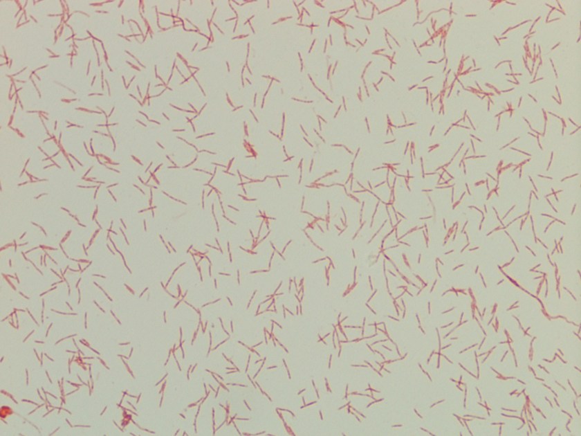
The blood culture was positive for long gram negative fusiform rods that tapered at both ends (Image 1). The organism grew as very small, whitish-yellow colonies on both blood and chocolate agars after 24 hours incubation in 5% CO2 at 37°C. Biochemical reactions for both catalase and oxidase were negative. The isolate was identified by addition biochemical reactions by the reference bench as Capnocytophaga sputigena. Her throat and viral cultures were negative for additional pathogens.
Discussion
The Capnocytophaga genus is comprised of nine species that grow as facultative anaerobes and have a characteristic fusiform appearance on Gram stain. Organisms from this genus make up the normal flora of the oral cavity of humans as well as the oral microbiota of dogs and cats. Capnocytophaga spp. contribute to periodontal disease in adolescents and adults and the majority of disseminated infections arise from this endogenous source. The individuals at most at risk for septicemia include those that are immunocompromised (mainly neutropenic patients), alcoholics, intravenous drug users or those that lack a spleen.
In the laboratory, Capnocytophaga spp. is often first recognized by its characteristic Gram stain which shows long, fusiform gram negative rods that taper at both ends. Organisms with similar appearing Gram stain morphology include Fusobacterium spp. and Leptotrichia buccalis, but both of these bacteria exhibit anaerobic growth in contrast to Capnocytophaga spp which grows aerobically. Capnocytophaga isolates tend to grow slowly and require enriched media and increased CO2 concentrations. The Capnocytophaga genus can further be broken down into a catalase- and oxidase-negative group and a catalase- and oxidase-positive group. Species in the first group include C. sputigena, C. gingivalis and C. granulosa. A notable species in the latter group includes C. canimorsus, which when it causes infection in humans it is most likely due to bites or contact with healthy dogs (25% colonization rate) or cats (15% colonization rate). Species differentiation can be challenging as some automated identification instruments can only identify to the genus level and many labs may not offer extensive biochemical work ups. However, the databases for the Bruker and Vitek MALDI-TOF MS currently include many of the species listed above.
In general, Capnocytophaga spp. are susceptible to broad spectrum cephalosporins, carbapenems, tetracyclines and fluoroquinolones. Resistance has been documented for aminoglycosides and colistin. In the case of our patient, her systemic infection was thought to be due to severe mucositis and the endogenous Capnocytophaga sputigena gained access to her blood stream via the numerous ulcers present. She responded well to antibiotic therapy and was discharged home.

-Lisa Stempak, MD, is an Assistant Professor of Pathology at the University of Mississippi Medical Center in Jackson, MS. She is certified by the American Board of Pathology in Anatomic and Clinical Pathology as well as Medical Microbiology. She is the director of the Microbiology and Serology Laboratories. Her interests include infectious disease histology, process and quality improvement, and resident education.
