Von Willebrand Disease

Von willebrand disease is the most common inherited bleeding disorder. Partial lack of VWF (von Willebrand factor) causes mild or moderate bleeding tendency. Patients typically present with menorrhagia, bruising, bleeding from gums or after surgery. It is typically autosomal dominant with variable penetrance. Laboratory investigation reveals defective platelet adherence (PFA-100) or long bleeding time, subnormal levels of von Willebrand antigen and factor VIII in plasma and low ristocetin cofactor activity or VWF activity.

Single very large molecules visualized by electron microscopy
Single very large molecules visualized by electron microscopy. Journal of Clinical Investigation, vol 76, October 1985
vWF unfolds under stress, the faster the blood flow, the stickier it gets. Blood, 1996; volume 88
vWF unfolds under stress, the faster the blood flow, the stickier it gets.
Blood, 1996; volume 88

Types of von Willebrand disease

  • Type 3: Patient has severe deficiency of vWF. Antigen, activity and factor VIII levels are all < 10%. Clinically patients have a hemophilia-like phenotype. It is inherited recessively.
  • Type 2: Patients have a qualitative defect (missense mutation) of the vWF. There are several different types. Usually there is a disproportionate decrease in vWF activity vs antigen.
  • Type 1: vWF antigen and activity are reduced proportionately. vWF levels range from < 20% to ~50%. Only 65% of cases are associated with VWF gene mutations. It has an autosomal dominant inheritance pattern with variable penetrance (affected by blood type, other factors).Defects in VWF processing, storage or secretion may account for cases lacking VWF gene mutation. Some cases are associated with accelerated VWF clearance.

Type-2 vWD

  • 2A: Deficiency of intermediate & large multimers due to either defective assembly (mutation in either of two domains involved in multimer formation), or increased susceptibility to proteolysis (mutation in domain cleaved by ADAMTS-13)
  • 2B: Largest multimers are missing. This is due to gain of function mutation in platelet Gp Ib binding domain of vWF.Largest multimers bind spontaneously to platelets and are cleared from blood. Rarely, a mutation in Gp Ib may have the same effect (“platelet-type” vWD).
  • 2M: Normal multimer pattern. There is loss of function mutation in GP Ib binding domain
American Society of Hematology.
Image: American Society of Hematology.

Laboratory analysis

Laboratory testing in vWD (Table 1)

Von Willebrand factor activity: Measures binding of patient VWF to latex beads coated with monoclonal Ab to GPIb binding site; sensitive to multimer size and platelet-binding ability

Platelet function screen (PFA): Measures time necessary for platelet plug to form in collagen coated tube under high shear conditions in the presence of ADP or epinephrine

Desmopressin (DDAVP) in VWD

DDAVP releases vWF from endothelial cells and can be given IV or intranasally ( 0.3 mcg/kg IV, or 150 mcg per nostril ). It typically causes 2-4 fold increase in blood levels of vWF (in type 1 vWD), with half-life of 8+ hours. Response to DDAVP varies considerably. Administration of a trial dose si necessary to ensure a given patient responds adequately.

vwd4

Indications for clotting factor concentrate administration in vWD include

  • Type 2 or 3 vWD with active bleeding, surgery or other invasive procedures or
  • Type 1 vWD with inadequate response to DDAVP.

Acquired von Willebrand Disease

This can happen in association with either monoclonal gammopathy ( vWF neutralized by paraprotein) , autoimmune disorders (autoantibody to vWF), myeloproliferative disorder (large multimers absorbed onto neoplastic cells), cardiovascular diseases (AS, VSD, etc, high shear stress causes unfolding/proteolysis of large multimers), or hypothyroidism (decreased release of vWF from endothelial cells). Treatment varies depending on the cause/mechanism in each case.

NEJM 2009;361:1887
NEJM 2009;361:1887


Vajpayee,Neerja2014_small

-Neerja Vajpayee, MD, is an Associate Professor of Pathology at the SUNY Upstate Medical University, Syracuse, NY. She enjoys teaching hematology to residents, fellows and laboratory technologists.

You Make the Diagnosis: A 62 Year Old Female with Hemoptysis and Fatigue

A 62-year-old female presents with hemoptysis and fatigue. A large mass is found adjacent to the right main bronchus. A representative field is shown here. What is the diagnosis?

smallcell

  1. Invasive adenocarcinoma
  2. Squamous cell carcinoma
  3. Adenocarcinoma in situ (formerly bronchoalveolar carcinoma)
  4. Small cell carcinoma
  5. Large cell carcinoma


The diagnosis in this case is small cell carcinoma. Also called “oat cells,” the malignant cells in this tumor tend to be round to somewhat elongated in shape, with the typical “salt-and-pepper” chromatin of neuroendocrine tumor cells. The cells are often so closely apposed that their nuclear contours show a characteristic “molding” effect. Small cell carcinoma is a fast growing tumor (note the large mitotic figure at 4 o’clock) with a poor prognosis.


Krafts

-Kristine Krafts, MD, is an Assistant Professor of Pathology at the University of Minnesota School of Medicine and School of Dentistry and the founder of the educational website Pathology Student.


Sweat Testing

August in Texas is a good time to write a blog post about sweat. In this case though, I’m going to specifically talk about testing collected sweat samples for chloride concentration. Sweat chloride concentrations are measured in people who are suspected of having Cystic Fibrosis (CF). Because CF has classically been considered a disease of childhood, sweat chloride testing is performed almost exclusively in pediatric institutions.

CF is a disease caused by mutations in the cystic fibrosis transmembrane conductance regulator (CFTR) gene. This is a large gene which codes for a large, transmembrane protein that acts as a chloride channel. More than 1500 mutations have been detected in the CFTR gene, not all of which are known to cause disease. Thus, even though the full gene has been sequenced, CF remains a diagnosis which is made by a combination of the presence of characteristic clinical features, or history of CF in sibling, or a positive newborn screen, PLUS identification of a disease-causing mutation in the gene or protein or laboratory evidence of chloride channel malfunction such as an elevated sweat chloride level.

Collecting a sweat sample for testing is an interesting manual process. The first step involves stimulating the sweat glands to produce sweat. This is accomplished by a process called iontophoresis, in which a sweat-gland-stimulating compound called pilocarpine is driven into the skin using a small electrical current between a set of electrodes applied to the skin. After a 5 minute stimulation, the electrodes are removed, the skin is cleaned, and the sweat that is subsequently produced in that stimulated area is collected for the next 30 minutes. The collection is either via absorption of the sweat into a piece of gauze or filter paper, or by a collection device which funnels the sweat into a small plastic tube as it’s produced. The amount of sweat collected after 30 minutes is determined by weight if gauze or filter paper is used, and by volume if the tubing is used. There is a lower acceptable limit for both cases, below which the sweat collection is insufficient (QNS) and must be repeated. The process sounds simple, however collecting a sufficient quantity of sweat can be problematic, and collecting too little may cause falsely elevated results.

After collection, the amount of chloride present in the collected sample is measured. In a normal sample, the amount of chloride present is well below the measurement range of the usual chloride ion-selective electrodes found in chemistry or blood gas instruments. For this reason, the chloride concentration in a sweat sample is most commonly measured using a method called coulometric titration in which a silver electrode placed in the sample gives off silver ions during a current flow. The silver ions complex with the chloride and precipitate as silver-chloride. This reaction continues until all the free chloride is gone, at which point a timer stops. Quantification is accomplished essentially by comparing the time necessary to complex all the patient’s chloride versus the time necessary to complex a known concentration of chloride in a standard. Calculations are performed using the time and the weight or volume of the sweat collected, among other parameters.

The entire test is very manual. Collection of appropriate sweat samples requires training and practice. In general the QNS rate – how often an adequate collection is not achieved – is carefully monitored by the lab, the CF clinic and the CF Foundation which accredits the clinic. In addition, measuring the chloride in the sweat by chloridometer is not an automated process of placing the sample on an instrument and pushing a button to go. For these reasons, the CFF recommends not performing sweat testing unless you perform a minimum number per year in order to stay proficient. In this day and age of increasing automation, sweat chloride testing remains the anomalous, old fashioned test requiring significant technologist time and expertise.

-Patti Jones PhD, DABCC, FACB, is the Clinical Director of the Chemistry and Metabolic Disease Laboratories at Children’s Medical Center in Dallas, TX and a Professor of Pathology at University of Texas Southwestern Medical Center in Dallas.

Microbiology Case Study: 58 Year Old Male with Right Upper Quadrant Pain

Case:

A 58 year old male with a history of pica presented to the emergency room complaining of bilious, non-bloody emesis associated with right upper quadrant pain, fatigue, decreased oral intake, and fevers. Initial labs revealed a lactate of 7.4, a white blood cell count of 22K, alkaline phosphatase 456, AST 373, ALT 444, total bilirubin 5.8, and a troponin of 3.4. Imaging showed a distended, gangrenous gallbladder with compression of the biliary system. Aerobic and anaerobic blood cultures were drawn and the anaerobic culture flagged positive at 20 hours with the following gram stain and colony morphology.


Large Gram-positive boxcar shaped bacilli.
Schaedler plate showing double zone of beta hemolysis.
Opacification of egg yolk agar around bacterial colonies.

Laboratory identification:

Clostridium perfringens was suspected due to the presence of large gram-positive boxcar shaped bacteria on the gram stain and obligate anaerobic growth with a double zone of hemolysis on the Schaedler plate. Opacification of the egg yolk agar demonstrated that the organism was lecithinase positive which is another characteristic of C. perfringens. Identification of the organism was confirmed by mass spectrometry.

Discussion:

Clostridium perfringens grows on the anaerobic blood agar plate as small gray to gray-yellow colonies with a glossy, dome- shaped appearance with a double zone of beta hemolysis. The organism is an obligate anaerobe and therefore the aerotolerance test is negative. Its biochemical characteristics including the following: lecithinase positive, lipase negative, and catalase negative.

C. perfringens is encountered in a number of clinical settings including wound infections, gas gangrene, bacteremia, septicemia, and food borne illness. The organism has a number of virulence factors including exotoxins and an enterotoxin. Although there are reports of resistance, penicillin is still recommended therapy in many cases.

C. perfringens is found in the biliary system and is associated with gangrenous cholecystitis.


-Lauren Pearson, D.O. is a 2nd year anatomic and clinical pathology resident at the University of Vermont Medical Center.

Wojewoda-small

-Christi Wojewoda, MD, is the Director of Clinical Microbiology at the University of Vermont Medical Center and an Assistant Professor at the University of Vermont.


Microbiology Case Study–A Very Sick 51 Year Old Man

Case history:

A 51 year old man with a medical history of liver abnormalities and long standing alcohol abuse presented with generalized weakness, hypoxemic respiratory failure, sputum production, significant hyperbilirubinemia, macrocytic anemia, and laboratory tests consistent with DIC. Chest X ray revealed a bronchopneumonia pattern. Sputum samples were sent for culture and blood cultures were obtained. Urine antigen legionella test was positive for Legionella pneumophila serogroup 1. The sputum was then plated on buffered charcoal yeast extract (BCYE) agar.

Laboratory identification:

Gram stain of colonies growing on BCYE revealed thin short to filamentous rod shaped organisms. The definitive diagnosis of Legionella pneumophila was made by isolating the organism on BCYE with confirmation on the MALDI-ToF. Colonies were speckled blue and

Legionella on BCYE agar plate
Gram negative, thin, short-to-filamentous rod shaped organisms

Discussion:

Legionella pneumophila was first identified and recognized during the outbreak that occurred during the American Legion Convention in Philadelphia in 1976. There are over 40 species of Legionella and 18 of those are human pathogens. Among those, L. pneumophila is an important cause of nosocomial and community-acquired pneumonia (CAP) and should be considered in the differential diagnosis in any patient who presents with pneumonia. Two clinical syndromes caused by this microorganism are: Legionnaires’ disease and Pontiac fever (acute, febrile, self-limited illness).

L. pneumophila is a thin faintly staining short to filamentous gram negative rod. Legionella is a fastidious organism and does not grow on standard media. Buffered charcoal yeast extract (BCYE) agar is the primary medium used for its isolation and it is the gold standard for its diagnosis although testing for the antigen in urine is more commonly performed. The urinary antigen assay only detects L. pneumophila serogroup 1 which cause 95-98% of community acquired Legionnaires’ disease.

The microorganism is found in natural water supplies and soil. It is also found in recirculating and water supply systems, where it breeds rapidly in favorable conditions (temperature of 35°C, range 25-45, stagnant water and water containing organic debris which can provide nutrients for growth). Macrolides (azithromycin or clarithromycin) or fluoroquinolones (levofloxacin or moxifloxacin) are the standard antibiotic drugs used to treat Legionnaires’ disease in humans.

Kossivi Dantey, M.D. is a 4th year anatomic and clinical pathology resident at the University of Vermont Medical Center.

Wojewoda-small

–Christi Wojewoda, MD, is certified by the American Board of Pathology in AP/CP and Medical Microbiology. She is currently the Director of Clinical Microbiology at the University of Vermont Medical Center and an Assistant Professor at the University of Vermont.

iridescent.

Naegleria fowleri

It’s summertime, which means that people are enjoying the outdoors by swimming in lakes, rivers, and ponds. It also means that those swimmers are at risk for Naegleria fowleri, an amoeba that causes a rare infection called primary amebic meningoencephalitis (PAM).

In this podcast, Dr. Lori Racsa discusses the laboratory identification of this amoeba. While most microbiology techs are unlikely to encounter a case, it’s best to be prepared.

Regulation of Laboratory Developed Tests (LDTs) – Revisited

Two years ago this coming September I posted a blog about the FDA’s intent to regulate LDTs and the need for laboratory professionals to both keep an eye on what happens and to be a part of it. I believe it’s time for an update on what has been happening and a further exhortation to stay involved.

The FDA is definitely going to regulate all LDTs. This is no longer a future possibility, but is now an approaching reality. In October of 2014, the FDA put out two new draft guidance documents for 120-day comment periods. One document, “Framework for Regulatory Oversight of Laboratory Developed Tests (LDTs)” lays out the FDA’s various risk categories and classifications for different LDTs and also lays out the FDA’s timeline for enforcing regulation of them. The second document, “FDA Notification and Medical Device Reporting for Laboratory Developed Tests (LDTs)” delineates how labs will report their LDT testing to the FDA and the protocol for adverse event reporting to the FDA, which all labs performing LDTs will be required to do.

During the 120-day comment period, many groups commented, weighing in on their perspective about the FDA regulation of LDTs. AACC and the Association for Molecular Pathology (AMP) published position statements. CAP Today did a comprehensive article. Although nearly everyone agrees that some form of LDT regulation is necessary, there is a wide range of opinions on what that regulation should entail, and even who should ultimately be responsible for it.

Despite many suggestions that perhaps the FDA should approach this regulation differently, they plan to move forward. Their “Framework . . .” document lays out about a nine-year timeline for regulating all LDTs, starting first with the highest-risk group. LDTs will broadly be classified into three groups: low-risk, which are also known as “traditional” LDTS, moderate-risk and high-risk. Traditional LDTs are those developed by a single lab for use on a single patient population. This classification will cover many hospital-based LDTs and it will have the least rigorous regulation by the proposed guidance documents. Moderate and high-risk LDTs will be tackled first by the FDA and will require pre-market review and approval as part of the regulatory requirements.

The FDA is perhaps listening to some of the comments being generated however. Most recently the FDA has announced that an interagency taskforce will be formed to deal with LDT regulation. Currently that task force includes the FDA and CMS, although many laboratory associations are hoping it will be expanded to include more groups. As laboratory professionals, it’s up to us to stay informed of this new regulation headed our way, and to do our best to be involved in the process.

-Patti Jones PhD, DABCC, FACB, is the Clinical Director of the Chemistry and Metabolic Disease Laboratories at Children’s Medical Center in Dallas, TX and a Professor of Pathology at University of Texas Southwestern Medical Center in Dallas.

Microbiology Case Study: 5 Year Old Male Immigrant with Rough Skin Lesions

Case:

A 5 year old boy who recently immigrated to the United States from Africa presented to his primary care physician with several rough, hypopigmented skin lesions and some hair loss on the scalp. The rash was treated with topical antifungal medication. The patient was treated with griseofulvin for tinea capitis and initially responded well. Six months later he developed pruritic macules on his body, for which clotrimazole was prescribed. The patient’s scalp at that time showed few lesions concerning for recalcitrant tinea capitis. He was prescribed weekly fluconazole and topical clotrimazole. Fungal cultures grew an organism with the following scotch tape prep and colony morphology.

Lactophenol analine blue tape preparation.
Colony morphology with deep purple red pigmentation.


Laboratory identification:

The scotch tape prep showed branched, tortuous hyphae and chlamydoconidia in chains but no microconidia or macroconidia were present. The colonies took over two weeks to grow and were initially cream-colored but later developed a port wine to deep violet color. Based on this information Trichophyton violaceum, Trichophyton rubrum, and Trichophyton soudanense were on the differential diagnosis. The specimen was sent to a referral lab for definitive speciation. The referral lab identified the organism as Trichophyton violaceum by MALDI-ToF. The organism was also sequenced for confirmation.

Discussion:

Trichophyton violaceum is a dermatophyte that can be recovered from hair, skin, and nails. The organism requires 14-18 days to grow. Its growth is enhanced by media containing thiamine, which helps differentiate it from other species within the Trichophyton genus. Infection typically causes “black dot” tinea capitis, tinea corporis, and onychomycosis. Infections with this particular Trichophyton species are seen primarily in persons living in Mediterranean region, the Middle East, and Africa. It is treated with oral griseofulvin.


-Lauren Pearson, D.O. is a 2nd year anatomic and clinical pathology resident at the University of Vermont Medical Center.

Wojewoda-small

-Christi Wojewoda, MD, is the Director of Clinical Microbiology at the University of Vermont Medical Center and an Assistant Professor at the University of Vermont.


Five Things to Know … Granulocyte Transfusions

This is the first in series of “5 Things to Know” updates on various Transfusion Medicine topics, posted on Lablogatory and the Blood Bank Guy website. Today, we will cover a topic that is a mystery to many: Granulocyte transfusions!

1. We aren’t totally sure that granulocytes actually work! In the nearly half-century that granulocyte concentrates have been transfused, numerous retrospective articles, case reports, and small series about granulocytes have been published. If you ask Hematology-Oncology docs, you will likely find a general consensus that granulocytes can be effective (including a really cool, almost miraculous-sounding story!). Sadly, stories are not evidence. A study begun in 2008 called The “RING study” (Resolving Infection in people with Neutropenia with Granulocytes) was designed to gather evidence by randomizing recipients into either a group that received granulocytes or one that did not. Unfortunately, the study failed to gather enough participants before closing in 2013 to be truly meaningful (though the data that was gathered did not show a substantial beneficial effect, according to data presented at the 2014 American Society of Hematology Annual Meeting). PByoukeepusingthatword-Product-2

So, does this mean that granulocyte transfusions are stupid? Not from my perspective! Honestly, if the standard for producing a product is proof positive that the product is effective, we blood bankers would have NOTHING on our shelves! Some of my colleagues disagree with me, but from my perspective, granulocytes have value in some limited settings (outlined below).

2. Modern granulocyte collections are a multiple day process. Historically in the United States, granulocytes were collected by apheresis either from “walk-in” unstimulated donors (more common) or from donors given a single dose of oral steroids to increase their white blood cell count. The unstimulated collections resulted in a product with a total granulocyte count of about 1.0 x 10¹⁰ (that number is the minimum requirement in at least 75% of collections, according to AABB Standards, and is a GREAT number to remember if you are studying for an exam!). However, if we have learned anything about granulocytes, we know that 1.0 x 10¹⁰ just isn’t enough! A “modern” U.S. granulocyte collection is from an apheresis donor stimulated the day before collection with an injection of Granulocyte Colony Stimulating Factor (“G-CSF”), in many cases supplemented by an oral dose of steroids (typically dexamethasone); this regimen typically results in a yield of 4.0 x 10¹⁰ granulocytes or more (please note that G-CSF is not FDA-approved for use in stimulating donors, so donors should have a formal informed consent prior to undergoing stimulation). As an aside, granulocytes may be produced in different ways outside of the US. The United Kingdom, in particular, does not permit G-CSF or steroid stimulation of granulocyte donors that are not family or friends of the patient. As a result, non-family member UK granulocyte products are produced from pooled buffy coats from ten random donors, with a yield of approximately 1.0 x 10¹⁰ granulocytes. These collections require lots of extra effort by numerous people (including our donors!), which leads us to number three…

3. Granulocytes should only be ordered in specific clinical settings Granulocyte collections are not easy, and they should not be ordered carelessly. While there is no universally agreed-upon set of conditions, granulocytes are most commonly utilized in one of the following clinical situations:

  • Patients with hematologic malignancies and low neutrophil counts due to chemotherapy
  • Stem cell transplant recipients during pancytopenic phase
  • Neonates with sepsis
  • Patients with chronic granulomatous disease

This is not to say that everyone in one of those situations will or should receive granulocytes. Instead, we are looking for some specific clinical and laboratory findings before agreeing to start the granulocyte collection process, especially in the first three groups of patients on the list above. Here’s what we like to see:

  • Proven or highly probable bacterial or fungal infection (NOTE: Available data suggests granulocytes work better with bacterial infections)
  • No response to appropriate antimicrobial therapy
  • Absolute neutropenia (<500 granulocytes/microliter)
  • A reasonable expectation that the patient will begin producing granulocytes soon

Traditionally, blood bankers have strongly resisted “prophylactic” granulocyte transfusions (for immunosuppressed patients without current evidence of infection but at high risk of acquiring one) or use in fever of unknown origin. There has been some debate about this recently, but most still believe (as do I) that granulocytes should only be given for patients who have an infection.

4. Granulocytes look funny, and have unusual storage, matching, and modification requirements. If you have never seen a granulocyte product, you might guess that granulocyte and platelet concentrates look alike, since both cells reside in the “buffy coat” and both are collected in the US primarily by apheresis technology. Your guess would be wrong! Apheresis granulocyte units typically contain between 30 and 50 mL of RBCs, because it is essentially impossible to get a good granulocyte yield without harvesting some RBCs as well (they are immediately adjacent to each other by density separation in the apheresis machine). 30-50 mL sounds like a small amount, but that quantity makes the granulocyte product look almost as “red” as a unit of RBCs! (see images below)

GrannieonhookGranniebag

Notice how the same 30 mL that makes the bottom of the bag look red during collection (on left) makes the whole bag look REALLY red after mixing!

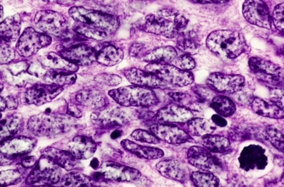
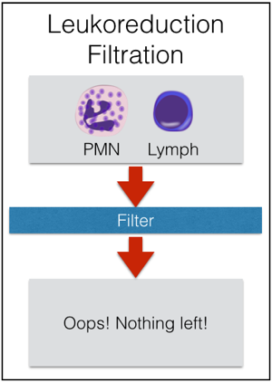
Granulocytes have pretty much the shortest shelf life of any product that we collect directly from donors. They should be transfused “as soon as possible” after collection, but definitely within 24 hours of collection. They are stored at room temperature, like platelets, but they should not be continuously agitated, unlike platelets. This out-of-the-ordinary storage is really another reason that granulocytes are only collected on an “as-needed” basis. Granulocytes are also unusual in that they are almost always issued prior to the availability of the infectious disease screening results! For that reason, essentially all unrelated apheresis granulocyte donors will be recent apheresis platelet donors who have had negative results on a donation within the previous 30 days. We still perform the testing, of course, but the results just aren’t available before the product has to be transfused. Because there are so many RBCs in each granulocyte product, the donor must be ABO compatible with the recipient, and the unit must be crossmatch-compatible with the recipient. By AABB Standards, if more than 2 mL of RBCs are present in any product, those RBCs must be compatible with the recipient’s plasma antibodies. In addition, if the recipient has an unexpected RBC antibody (like anti-D, anti-K, or any others), the granulocyte unit must come from someone who is negative for that antigen, as well. To make matters even more challenging, if the patient has developed anti-HLA antibodies (most commonly in previously pregnant females), then the donor should be HLA matched or at least HLA compatible with the patient’s antibodies. Finally, to complete the weirdness, it is essential to remember which modifications CAN and CAN’T be done to granulocytes. First, granulocytes CAN (and must) be irradiated to prevent Transfusion-associated Graft vs Host Disease (TA-GVHD). This is an extremely fresh product, full of highly active T-lymphocytes in addition to the granulocytes, and the recipient is immunocompromised, by definition. As a result, irradiation is essential to deactivate those donor T-lymphocytes and protect the patient (see image below). Second, granulocytes CAN’T be leukocyte-reduced. Every now and then, people will ask me about leukoreducing granulocytes for prevention of Cytomegalovirus (CMV) transmission. Rather than make fun of them, I usually just sit quietly while they work it out themselves (“OK, let’s see: Granulocytes are WBCs, which some people call ‘leukocytes,’ so if I leukocyte-reduce a unit of a product that is primarily composed of leukocytes, I would be left with…nothing! OHHHH!”). As a result of our inability to make a granulocyte product “CMV-safe” by leukoreduction, CMV-seronegative donors will be recruited to provide products for CMV-seronegative patients whenever possible. Note that it is totally fine to run granulocyte concentrate through a “standard” transfusion filter, just not a leukoreduction or microaggregate filter.

IrradOKNoFilter

Irradiation is not only OK for granulocyte concentrate, it is essential! Leukocyte reduction, on the other hand, makes no sense for granulocytes, and shouldn’t be done.

5. Granulocytes very commonly cause reactions in recipients. Granulocytes are famous for causing transfusion reactions much more often than other components. The two issues that are seen most often are fever and chills (without hemolysis) and pulmonary reactions. I won’t discuss the self-explanatory fever and chills issue, but the pulmonary reactions are worth mentioning here. Considering that current thought suggests that neutrophils are the primary cell involved in the pathogenesis of Transfusion-related Acute Lung Injury (TRALI), it is not surprising that granulocyte transfusions are thought to cause pulmonary compromise quite often (5% in one study). Granulocytes LOVE the lungs, and they localize there very quickly (especially when the infection being treated is pneumonia). So, even if full-blown TRALI does not occur (which certainly CAN happen!), granulocyte transfusions are notorious for at least transient pulmonary compromise. Clinicians should be prepared to manage dypsnea and hypoxia during granulocyte transfusion. Note that the previously suggested “link” between more pulmonary reactions when granulocytes were given with the antifungal Amphotericin B seems to be disproven (though many still advise avoiding transfusing granulocytes within a few hours of administration of that medication). As mentioned in item 4, TA-GVHD is a risk for a fresh product, so irradiation is required. Further, HLA alloummunization can certainly occur as a result of granulocyte transfusion. Final thoughts: There is no question to me that granulocytes CAN, in some circumstances, appear to make a difference for patients. Unfortunately, there’s not a lot of objective proof that they are a predictably helpful treatment. However, given our current ability to collect granulocyte products with substantially higher yields than in years past, I expect that interest in this product will continue despite the lack of “proof” of its effectiveness.

I hope that this quick “5 Things to Know” has been useful for you!

Chaffin

-Joe Chaffin, MD, is the new Vice President and Chief Medical Officer for LifeStream, a Southern California blood center headquartered in San Bernardino, CA. He has a long history of innovative educational efforts and is most widely known as the founder and chief author of “The Blood Bank Guy” website (www.bbguy.org).

The Law of Unintended Consequences

Has the guidelines that encourage less Pap testing over the course of a woman’s life contributed to less women being screened for STDs such as Chlamydia? According to a study published this week, yes.  The cohort is rather small–3000 teenagers and young women–but even so, the results are striking. Before 2009, 30 percent of patients were screened for Chlamydia; after 2009, only 1 percent were.

While this is discouraging, the CDC found that teenagers are having less sex than they were twenty-five years ago, which some attribute to the HPV vaccine and its accompanying education.

Further reading on the relationship between HPV vaccines, Pap screening, and STD testing:

Chicago Tribune

NPR

Healthday

Swails

Kelly Swails, MT(ASCP), is a laboratory professional, recovering microbiologist, and web editor for Lab Medicine.