Microbiology Case Study: A 41 Year Old Male with Pneumonia

Case Description

A 41 year old male with a past medical history significant for HIV presented to the emergency department with complaints of a fever, shortness of breath, cough, myalgias, diarrhea, and dark urine for five days. Upon presentation, he was found to meet sepsis criteria for fever of 105° F, white blood cell count of 19 x 109 /L, and tachycardia. Physical exam was unrevealing. Chest x-ray revealed a lobar pneumonia and a CT chest showed ground-glass opacities with superimposed interlobular thickening and intralobular septal thickening, commonly referred to as “crazy-paving” (Image 1). Initial laboratory assessments included CBC, CMP, blood and sputum cultures, and a T-spot. A legionella urine antigen test was ordered a short time later following an infectious disease consult.

CBC and CMP were significant for leukocytosis (19 x 109 /L), hyponatremia (130 mmol/L), and transaminitis (AST: 97, ALT: 91). Blood and sputum cultures were negative as was the T-spot. Ultimately, the diagnosis of Legionnaire’s Disease was made by the positive urine antigen test.

Image 1. Computed tomography images of the chest without contrast demonstrates consolidation and “crazy paving” patterns of radiographic pathology.
Image 2. Representative example of Legionella pneumophila on Buffered Charcoal Yeast Extract (BCYE) media with small wet gray colonies.

Discussion

Legionella is a genus of aerobic, gram negative, intracellular pathogens that are most often found in soil and water.1 There are over 60 known species of Legionella with each consisting of a varied number of serogroups. At least 26 of these species are pathogenic in humans, however only a few are responsible for the vast majority of known cases.2 In North America, upwards of 90% of cases are caused by L. pneumophila, and more specifically its serogroup 1. In Australia and New Zealand, L. longbeachae is the predominant human pathogen.3 Legionella infections most commonly cause community-acquired pneumonia after inhalation of aerosols and can less frequently cause a self-limited febrile syndrome known as Pontiac Fever. Characteristic signs that may cause a treating physician to think of Legionella infection include a constellation of symptoms that include diarrhea, hyponatremia, and elevated liver function tests.4

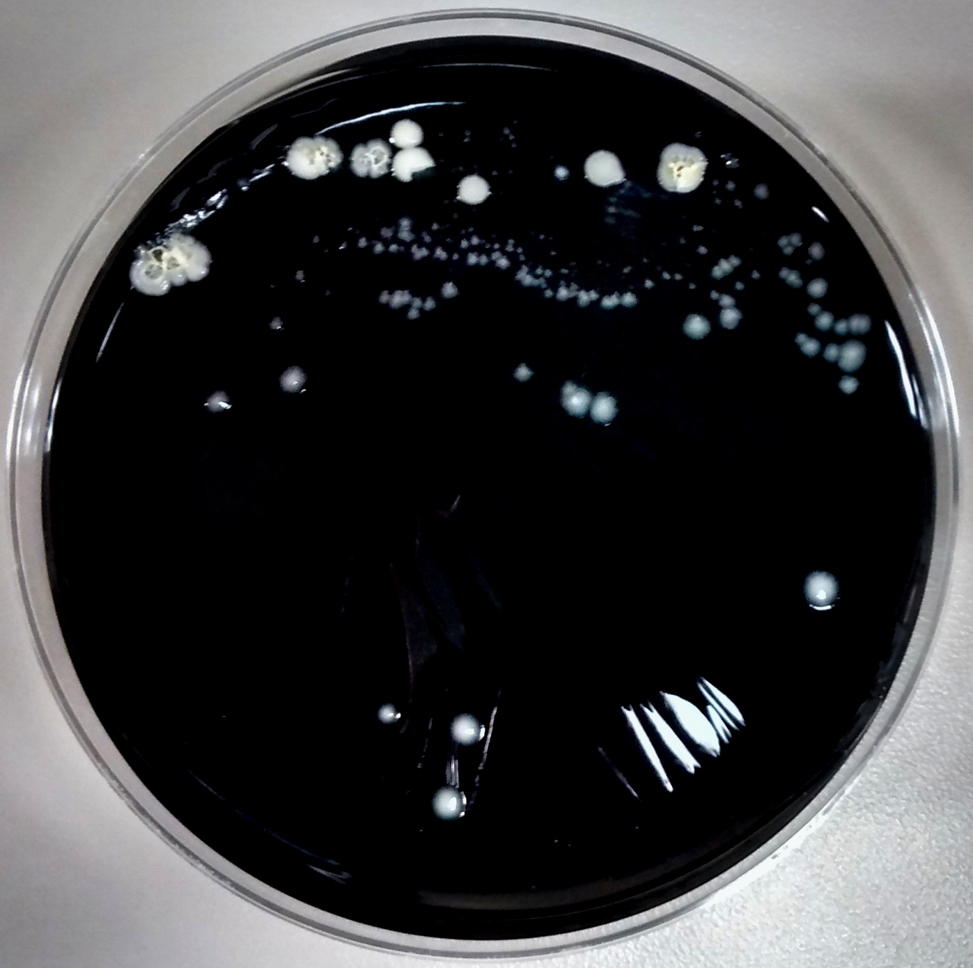
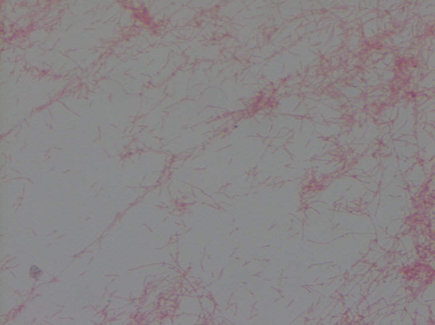
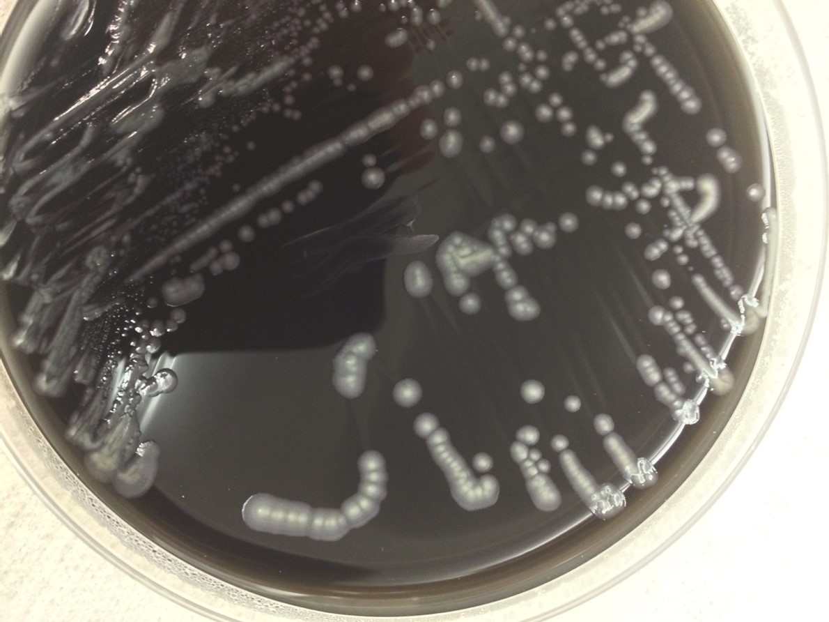
This case reviews a typical presentation of Legionnaire’s disease and highlights several diagnostic pearls worth remembering. Despite commonly being thought of as an exotic pathogen, Legionella is known to cause between 2%-15% of community-acquired pneumonia cases in the United States and Europe.5 The gold standard for diagnosis is culture of lower respiratory secretions, however it is a fastidious organism that is not easily picked up on gram stain or grown on standard media. When culture is attempted, nutrient enriched BCYE agar is required and the timeframe for growth must be increased to 5 to 7 days for L. pneumophila and 14 days for non-pneumophila strains (Image 2).6 Adding to the difficulty in detection, Legionella is easily treated by empiric therapies, such as macrolides, that cover atypical infections; therefore, delays in testing further reduce sensitivity. The urine antigen test does help to overcome this problem as it can detect infection within 2-3 days of symptom onset and remains positive for at least 1 month following resolution of the illness.6

References

  1. Edelstein PH and Roy CR. Legionnaires’ Disease and Pontiac Fever. In: Mandell, Douglas, and Bennett’s Principles and Practice of Infectious Diseases, 8, Bennet JE, Dolin R and Blaser MJ (Eds), Elsevier, Pennsylvania 2015.
  2. National Center for Biotechnology Information Taxonomy Browser. https://www.ncbi.nlm.nih.gov/Taxonomy/Browser/wwwtax.cgi?mode=Tree&id=444&lvl=3&keep=1&srchmode=1&unlock (Accessed on April 14, 2021).
  3. Yu VL, Plouffe JF, Pastoris MC, et al. Distribution of Legionella species and serogroups isolated by culture in patients with sporadic community-acquired legionellosis: an international collaborative survey. J Infect Dis 2002; 186:127.
  4. Robert R. Muder, L. Yu Victor, Infection Due to Legionella Species Other Than L. pneumophilaClinical Infectious Diseases. 2002; 35(1):990-998. doi.org/10.1086/342884
  5. Chahin A, Opal SM. Severe pneumonia caused by Legionella pneumophila: differential diagnosis and therapeutic considerations. Infect Dis Clin North Am. 2017;31(1):111-121. doi:10.1016/j.idc.2016.10.009
  6. Mercante JW, Winchell JM. Current and emerging Legionella diagnostics for laboratory and outbreak investigations. Clin Microbiol Rev. 2015;28(1):95-133. doi:10.1128/CMR.00029-14

-Allen Green is a first year Clinical Pathology resident at UT Southwestern. He has broad interest in laboratory medicine and Transfusion medicine.

-Dominick Cavuoti is a full professor at UT Southwestern and practices both Medical Microbiology, Infectious Disease Pathology and Cytology.

-Clare McCormick-Baw, MD, PhD is an Assistant Professor of Clinical Microbiology at UT Southwestern in Dallas, Texas. She has a passion for teaching about laboratory medicine in general and the best uses of the microbiology lab in particular.

Microbiology Case Study: A 65 Year Old Man with Pneumonia

Case History

A 65 year old male with a history of systolic heart failure secondary to non-ischemic (alcohol-induced) dilated cardiomyopathy underwent cardiac transplantation on 10/11/2016. He was hospitalized between 3/1/17 and 4/15/17 for neutropenia and CMV viremia. Two days after discharge, he presented to the hospital with a gradual-onset of left-sided sharp chest pain described as “soreness” over his rib cage and exacerbated by breathing. Associated symptoms included fever, malaise, and fatigue. In the emergency department, vital signs included: BP 144/75 mmHg, T 40.2°C, RR 24/min, HR 101 bpm, SpO2 97% on room air. A CBC revealed a normal white blood cell count and a chest X-ray demonstrated a lingular opacity. The immunocompromised patient was admitted for sepsis secondary to presumed pneumonia following recent hospitalization. He was treated empirically for hospital-acquired pneumonia with vancomycin and piperacillin-tazobactam. After a urine antigen test detected the presence of Legionella pneumophila serogroup 1, antibiotic treatment was changed to levofloxacin and an induced sputum culture was obtained for Legionella surveillance.

legion1
Image 1. Sputum culture on BCYE agar with PAV shows Legionella pneumophila colonies that are circular with smooth edges, grey-white, and glistening in addition to few usual oropharyngeal flora.

Discussion

Legionnaires’ disease, caused by Legionella bacteria, is a cause of 1-9% of both community-acquired and hospital-acquired pneumonias. Symptoms of fever, chills, cough, and chest pain are similar to other causes of pneumonia; however multiple organ systems may be involved, producing additional symptoms including gastrointestinal (diarrhea, nausea, and vomiting) and central nervous system (headache and confusion) findings. Legionella was first discovered after a 1976 outbreak of pneumonia among Pennsylvania State American Legion members who attended a convention at a Philadelphia hotel that had infected water in the air conditioning system; it is reported that 29 out of 182 infected people died. At present, the mortality rate of Legionnaires’ disease ranges from less than 10% in treated community-acquired cases to approximately 30% for hospital-acquired cases.

The genus Legionella contains greater than 60 species of which approximately 20 are human pathogens. Legionella pneumophila (consisting of serogroups 1-16) is the most common cause of Legionnaires’ disease and, in particular, L. pneumophila serogroup 1 causes 70-90% of cases. The organisms are ubiquitous in nature, particularly in warm freshwater environments including lakes and streams, where they infect and multiply within single-celled host organisms. Of pathogenic concern, they can be present in high numbers in human-made complex water systems (such as cooling towers, whirlpool spas, humidifiers, and decorative fountains). After environmental aerosols are inhaled or contaminated water is aspirated into the lungs, alveolar macrophages are infected by the obligate intracellular bacteria. Host risk factors for developing Legionnaires’ disease include organ transplantation, immunocompromised state, immunosuppresion, age greater than 60 years, chronic lung disease, and smoking.

In the microbiology laboratory, Legionella are mesophilic (20-45 °C) obligate aerobes. The small, thin gram negative rods react poorly with Gram stains and are not usually stained in direct clinical samples. The patient’s Gram smear revealed moderate neutrophils, few squamous epithelial cells, and mixed gram positive and gram negative organisms present. Sensitivity for detecting the biochemically inert and fastidious bacteria is increased with culture on buffered charcoal yeast extract (BCYE) agar. For sputum samples that are likely contaminated with usual oropharyngeal flora, BCYE agar with polymyxin B, anisomycin, and vancomycin (PAV) media are used. After 3-5 days of incubation, Legionella colonies appear convex, circular, 3-4 mm in diameter, grey-white to blue-green, and glistening. This identification was confirmed by MALDI-TOF MS. Laboratory in vitro susceptibility studies are not recommended on individual isolates, as they do not correlate with clinical responses. Monotherapy with a fluoroquinolone (Levofloxacin) or macrolide (Azithromycin) is active against Legionella.


-Adina Bodolan, MD is a 1st year anatomic and clinical pathology resident at the University of Vermont Medical Center.

Wojewoda-small

-Christi Wojewoda, MD, is the Director of Clinical Microbiology at the University of Vermont Medical Center and an Associate Professor at the University of Vermont.

Microbiology Case Study–A Very Sick 51 Year Old Man

Case history:

A 51 year old man with a medical history of liver abnormalities and long standing alcohol abuse presented with generalized weakness, hypoxemic respiratory failure, sputum production, significant hyperbilirubinemia, macrocytic anemia, and laboratory tests consistent with DIC. Chest X ray revealed a bronchopneumonia pattern. Sputum samples were sent for culture and blood cultures were obtained. Urine antigen legionella test was positive for Legionella pneumophila serogroup 1. The sputum was then plated on buffered charcoal yeast extract (BCYE) agar.

Laboratory identification:

Gram stain of colonies growing on BCYE revealed thin short to filamentous rod shaped organisms. The definitive diagnosis of Legionella pneumophila was made by isolating the organism on BCYE with confirmation on the MALDI-ToF. Colonies were speckled blue and

Legionella on BCYE agar plate

Gram negative, thin, short-to-filamentous rod shaped organisms

Discussion:

Legionella pneumophila was first identified and recognized during the outbreak that occurred during the American Legion Convention in Philadelphia in 1976. There are over 40 species of Legionella and 18 of those are human pathogens. Among those, L. pneumophila is an important cause of nosocomial and community-acquired pneumonia (CAP) and should be considered in the differential diagnosis in any patient who presents with pneumonia. Two clinical syndromes caused by this microorganism are: Legionnaires’ disease and Pontiac fever (acute, febrile, self-limited illness).

L. pneumophila is a thin faintly staining short to filamentous gram negative rod. Legionella is a fastidious organism and does not grow on standard media. Buffered charcoal yeast extract (BCYE) agar is the primary medium used for its isolation and it is the gold standard for its diagnosis although testing for the antigen in urine is more commonly performed. The urinary antigen assay only detects L. pneumophila serogroup 1 which cause 95-98% of community acquired Legionnaires’ disease.

The microorganism is found in natural water supplies and soil. It is also found in recirculating and water supply systems, where it breeds rapidly in favorable conditions (temperature of 35°C, range 25-45, stagnant water and water containing organic debris which can provide nutrients for growth). Macrolides (azithromycin or clarithromycin) or fluoroquinolones (levofloxacin or moxifloxacin) are the standard antibiotic drugs used to treat Legionnaires’ disease in humans.

Kossivi Dantey, M.D. is a 4th year anatomic and clinical pathology resident at the University of Vermont Medical Center.

Wojewoda-small

–Christi Wojewoda, MD, is certified by the American Board of Pathology in AP/CP and Medical Microbiology. She is currently the Director of Clinical Microbiology at the University of Vermont Medical Center and an Assistant Professor at the University of Vermont.

iridescent.