An 83 year old man presents to an emergency department after an unwitnessed fall and being found down for an undetermined amount of time. His past medical history includes dementia and type II diabetes mellitus. A tick is found in his left groin. Workup reveals mild rhabdomyolysis, mild transaminitis, thrombocytopenia, and multiple infiltrates on chest x-ray, concerning for aspiration pneumonia. His hospital course is complicated by fever, hypotension, and atrial fibrillation with rapid ventricular response. Representative images from his peripheral blood smear are shown below.
PCR confirmed the diagnosis of Anaplasma phagocytophilum. The patient recovered after a 10-day course of doxycycline.
The clinical findings of recent tick exposure, thrombocytopenia, transaminitis, and acute illness are consistent with acute infection with either Ehrlichia or anaplasma. The images depict morulae (microcolonies of bacteria) within the cytoplasm of neutrophils. A. phagocytophilum, Ehrlichia chaffeensis, and Babesia microti can all be seen intracellularly on peripheral blood smears. A. phagocytophilum preferentially infects neutrophils, while E. chaffeensis infects monocytes and B. microti infects erythrocytes. Unlike A. phagocytophilum and E. chaffeensis, which both appear as intracellular morulae (clusters of bacteria), B. microti is a protozoa whose trophozoite forms are seen in red blood cells as delicate rings or grouped in tetrads (the characteristic “Maltese cross”).
The finding of morulae in granulocytes has a sensitivity of 20-80% for anaplasmosis within the first week of infection. However, morulae within granulocytes are also found in infection with Ehrlichia ewingii (human granulocytotropic Ehrlichiosis, distinct from human monocytotropic Ehrlichiosis caused by E. chaffeensis), a less common relative of E. chaffeensis that is also transmitted by the Lone Star tick (Amblyomma americanum) and appears to preferentially infect immunocompromised hosts. For E. chaffeensis, the sensitivity of finding morulae in monocytes is only 7-17% amongst immunocompetent hosts.
References
Chapman, AS, JS Bakken, SM Folk, et al. “Diagnosis and management of tickborne rickettsial diseases: Rocky Mountain spotted fever, ehrlichioses, and anaplasmosis–United States: a practical guide for physicians and other health-care and public health professionals.” MMWR Recomm. Rep. 2006; 55(RR-4):1.
McPherson, R, and M Pincus. (2011). Henry’s Clinical Diagnosis and Management By Laboratory Methods (22nd Edition, pp. 1073-1074, 1205). Philadelphia, PA: Elsevier Saunders.
Paddock, CD, SM Folk, GM Shore, et al. “Infections with Ehrlichia chaffeensis and Ehrlichia ewingii in persons coinfected with human immunodeficiency virus.” Clin Infect Dis. 2001; 33(9):1586-94.
Schotthoefer, AM, JK Meece, LC Ivacic, et al. “Comparison of a Real-Time PCR Method with Serology and Blood Smear Analysis for Diagnosis of Human Anaplasmosis: Importance of Infection Time Course for Optimal Test Utilization.” J Clin Microbiol. 2013 Jul; 51(7): 2147-2153.
-Frederick Eyerer, MD is a 3rd year anatomic and clinical pathology resident at the University of Vermont Medical Center.
-Christi Wojewoda, MD, is the Director of Clinical Microbiology at the University of Vermont Medical Center and an Associate Professor at the University of Vermont.
(No. Not that kind of critical value; your patient’s hemoglobin is absolutely fine…)
Hello again everyone! Back from a relevant and important case-study last month, I’d like to pivot to highlighting an important annual celebration that happens every May 6-12th around the world: International Nurses’ Day.
I know, I know. This is a blog for medical laboratory professionals, what gives? We get lab week, they get nurses’ week…separate but equal? In my opinion, not exactly the best modus operandi for collaborative medicine. In fact, nursing and laboratory medicine have both had a rich, connected, and parallel history for decades and the professional intersections of their efforts is something to be celebrated!
A lot of current published literature focuses on nursing in the pre-analytical realm of specimen collection and processing. That’s totally fine, and quite important to diagnostic testing, but if you go back a few decades with me it’s clear to see we’re both frontline medical professionals—and that’s without a global viral pandemic!
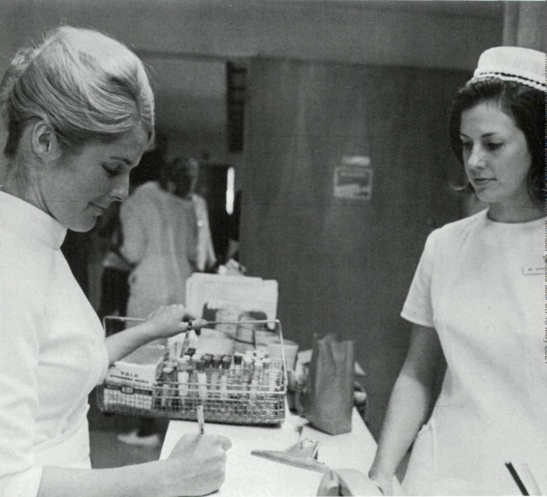
Image 1. I’m sorry but the label needs to be dated and initialed at bedside—KIDDING. That happens a lot I’m sure (I would know), but nurses and medical laboratory scientists (technologists) have been counterparts for decades. The present is no different. We are two parts of a strong interdisciplinary, collaborative effort to improve healthcare for our patients. After all, we’re always #StrongerTogether. (Source: Laboratory Medicine, June 1970)
In the very first issue of Laboratory Medicine¸ one of ASCP’s main journals, an article discussed the critical communication between nursing and the laboratory. The bottom line (from 1970)? Doctors and nurses might use “stat” too much, and medical technologists are very busy folks—has anything changed? In another piece in the same journal from 1984, the topic of MLS (then MT) recognition was examined. Almost 14 years apart, this article highlighted that the lab wished for recognition, and that doctors and nurses valued their MT’s experience and knowledge, despite their work behind the scenes. Fast forward to the present, professional societies like ASCLS have established national celebrations of our hard work with Medical Laboratory Professional’s Week in April. In May, the International Council of Nurses (and our American Association of Nursing) promotes Nurses’ Week. Our super-hero themed celebration of diagnostic excellence and commitment is matched by their theme of nursing the world back to health. The World Health Assembly designated 2020 as the year of the nurse for obvious reasons. None of us would be able to thrive, survive, or work in or out of healthcare without nurses. So for this year’s Nurses’ Week, Nurses’ Day, or even the Year of the Nurse, this pathology postgraduate-trainee is happy to celebrate our clinical friends and colleagues.
Image 2. Melizza, Nataliya, Donna, and Roksana are the nurses that manage Loyola’s robust apheresis clinic service. I don’t think it would run without them. They effectively demonstrate that nursing is critical across any specialty, and patient safety and outcomes are directly related to the care they receive.
Significant parts of pathology and laboratory medicine rely heavily on nurses. In transfusion medicine, you wouldn’t be able to have any significant apheresis clinic activity without the compassion and attention of nurses. Clinical Pathologists really lean on the knowledge, skill, talent, grace, compassion, and dedication of the nurses that care for patients receiving this highly specific and specialized treatment modality. Think about how other “interventional pathology” specialties like cytopathology or hematopathology would suffer without the commitment of nurses to keep our patients safe during a fine-needle aspiration, or bone marrow collection.
I write a lot about our work in pathology and laboratory medicine, from bench to bedside, but considering our nursing colleagues:
Nurses are kind. Nurses are brilliant. Nurses are skilled. Nurses work in all kinds of roles, jobs, and settings. Whether they work in an office, clinic, or hospital unit, care for you or a loved one, close the learning gaps for a medical student or resident, champion for better outcomes for one patients or thousands or people at once; our lives are all better for it. Their work never ends, and our need for them never will.
Image 3. Mini-brag: my wife (who has secretly appeared in a few ASCP-related media as she accompanies me to meetings—and even roundtables!) is a nurse and a fantastic one. An INF Top 40 Under Forty Nursing Leader, a former community-based health clinic non-profit chief of staff, an internationally renowned lecturer on emergency and disaster mitigation strategies, and a very important Chicago region disaster response manager who’s inches from finishing her DNP in Public Health so, like, what am I even doing…?
I obviously see the value and impact of nursing every day. Doesn’t hurt that I’m surrounded by great ones at work and at home.
Happy (belated) Nurses Week!
Thanks for reading, see you next time!
–Constantine E. Kanakis MD, MSc, MLS(ASCP)CM is a first-year resident physician in the Pathology and Laboratory Medicine Department at Loyola University Medical Center in Chicago with interests in hematopathology, transfusion medicine, bioethics, public health, and graphic medicine. He is a certified CAP inspector, holds an ASCP LMU certificate, and xxx. He was named on the 2017 ASCP Forty Under 40 list, The Pathologist magazine’s 2020 Power List and serves on ASCP’s Commission for Continuing Professional Development, Social Media Committee, and Patient Champions Advisory Board. He was featured in several online forums during the peak of the COVID pandemic discussing laboratory-related testing considerations, delivered a TEDx talk called “Unrecognizable Medicine,” and sits on the Auxiliary Board of the American Red Cross in Illinois. Dr. Kanakis is active on social media; follow him at @CEKanakisMD.
In the 1962 Japanese release of King Kong vs. Godzilla, it was pretty easy to determine who the winner of the epic battle was (it was Kong, if you’re keeping track). Almost 60 years later, the two famous giant behemoths met again on the big screen, this time in an American version. This second battle, while fun to watch, had a less-obvious outcome. Those who sided with either monster had moments to cheer during the movie, but in the end, (spoiler alert!) a secondary issue overshadowed the much-hyped monster match. It seemed obvious to me that the script writers tried their best to satisfy audiences on both sides of the aisle. In doing this, however, the movie lost a little bit of its punch. That can happen in lab safety as well. If the message isn’t strong or clear, safety issues will ensue.
The lab manager noticed an increase in employee injuries on the job. Someone cut their finger on a microtome blade. Two different specimen processors splashed serum into their eyes. A night shift tech kneeled onto the floor to pick up a box, and her knee landed on broken glass that hadn’t been swept up. It was clear people were not paying close attention while they were performing their duties. The manager held a meeting in the lab to raise safety awareness. He delivered his message, but staff noticed he was chewing gum and that he did not put on a lab coat even though he was in the lab for over 20 minutes. The message was not clear. He said he was for a safer lab, but he didn’t look to anyone like he meant it.
Jake was new to the histology lab and he was excited to make a good impression as he began his career. During his departmental orientation, the supervisor stressed the importance of chemical safety. Formaldehyde and xylene were in use in the department, and they were potentially dangerous. Jake got the message and was ready to show the department he could work there safely. After orientation was done, Jake came to work to begin cutting tissue. When he sat down at his station, he opened the drawer for supplies and saw a packet of oatmeal a spoon, and a coffee cup. The medical director came in to the lab to welcome Jake, and he noticed she was drinking tea. The message Jake received about safety was no longer clear. The supervisor spoke about safety, but it was clear no one enforced it.
When the message we send to staff about lab safety is mixed, we really can’t blame them when the culture is bad. The problem is that many leaders are not aware they are sending this confusing communication. If you’re not sure if you are one of those leaders, take a step back and look in the mirror. What kind of message do you send? Do you support safety? Do you do it with your words AND your actions? What sort of example do you set?
If you’re not in lab leadership, you still have a responsibility to represent safety with your lab practices as well. Everyone has an impact on the overall safety culture, not just leaders. What do you do to promote safety in your department?
Maybe you are an employee and it’s your leader who is sending mixed messages. First, make sure you’re choosing the side of safety in your work no matter what others are doing. Second, it may be time to “manage up” and ask leadership why certain unsafe practices occur. If the leader is part of the problem, it is acceptable to point that out, provided you do so with purpose, tact and professionalism.
Leading by example for safety is vitally important no matter your role in the department. Choose your side, stick to it in all circumstances, and over time you will be able to be declared the decisive winner. Those mixed safety messages usually lead to a draw, or worse, a loss for the team.
Because I have been a Godzilla fan for decades, I thoroughly enjoyed this latest film entry. In truth, I was able to discern a clear winner of his fight with Kong despite the writer’s intentions, but that may be because I had support for my favorite going in. That might be your way to victory as well. Root for “Team Safety,” and your support will be noticed and followed for the win!
–Dan Scungio, MT(ASCP), SLS, CQA (ASQ) has over 25 years experience as a certified medical technologist. Today he is the Laboratory Safety Officer for Sentara Healthcare, a system of seven hospitals and over 20 laboratories and draw sites in the Tidewater area of Virginia. He is also known as Dan the Lab Safety Man, a lab safety consultant, educator, and trainer.
A 41 year old male with a past medical history significant for HIV presented to the emergency department with complaints of a fever, shortness of breath, cough, myalgias, diarrhea, and dark urine for five days. Upon presentation, he was found to meet sepsis criteria for fever of 105° F, white blood cell count of 19 x 109 /L, and tachycardia. Physical exam was unrevealing. Chest x-ray revealed a lobar pneumonia and a CT chest showed ground-glass opacities with superimposed interlobular thickening and intralobular septal thickening, commonly referred to as “crazy-paving” (Image 1). Initial laboratory assessments included CBC, CMP, blood and sputum cultures, and a T-spot. A legionella urine antigen test was ordered a short time later following an infectious disease consult.
CBC and CMP were significant for leukocytosis (19 x 109 /L), hyponatremia (130 mmol/L), and transaminitis (AST: 97, ALT: 91). Blood and sputum cultures were negative as was the T-spot. Ultimately, the diagnosis of Legionnaire’s Disease was made by the positive urine antigen test.
Image 1. Computed tomography images of the chest without contrast demonstrates consolidation and “crazy paving” patterns of radiographic pathology.Image 2. Representative example of Legionella pneumophila on Buffered Charcoal Yeast Extract (BCYE) media with small wet gray colonies.
Discussion
Legionella is a genus of aerobic, gram negative, intracellular pathogens that are most often found in soil and water.1 There are over 60 known species of Legionella with each consisting of a varied number of serogroups. At least 26 of these species are pathogenic in humans, however only a few are responsible for the vast majority of known cases.2 In North America, upwards of 90% of cases are caused by L. pneumophila, and more specifically its serogroup 1. In Australia and New Zealand, L. longbeachae is the predominant human pathogen.3Legionella infections most commonly cause community-acquired pneumonia after inhalation of aerosols and can less frequently cause a self-limited febrile syndrome known as Pontiac Fever. Characteristic signs that may cause a treating physician to think of Legionella infection include a constellation of symptoms that include diarrhea, hyponatremia, and elevated liver function tests.4
This case reviews a typical presentation of Legionnaire’s disease and highlights several diagnostic pearls worth remembering. Despite commonly being thought of as an exotic pathogen, Legionella is known to cause between 2%-15% of community-acquired pneumonia cases in the United States and Europe.5 The gold standard for diagnosis is culture of lower respiratory secretions, however it is a fastidious organism that is not easily picked up on gram stain or grown on standard media. When culture is attempted, nutrient enriched BCYE agar is required and the timeframe for growth must be increased to 5 to 7 days for L. pneumophila and 14 days for non-pneumophila strains (Image 2).6 Adding to the difficulty in detection, Legionella is easily treated by empiric therapies, such as macrolides, that cover atypical infections; therefore, delays in testing further reduce sensitivity. The urine antigen test does help to overcome this problem as it can detect infection within 2-3 days of symptom onset and remains positive for at least 1 month following resolution of the illness.6
References
Edelstein PH and Roy CR. Legionnaires’ Disease and Pontiac Fever. In: Mandell, Douglas, and Bennett’s Principles and Practice of Infectious Diseases, 8, Bennet JE, Dolin R and Blaser MJ (Eds), Elsevier, Pennsylvania 2015.
Yu VL, Plouffe JF, Pastoris MC, et al. Distribution of Legionella species and serogroups isolated by culture in patients with sporadic community-acquired legionellosis: an international collaborative survey. J Infect Dis 2002; 186:127.
Robert R. Muder, L. Yu Victor, Infection Due to Legionella Species Other Than L. pneumophila. Clinical Infectious Diseases. 2002; 35(1):990-998. doi.org/10.1086/342884
Chahin A, Opal SM. Severe pneumonia caused by Legionella pneumophila: differential diagnosis and therapeutic considerations. Infect Dis Clin North Am. 2017;31(1):111-121. doi:10.1016/j.idc.2016.10.009
Mercante JW, Winchell JM. Current and emerging Legionella diagnostics for laboratory and outbreak investigations. Clin Microbiol Rev. 2015;28(1):95-133. doi:10.1128/CMR.00029-14
-Allen Green is a first year Clinical Pathology resident at UT Southwestern. He has broad interest in laboratory medicine and Transfusion medicine.
-Dominick Cavuoti is a full professor at UT Southwestern and practices both Medical Microbiology, Infectious Disease Pathology and Cytology.
-Clare McCormick-Baw, MD, PhD is an Assistant Professor of Clinical Microbiology at UT Southwestern in Dallas, Texas. She has a passion for teaching about laboratory medicine in general and the best uses of the microbiology lab in particular.
A middle aged male presented to the emergency department with a several day history of altered mental status, insomnia, and lethargy. His family stated that he also had possibly had a seizure. Upon arrival to the emergency department he was febrile to 102°F, hypoxic, but denied shortness of breath, cough, nausea, and vomiting. While in the care of the emergency department he had several witnessed seizures requiring Ativan treatment, so the decision was made to admit the patient for 24-hour EEG monitoring. Past medical history was unremarkable. The patient is a smoker. His hobbies include working as a mechanic and working outdoors in his garden.
Initial brain imaging on MRI demonstrated cortical thickening/gyral swelling with associated signal abnormality within bilateral mesial temporal lobes that is seen with mild associated diffusion abnormality. A lumbar puncture was ordered and his CSF analysis demonstrated normal glucose (73 mg/dL [reference range: 40-80 mg/dL]), normal protein (32 mg/dL [reference range: 15-45 mg/dL]), and normal nucleated cells [reference range: 0-8/mm3]. Bacterial, fungal, and AFB culture as well as PCR for herpes simplex virus and varicella zoster virus were ordered. The CSF bacterial, fungal, and AFB cultures showed no growth of any microorganisms, and PCR results were negative for HSV and VZV. What additional infectious etiologies would you like to test for?
Discussion
Serology studies were ordered which demonstrated positive IgM and IgG antibodies for West Nile virus.
West Nile virus is a member of the Flavivirus genus. It is spread through mosquito bites, and birds are the primary reservoir for this virus. The incubation period for West Nile virus is 4-10 days. Typically, about 80% of those infected with the virus will be asymptomatic. In 20% of cases, the patient will develop a febrile illness, with possible symptoms of headaches, body aches, weakness, joint pain, and fatigue. About 1 in 150 of those infected will develop illness involving the central nervous system. In these cases, symptoms can include high fever, headache, neck stiffness, confusion, seizures, and coma. Death occurs in 10% of those with involvement of the central nervous system. The most important risk factor for death is age with patients over 70 years of age being most at risk.
Diagnosis of West Nile virus generally made by detection of IgM and IgG in the serum and/or CSF. IgM can be detected 3-8 days post symptom onset and remains positive for 2-3 months in the CSF, and occasionally longer in the serum. Diagnosis is made by detection of IgM antibodies or conversion of IgG antibodies, while detection of IgG antibodies in isolation indicates a prior infection. False-positive results can occur in CSF specimens contaminated with blood that is IgM antibody positive. Patients who have been infected with or vaccinated against other flaviviruses can have false-positive serum antibodies. Plaque reduction neutralization test (PRNT) assay can be performed to rule out cross-reactivity with other flaviviruses. RT-PCR testing on CSF is also available for detection of West Nile virus RNA. The sensitivity of CSF RT-PCR is low in immunocompetent patients, but increased in immunocompromised patients due to prolonged viremia.
West Nile virus cases could increase in the future as a consequence of climate change. Currently, most cases of West Nile virus occur between June and September and cases have been reported in all 48 lower states. As climate change progresses, average temperatures are expected to increase around the world. As temperature increases, summers become longer, which means a longer season for mosquitoes. Laboratory studies also demonstrate that the virus replicates faster in warmer temperatures. Precipitation patterns are also expected to change in the future. The effect of precipitation is not as clear as that of temperature, but it can still play a role. In areas with more precipitation, increasing amounts of water could provide new breeding grounds for mosquitoes. On the other hand, extra rain can dilute some of the nutrients that the mosquitoes need. In areas with less precipitation, drought conditions can cause rivers to dry up, creating the pools of standing water that give rise to mosquitoes. In addition, as bodies of water become smaller, birds and mosquitoes will be in closer proximity, facilitating faster spread of the virus. We must be prepared for the possibility that West Nile virus cases may be on the rise in the future.
The patient continued to suffer from seizures and required continuous sedation with phenobarbital. For seizure prophylaxis he was given a combination of Keppra and Vimpat, with the dosage adjusted as needed. Throughout his entire hospital course he was monitored on EEG. For the first week of his stay, his EEG results demonstrated seizure like activity, requiring continued use of anti-seizure medication and sedation. After about one week, his seizure activity began to show improvement, and the process of decreasing his medication had begun.
After several days of improvement, his recover was complicated by abdominal compartment syndrome requiring laparotomy. After this, the patient’s seizure activity began to worsen. Within a few days, the patient became hypotensive, and broad spectrum antibiotics were given to protect against infection. Cultures were taken but no infection was identified. The patient ultimately developed shock and passed away.
Murray, P. R., Rosenthal, K. S., & Pfaller, M. A. (2021). Togaviruses and Flaviviruses. In Medical microbiology. Edinburgh: Elsevier.
Miller, J. Michael, et al. “A guide to utilization of the microbiology laboratory for diagnosis of infectious diseases: 2018 update by the Infectious Diseases Society of America and the American Society for Microbiology.” Clinical Infectious Diseases 67.6 (2018): e1-e94.
-Robert Toelke, MD is a 1st year clinical and anatomic pathology resident at University of Chicago (NorthShore).
-Erin McElvania, PhD, D(ABMM), is the Director of Clinical Microbiology NorthShore University Health System in Evanston, Illinois. Follow Dr. McElvania on twitter @E-McElvania.
Hello again everyone and welcome back! Pardon the absolutely cheesy title, but this month I’d like to share parts of a clinical case and discuss some pretty important topics in pathology practice in 2021. While last month’s brief primer on the Cures Act is a definite part of the future of medicine, there are a lot of things developing in our profession—and we should all keep up!
*** Disclaimer: Please note that if/when I share cases or clinical content, they are not current, already signed out, completed and done in compliance full patient privacy guidelines. Always. ***
So let’s dive right in. A rather young male patient was sent to us from an outside facility for mildly painful, uncomfortable bilateral breast lumps. He was noted to have bilateral gynecomastia clinically and was to be evaluated via mammogram and localized needle biopsy. An ultrasound guided core needle biopsy of the left breast was collected from a mass identified on mammography.
Figure 1. Not the typical visual presentation noted on mammography. Biopsy clip marking the location of where the core biopsies were obtained. Male patients can have histologically identical cases of breast carcinoma when compared to females and are often associated with germline mutations, high-risk BRCA1 & 2 status, and Klinefelter syndromes.
Most commonly, when evaluating gynecomastia in a male and considering the diagnostic algorithms including malignancy one worries most about ductal carcinoma: invasive, unspecified type and ductal carcinoma in situ (DCIS) pretty much round off the differential for male breast cancer. Important rule outs include clinical, hormone-related gynecomastia or otherwise relating to germ-line or chromosomal abnormalities (i.e. XXY in Klinefelter), myofibroblastoma, or unrelated distant metastases. So without too much clinical information or history—since this was an outside referral—the differential of potential considerations was pretty short therein.
The biopsy was completed shortly after mammography and was signed out as: fibroadenoma.
Wait. What?
Fibroadenomas are benign and, along with potentially malignant phyllodes tumors, come from mammary tissue, demonstrate biphasic cellular components of benign epithelium (or low-grade stroma).
Translation: to have one of these entities you need mammary tissue, specifically terminal ductal/lobular units to form the necessary architecture changes. Simpler translation: fibroadenomas need breast tissue to be fibroadenomas.
Okay, there definitely appears to be breast tissue here to form this entity, however, it is incredibly rare for a male patient (outside of the previously mentioned conditions) to have anatomically functional lobular units. With the other etiologies ruled out, further investigation into the patient’s history was warranted. It was discovered that this patient, although male by indication on referring paperwork, actually identified as female and was on gender affirming hormone therapy for several years. A considerably simple explanation for the confounding biopsy result.
Transgender medicine and transgender pathology considerations have been talked about more in recent years than ever before. While these patients have always existed, societal changes in the last decade alone have brought to light many disparities in medical care and practice habits overall. Here on Lablogatory, there have been between 20-30 mentions of transgender patients since 2018 with entire pieces dedicated to transgender endocrinology, clinical chemistry, etc. Specific examples have included the complicated intersection of transgender care and electronic medical records, various lab values in trans patients, and an excellent overview of laboratory medicine and trans patients by Dr. Jeff SoRelle. There he discusses approaches, literature, and—most importantly—the purposeful vocabulary of transgender medicine as an empowering and informative practice.
Image 2. A 2016 paper in Laboratory Medicine highlights this patient population’s challenges to quality and equity in healthcare and establishes that gatekeepers to data—including us—should consider new ways to create inclusive laboratory medicine practices to maintain our high standard of care. Read it here.
While, this case was rather straightforward after the discovery of the patient’s history of gender affirming therapy, there are still several critical points to highlight here. First, it’s critically important in the setting of medical care to correctly and compassionately include gender identity in patient records—within the caveat of appropriate confidentiality and patient autonomy. Patient care, health management, and diagnostic efforts like ours may rely on a simple biological indication of how this patient presents and experiences their identity with relation to their clinical course. This further strengthens relationships between trans patients and their primary care providers, as well as non-patient-facing specialists like us who often put pieces of the puzzle together without an entire history at our disposal. How does this impact patient care? Simple: a trans male patient may always carry a residual indolent risk of breast cancer, while a trans female patient may need to be included in future mammogram screening. The bottom line should consider treating who you have, and screening with what you have to. Since this patient was sent because of breast lumps, as a male patient a biopsy report of “fibroadenoma” would be exceedingly rare and confounding because of normal physiology outside of specific genetic conditions. But with the right information at the right time, better patient-centered care is possible.
Finally, as a note about proper terminology, vocabulary, and available guidelines in the literature, there is both a paucity of effective transgender medical guidelines as well as a poor grasp on the terminology. Dr. SoRelle does a fantastic job with a short vocabulary outline, but we should all do better. The experiences of our trans patients is challenging to say the least, so as we continue to become stronger and closer advocates for patients in pathology and laboratory medicine, we must become familiar with their experience and advocate for the progress of inclusive medical care.
Trans patients are special, and we are specialists.
Making sure they received the highest level of care alongside all our other patients should be guaranteed. Whether in our clinical chemistry labs or surgical pathology sign outs, we already know how to create an environment of accuracy and enduring inclusion for all our patients—let’s keep it going!
Thank you for reading, I’ll see you next time!
–Constantine E. Kanakis MD, MSc, MLS(ASCP)CM is a first-year resident physician in the Pathology and Laboratory Medicine Department at Loyola University Medical Center in Chicago with interests in hematopathology, transfusion medicine, bioethics, public health, and graphic medicine. He is a certified CAP inspector, holds an ASCP LMU certificate, and xxx. He was named on the 2017 ASCP Forty Under 40 list, The Pathologist magazine’s 2020 Power List and serves on ASCP’s Commission for Continuing Professional Development, Social Media Committee, and Patient Champions Advisory Board. He was featured in several online forums during the peak of the COVID pandemic discussing laboratory-related testing considerations, delivered a TEDx talk called “Unrecognizable Medicine,” and sits on the Auxiliary Board of the American Red Cross in Illinois. Dr. Kanakis is active on social media; follow him at @CEKanakisMD.
A patient presents with a worm they found in the toilet.
Image 1. The offender. Image 2. Mouth parts of the worm.
Discussion
This is Ascaris lumbricoides, a roundworm. Distinctive morphologic features include tapered ends, mouthparts consisting of three prominent lips (pictured in image 2), and a length of up to 35cm for females. The adults live in the duodenum and proximal jejunum. The eggs have an irregular external mamillated outer shell that gives them a roughened outer surface. Clinically, infection can range from asymptomatic to severe disease, in which the larvae, hatched from ingested eggs, migrate from the small bowel through the circulatory system to the lungs, where they mature in the alveolar capillary bed and cause Ascariasis pneumonitis (Löffler syndrome).
Other diagnostic considerations include Enterobius vermicularis (pinworm), Lumbricus terrestris (earthworm), Trichuris trichiura (whipworm), and the hook worms, Necator americanus and Ancylostoma duodenale.
Enterobius vermicularis, the pinworm, is the most common helminth infection in the United States. Clinically, the classic presentation is a child with pruritus ani. Females measure up to 1.3 cm in length and have a pointed posterior end, and both sexes have lateral alae and a prominent esophageal bulb. The worm in this case is far too large to be a pinworm.
The earthworm, Lumbricus terrestris, is soil-dwelling and non-pathogenic but occasionally encountered in the laboratory for identification purposes. Key morphologic features include a segmented body with no distinctive mouthparts and a clitellum (a mating organ that is a non-segmented portion of the body and often a different color from the rest of the body).
Trichuris trichiura, the whipworm, have a classic whip-like appearance with long, narrow anterior ends that anchor the worm to the large intestine, where they can remain for up to 10 years. Both males and females measure up to 5.0 cm in length, and diagnosis is often made by identification of the eggs, which are football-shaped and have polar plugs at both ends. Clinically, trichuriasis can cause dysentery-type symptoms and, in heavily infected children, can lead to rectal prolapse.
Necator americanus and Ancylostoma duodenale are the hookworms. Adult females measure up to 1.2 cm, and these two species are differentiated by examination of the mouthparts: Necator americanus has cutting plates, while Ancylostoma duodenale has cutting teeth. In addition to the large size difference between hookworms and roundworms, the lamprey-like appearance of these mouthparts is notably different from the “fleshy lips” of Ascaris. Hookworms and roundworms, however, are similar in that their larvae have the ability to migrate through tissue to the blood stream then the lungs, where they can cause Löffler syndrome and are expectorated then swallowed before reaching the small bowel. Unlike Ascaris, the larvae of which hatch from ingested eggs and penetrate the host through the bowel wall to get to the lung capillary beds where they can mature, hookworm larvae hatch outside the body and, on contact with a host (once again, lamprey-style), directly penetrate the skin, enter the circulation, travel to the lungs, then migrate up the bronchial tree to be swallowed. If ingested, Ancylostoma larvae can mature into adults in bowel without needing to migrate through the lungs.
McPherson, R, and M Pincus. (2011). Henry’s Clinical Diagnosis and Management By Laboratory Methods (22nd Edition, pp. 1218-1220). Philadelphia, PA: Elsevier Saunders.
-Frederick Eyerer, MD is a 3rd year anatomic and clinical pathology resident at the University of Vermont Medical Center.
-Christi Wojewoda, MD, is the Director of Clinical Microbiology at the University of Vermont Medical Center and an Associate Professor at the University of Vermont.