Vitamin Deficiency or Acute Leukemia?

67 year old patient with a history of uterine carcinoma (leiomyosarcoma), presented with pancytopenia and history of B-12 deficiency. CBC showed

  • WBC 4.1 K/ul
  • RBC *2.37 M/ul
  • Hgb *7.2 g/dl
  • MCV 91.1 fl
  • MCH 30.4 pg
  • MCHC 33.3 %
  • Platelets *25 K/ul

Peripheral blood differential count showed 3.5 % bands, 68.5 % Neutrophils, 3.5 % Eosinophils, 11.5 % Lymphocytes and 13.0 % Monocytes

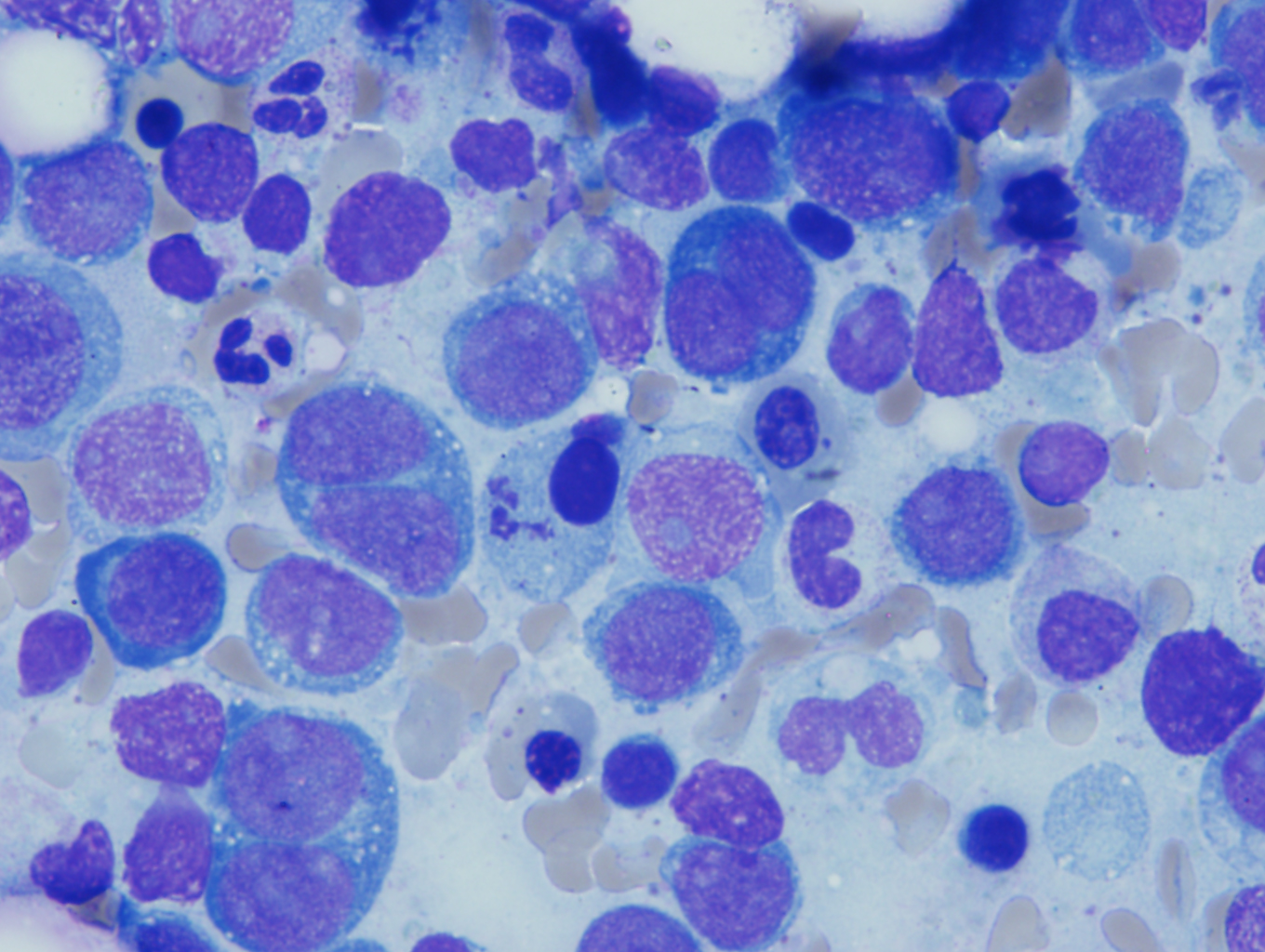
Bone marrow differential count of the bone marrow showed 65.0 % Erythroid Precursors with 48.4% erythroblasts and 7% myeloblasts

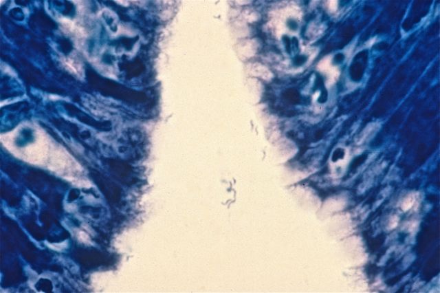
Several erythroblasts were seen, which often had overlapping morphological features with myeloblasts. Erythroblasts had slightly coarser nuclear chromatin compared to myeloblasts and often had deeply basophilic vacuolated cytoplasm. Erythroid maturation was markedly megaloblastic /dysplastic and left shifted with marked preponderance of erythroblasts. Dysplastic forms characterized by presence of precursors with irregular nuclear borders along with few multinucleated forms and gigantoblasts were present.

Cells counted as myeloblasts had high N/C ratio, finer nuclear chromatin with occasionally distinct 1 to 2 nucleoli and scant cytoplasm.

Bone marrow with erythroid hyperplasia
Megaloblastic erthroid precursors with binucleate forms

Discussion:

The current WHO classification subtypes acute erythroid leukemia into two categories based on the presence or absence of significant myeloid component.

Erythroleukemia or Erythroid/Myeloid (FAB subtype A – M6a) comprises of more than 50% erythroid precursors among all nucleated cell population of bone marrow and more than 20% myeloblasts among non erythroid cells.

Pure erythroid leukemia (FAB subtype B – M6b) comprises of more than 80% immature cells of erythroid lineage with no evidence of a significant myeloid component

The most common reactive process that can mimic acute erythroid leukemia is megaloblastic anemia caused by vitamin B12 and folate deficiency. Features associated with pernicious anemia are hemolysis with increased mean corpuscular volume (MCV), hypersegmented neutrophils, leukopenia and thrombocytopenia increased LDH and urobilinogen. Bone marrow findings show hypercellular marrow witn marked erythroid hyperplasia. Other non-neoplastic diseases mimicking acute erythroid leukemia are post-chemotherapy recovery, parvovirus infection, drug effect, heavy metal intoxication and congenital dyserythropoiesis. A detailed clinical history, laboratory work up, peripheral blood and bone marrow examination, cytochemical, immunoshistochemical, flow cytometry, cytogenetic and molecular studies are required for the diagnosis of acute erythroid leukemia.

The oncologist was contacted and it was confirmed that B12 was repleted before the bone marrow study was performed. Diagnosis of acute erythroid /myeloid leukemia was only made after it was confirmed with the oncologist that patient was not B12 deficient at the time of the study.

Vajpayee,Neerja2014_small

-Neerja Vajpayee, MD, is an Associate Professor of Pathology at the SUNY Upstate Medical University, Syracuse, NY. She enjoys teaching hematology to residents, fellows and laboratory technologists.

Are Feasibility Limits Feasible?

In the laboratory we’re constantly seeking ways to check that the test results that we put out are accurate. Our primary reason for doing this is that we want to make sure the patient is treated appropriately based on the results of the tests we run. Also, it’s nice not to release values that appear to be nonsense. A tool that is sometimes used to help us check results is something called feasibility limits.

Lab computer systems often allow you to enter feasibility limits for your test results or for your test parameters. These are values outside of which you would not expect to find an analyte concentration in a living person. For example, you might expect a serum creatinine of 200 mg/dL (17,680 µmol/L) in a zombie, but you wouldn’t expect to find one in a living human being. Setting feasibility limits helps you catch things that make no sense before the physician calls you on them, for instance if you have a decimal malfunction and mistakenly try to report a plasma calcium value of 90 mg/dL (22.5 mmol/L) instead of 9 mg/dL (2.25 mmol/L). The trick to feasibility limits is deciding on the highest or lowest value you might expect to see in a living human being. In the case of calcium, upper feasibility limits of 20 mg/dL (5 mmol/L) may give you wiggle room without letting you report nonsense. However, feasibility limits have their drawbacks also. One of those drawbacks is that I’ve found that with human beings, nearly anything is feasible, especially in the pediatric realm. I finally removed the feasibility limits from the LIS in my institution, after a couple of different episodes led me to that conclusion.

One was a body temperature on a blood gas analysis. Under normal circumstances one would not expect to encounter a body temperature much below 90° F (32.2 °C) ever. But of course hospitals are not known for their populations being “under normal circumstances”. The body temperature of the patient in question was 70° F (21°C) on a patient who had been cooled down for surgery. The blood gas instrument accepted the temperature, but the lab computer system would not because of the feasibility limits set in the computer. The patient’s blood gas results could not be released in the computer until we took the temperature feasibility limits out.

Another example was with sodium. It would seem reasonable to have an upper feasibility limit of 180 mmol/L for sodium. Yet we had a patient whose sodium was 199 mmol/L when he arrived in the ED. Reasonably expecting some sort of contamination issue, we requested another sample, which had a sodium of 204 mmol/L, followed by 200 mmol/L in the next sample. These were real sodium results and over the course of several days’ time the physicians managed to get the patient’s electrolytes normalized. Again, feasibility limits interfered with result reporting and had to be removed from the computer.

These episodes caused us to remove most of our feasibility limits from the computer. They also helped me to remember and important point: Tools are fine, but you must understand their uses and their limitations in order to use them appropriately. Feasibility limits can be useful as long as you keep in mind that with humans, you often see the unfeasible.


-Patti Jones PhD, DABCC, FACB, is the Clinical Director of the Chemistry and Metabolic Disease Laboratories at Children’s Medical Center in Dallas, TX and a Professor of Pathology at University of Texas Southwestern Medical Center in Dallas.

Education Proposals for ASCP’s 2016 Annual Meeting

Are you interested in presenting an education course at ASCP’s 2016 Annual Meeting? If so, the call for proposals is now out. You can find it at the direct link below.

ASCP’s 2016 Annual Meeting will be held at the Mandalay Bay Hotel & Casino in Las Vegas, NV on September 14-16, 2016.

Click here to access the 2016 Call for Proposals submission site

Do Children of Teenage Fathers Have an Elevated Rate of DNA Mutations?

If you recall one of my previous posts about DNA mutations, I discussed briefly the difference between somatic and germ-line mutations as well as the various types of mutations and resulting consequences.

Recently, I came across a research article that suggests an elevated germ-line mutation rate in teenage fathers, thus leading to an unexpectedly high level of DNA mutations in the children born to teenage fathers.

Many studies have been conducted on the theory that male germ cells go through a higher number of cell divisions when compared to that of female germ cells, and that the higher number of paternal cell divisions leads to an increased DNA mutation rate. The paper suggests that the increased presence of DNA mutations in sperm cells of teenage boys could explain why their offspring might be at higher risk for a spectrum of disorders when compared to parents in their twenties.

It’s an interesting read! More information as well as the full article can be found at http://rspb.royalsocietypublishing.org/content/282/1803/20142898


L Noll Image_small

-LeAnne Noll, BS, MB(ASCP)CM is a molecular technologist at Children’s Hospital of Wisconsin and was recognized as one of ASCP’s Top Five from the 40 Under Forty Program in 2015.

AMP Submitts Written Testimony about LDTs

From the press release:

“The Association for Molecular Pathology (AMP), the premier global, professional society serving molecular diagnostics professionals, yesterday submitted written testimony to the House Energy and Commerce Subcommittee on Health for their hearing on “Examining the Regulation of Diagnostic Tests and Laboratory Operations.” AMP urged the Committee to use AMP’s proposal to modernize the Clinical Laboratory Improvement Amendments (CLIA) at the Centers for Medicare & Medicaid Services (CMS) as the basis for legislation that would preserve innovative patient care by building upon the current CMS-based system for oversight of laboratory developed procedures (LDPs).

‘Molecular pathologists are highly trained professionals and our professional judgment is used throughout the design, validation, performance, ongoing monitoring, and interpretation of test results. It is our mission to ensure that patients have access to innovative, accurate, reliable, and medically useful laboratory testing procedures,’ said Roger D. Klein, MD, JD, AMP Professional Relations Chair. ‘The AMP CLIA Modernization proposal preserves patient access to essential laboratory services that would no longer be offered if a costly FDA-based regulatory system were imposed upon academic medical centers, cancer centers, hospitals and small independent laboratories,’ he added.

To read the press release in full, visit www.amp.org.

CLSI Publishes a Revised Microbiology Document

From the press release:

“The Clinical and Laboratory Standards Institute (CLSI) has published a revised document titled Methods for Antimicrobial Dilution and Disk Susceptibility Testing of Infrequently Isolated or Fastidious Bacteria (M45-Ed3).This guideline informs clinical, public health, and research laboratories on susceptibility testing of infrequently isolated or fastidious bacteria that are not included in CLSI documents M02, M07, or M100. Antimicrobial agent selection, test interpretation, and quality control are discussed.
“Susceptibility testing is particularly necessary in situations in which the etiological agent belongs to a bacterial species for which resistance to commonly used antimicrobial agents has been documented, or could arise. The intent of this document revision is to assist labs in determining an approach for testing that is relevant to their individual practice settings.”

To order the document go to shop.clsi.org/m45.

Microbiology Case Study: 22 Year Old Male with Dysuria

A 22 year old male, treated for gonorrhea one month ago, presents with two days of dysuria and white penile discharge. He reported complete resolution of his symptoms with treatment. He has had sexual contact with 2 partners in the past month.

Cultures are taken and the following is seen on gram stain.

N. gonorrhoeae is a gram negative diplococci responsible for the disease of gonorrhea. Though most specimens for gonorrhea are received from sources suspected of having the disease, it should be noted that whenever dealing with a gram negative diplococci of unknown source, care should be taken, and the specimen should be handled under a hood for possible exposure to N. meningitidis.

Seems like a clear cut case of gonorrhea, but given the patient’s history is this treatment failure, or likely reinfection?

The CDC has warned that rates of antibiotic resistant gonorrhea are on the rise. A once easily curable disease, is now becoming resistant to our mainstay treatment of ceftriaxone and azithromycin. Although cases have been reported, the majority of ‘failure of treatment’ scenarios are likely re-infection. Patients likely do not abstain until they are symptom free, or the partner may not be treated, leading to a reinfection of the patient. In the event of treatment failure, the CDC even recommends re-treating with the same ceftriaxone/azithromycin regiment. It is still recommended that a sample be taken for culture in the event that there is any concern over failure of treatment and possible antibiotic resistance.

Historically, testing for N. gonorrhoeae consisted of culture and gram stain, but the CDC now recommends testing for gonorrhea with nucleic acid amplification testing (NAAT). This test has improved sensitivity, less subjectivity and a much faster turnaround time than gram staining and culturing. It cannot, however, be used for test of cure, as the NAAT tests for DNA, which still may be present, even if the organisms have been killed within the host, leading to a false positive result.

When a clinician suspects gonorrhea, he or she should take care with what site the specimen is being collected from, and what sample is specifically needed. This lab is validated for only certain types of specimens, and it is important to communicate with the clinicians to ensure the proper specimens are being submitted.

The CDC states that the optimal samples should be vaginal swabs for women, and first catch urine for men. Great care should be used when collecting the ‘first catch urine’ as this is a dirty catch, and that some tests have specific requirements about quantity. For instance, at this lab, we require no more than 30 ml of urine. Any more volume could dilute the specimen which could cause a false negative result.

If a culture is required in the event of treatment failure, it is best if the clinician innoculates the sample directly onto Thayer Martin agar (or chocolate agar if that is all that is available). Given that N. gonorrhoeae is such a labile organism, it need special media to grow, and great care must be taken with transport. This lab recomends inoculating onto the Jembec plates (Thayer Martin media with a CO2 pellet) allowing survival during transport.

N. gonorrhoeae inoculated directley onto the Jembec plate
N. gonorrhoeae inoculated directly onto the Jembec plate
Cultures can also be innoculated onto chocolate agar. Notice the smaller grey colonies, which is the N. gonorrhoeae.  Notice the other colonies on the plate, as chocolate agar is not a selective medium.
Cultures can also be innoculated onto chocolate agar. Notice the smaller grey colonies, which is the N. gonorrhoeae. Notice the other colonies on the plate, as chocolate agar is not a selective medium.

In conclusion, although the majority of cases of gonorrhea treatment failure are likely to be due to re-infection, antibiotic resistance is still of growing concern and clinicians should know when a culture is necessary for antibiotic sensitivity testing. N. gonorrhoeae requires nutrient supplementation for growth, either on Thayer Martin media, or chocolate agar. Care must be taken when sampling and transporting the specimen. Clinicians also need to be aware of the requirements for sample collection for NAATs, as it is not always clear as to why one can’t simply run a test on a sample greater than 30 ml, or a clean catch urine.

-Rich Smith is a Pathology Student Fellow at University of Vermont Medical Center.

Wojewoda-small

-Christi Wojewoda, MD, is the Director of Clinical Microbiology at the University of Vermont Medical Center and an Assistant Professor at the University of Vermont.

Radiation in the Lab

Radioactivity is no longer common in most clinical laboratories. At one point in my career, radioimmunoassays were commonly found in laboratories, and most labs had institutional radiation safety plans and carefully followed the CAP checklist for handling and dealing with radioactivity. With the advent of enzyme-linked immunoassays, sensitive nephelometers, various fluorescent and chemiluminescent technologies, and then mass spectrometry, radiation was quickly replaced in most clinical labs. The general prevailing thought was: Why deal with radiation if you don’t have to? Now days, radioimmunoassays are essentially only found in reference labs and utilized for esoteric analytes or those which cannot be measured any other way.

Despite that being true, it’s important for a lab to know what to do if radioactive materials should appear in the lab. How likely is that to happen? Perhaps more likely than you may think. Last week a sample shipped to us from an outside institution set off the radioactivity monitor on our hospital loading dock. The package was on a delivery cart with other packages so per protocol, the whole cart went to nuclear medicine where it was determined that the radioactive package was for the lab and it was brought to us. The radioactive sample turned out to be a urine sample for VMA/HVA analysis from a patient on a new cancer treatment protocol. The urine was indeed radioactive.

The shipping institution was contacted and the packaging personnel had no idea either that the sample was radioactive, although they were aware a new protocol was going into place. Working with our nuclear medicine department and the institutional radiation safety group, we have now once again put appropriate processes in place to handle and deal with radioactivity. And we’ve dusted off our old CAP checklist regulations as well.

This episode actually turned out to be a benefit to us, as we discovered that our own nuclear medicine department will be starting this new protocol soon, and had not thought ahead to possible radioactive samples sent to the lab. We are now working closely with them to ensure proper procedures and safeguards, and have a plan of action clearly in place. We also continue to work with the institution that sends us samples. The very next sample from them was properly labeled as potentially radioactive.

-Patti Jones PhD, DABCC, FACB, is the Clinical Director of the Chemistry and Metabolic Disease Laboratories at Children’s Medical Center in Dallas, TX and a Professor of Pathology at University of Texas Southwestern Medical Center in Dallas.

Times are Definitely Changing

Just returning from the ASCP 2015 conference in Long Beach, California, I can’t help but reflect on what a wonderful experience I had. The weather was picturesque, attendees at an all time high, and a variety of educational offerings and speakers on point.

The highlight for myself (and I know I am not speaking out of line when I mention the four other amazing ladies) was being recognized as ASCP’s Top Five from the 40 Under Forty program this year. I cannot begin to tell you how refreshing and energizing it is to be a part of an amazing group of five women who are all dedicated to advancement in Pathology and Laboratory Science.

To be honest, I was a little nervous going into the meeting… What would the other honorees be like? What would they think of me? Am I going to be completely out of my league there? Well let me tell you, everyone that I met was wonderful! We discussed a variety of topics, and even tossed around an idea for future collaboration.

Yes, I could go on and on about my new friends, but I think what I want to point out is that it cannot, and should not go unnoticed that the Top 5 this year consisted of all women, all WELL deserving women at that. How were we chosen? Yes, we blogged, but don’t forget that we also submitted CVs and biographies. We wrote essays as well as recorded videos (some of us spending hours re-shooting and cringing at ourselves). Not to mention, votes were cast by a dedicated panel as well as online voters. Your 40 Under Forty, including the Top Five, came from all over the country and represent various specialties in our field.

Unfortunately, at some point I had to come down from the “girl power” high. I returned to Milwaukee and thought to myself, I wonder what the ratio of men to women is in our laboratory alone? A few short minutes later, I crunched the numbers and it was easy to see that women make up 85% of laboratory staff at our organization. The totals are inclusive of all laboratory departments and shifts as well as administrative support, Pathologists, and Directors at Children’s Hospital of Wisconsin.

womeninthelab

It was during these thoughts on women in science and recognition, that I remembered an article I had read quite a while back. The article had discussed how historically, women occupied most of the laboratory jobs (the strange term, “lab gals” sticks out in my mind). This was thought to be the case because it was believed that women had more patience, were more detail oriented, and therefore were trained to perform the work that doctors did not want to do. At the same time, men typically occupied the higher-level decision making positions (those that required an advanced degree, PhD, and MD). I thought to myself that even today, it sure does seem that there are more women in the laboratory profession. However, we have come a long way and are seeing an increase in women being honored for their education, professional achievements, and advancements in the field. Every single lead technologist and laboratory manager at our institution as well as the CEO of our health system currently is female. Interestingly, more than HALF of the 40 Under Forty Honorees this year are highly educated women with advanced degrees!

We all know full well that more than 70% of critical medical decisions are based on laboratory results. Therefore, if the field of laboratory professionals is made up of mostly women, it appears that our attention to detail is instrumental in making some major decisions. Yes, there still may be gender gaps when comparing men to women in academia however, what once was a field dominated by the male PhDs and MDs, appears to be shifting majorly as more and more women are making their presence known in Pathology and Laboratory Science.

I applaud everyone who was honored this year as one of ASCPs 40 Under Forty. Women are the past, present, and the future of laboratory science and medicine and it brings a little extra smile to my face to know that so many well deserving women are being recognized by ASCP this year.

2015 ASCP 40 Under Forty Top Five: Amanda Wehler, Tiffany Channer, Jennifer Dawson, LeAnne Noll, and Kimberly Russell

L Noll Image_small

-LeAnne Noll, BS, MB(ASCP)CM is a molecular technologist at Children’s Hospital of Wisconsin and was recognized as one of ASCP’s Top Five from the 40 Under Forty Program in 2015.

You Make the Diagnosis: A 68 Year Old with Epigastric Pain

A 68 year old male presents with chronic, gnawing epigastric pain which has been getting worse over the past year. An endoscopy is performed and a representative gastric biopsy section, stained with Giemsa stain, is shown here. What organism is responsible for this patient’s symptoms?

gastric biopsy Giemsa stain
A. Candida albicans
B. Helicobacter pylori
C. Bacillus cereus
D. Staphylococcus aureus
E. Campylobacter jejunum

The diagnosis in this case is Helicobacter pylori. Described by Warren and Marshall in 1983, Helicobacter pylori is now known to be the cause of most cases of chronic gastritis and peptic ulcer. Helicobacter is a tiny, corkscrew-shaped bacillus, and is easily missed on routine H and E-stained histologic sections. Giemsa staining, as seen in the image above, can be helpful, as can silver staining, as seen in this image:

H. pylori

Helicobacter does not invade the gastric mucosa, but produces its symptoms through continual stimulation of the host immune response. Treatment involves triple therapy (e.g., omeprazole, amoxicillin and clarithromycin), and prognosis is excellent. A small number of patients, however, develop gastric adenocarcinoma or MALT lymphoma.

Krafts

-Kristine Krafts, MD, is an Assistant Professor of Pathology at the University of Minnesota School of Medicine and School of Dentistry and the founder of the educational website Pathology Student.