Microbiology Case Study: Immunocompromised Boy with Skin Nodules

An elementary school aged boy with a history of pre-B cell acute lymphocytic leukemia with a failed bone marrow transplant was transferred to a regional children’s hospital for leukodepletion and participation in an experimental clinical trial. At that time, his CBC was significant for 10% polymorphonuclear cells and 50% blasts. He was subsequently transferred to the ICU in respiratory failure and developed papulonecrotic lesions on his face, trunk, and bilateral legs. Prior to this, he was pancytopenic with no blasts present with cell counts of 100 WBC, hemoglobin 8.3 and 37,000 platelets. His Fungitell assay, which detects (1-3)-β-d-glucan, was positive.

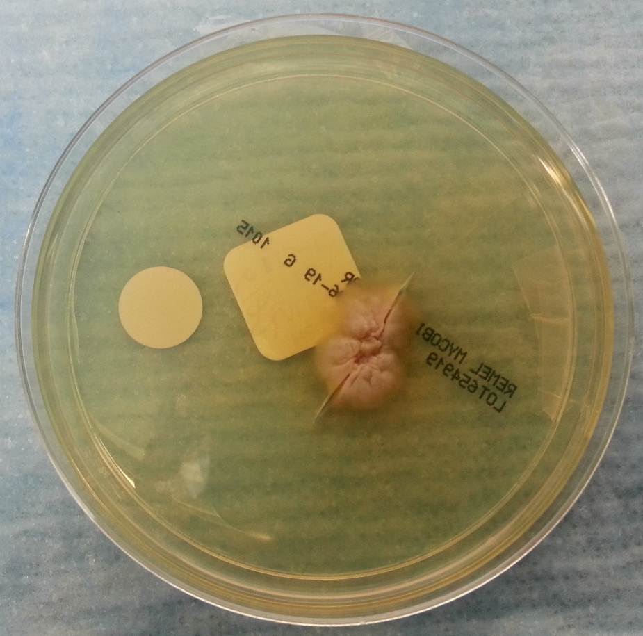
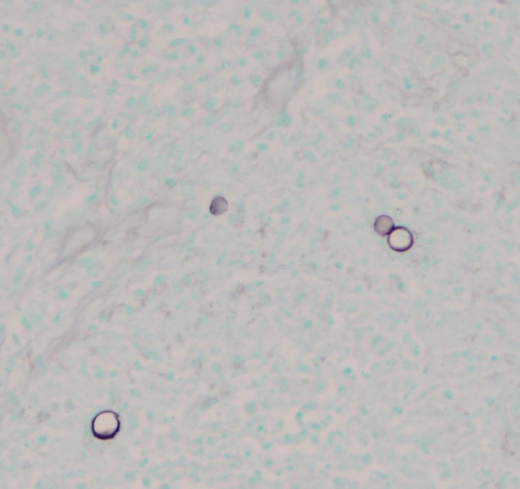
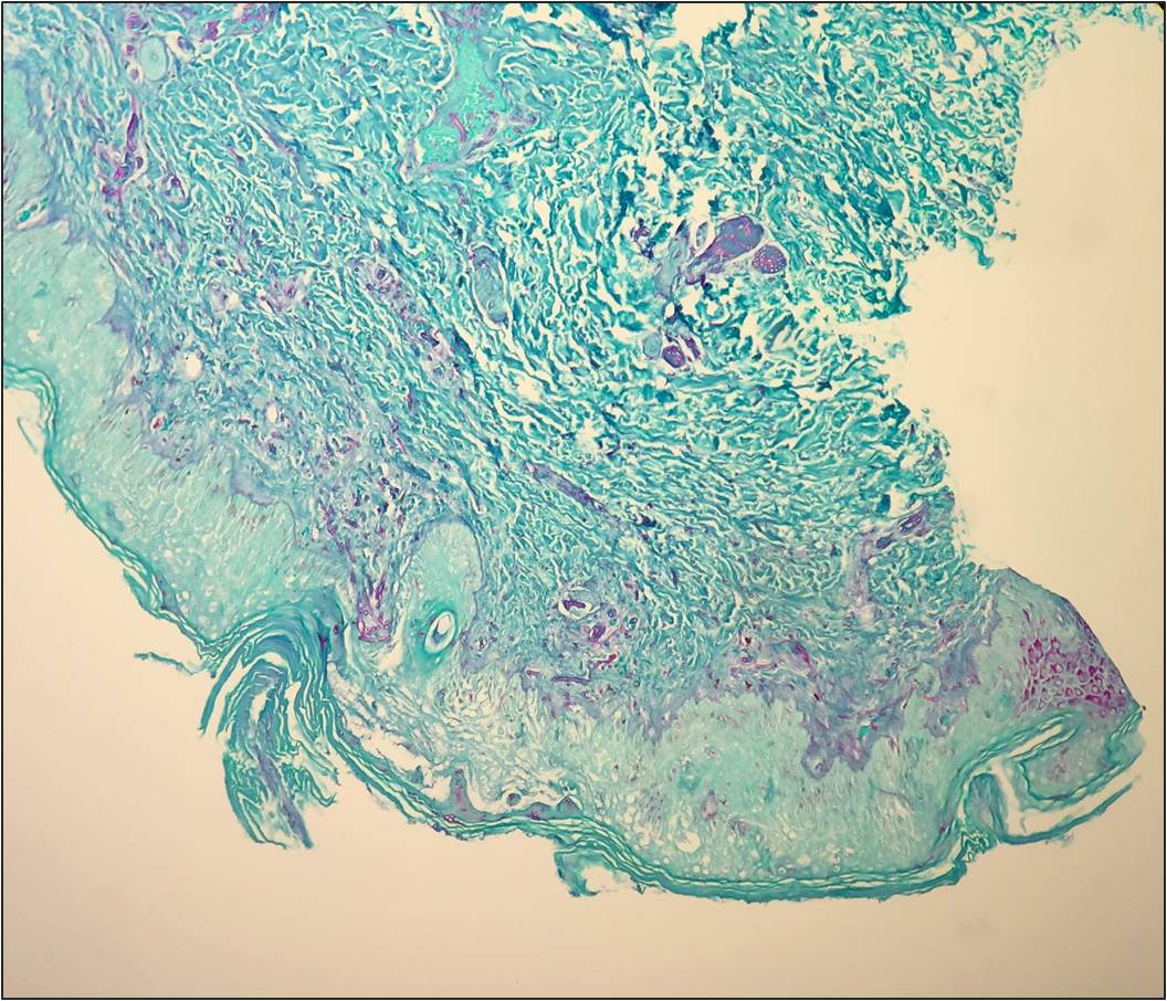
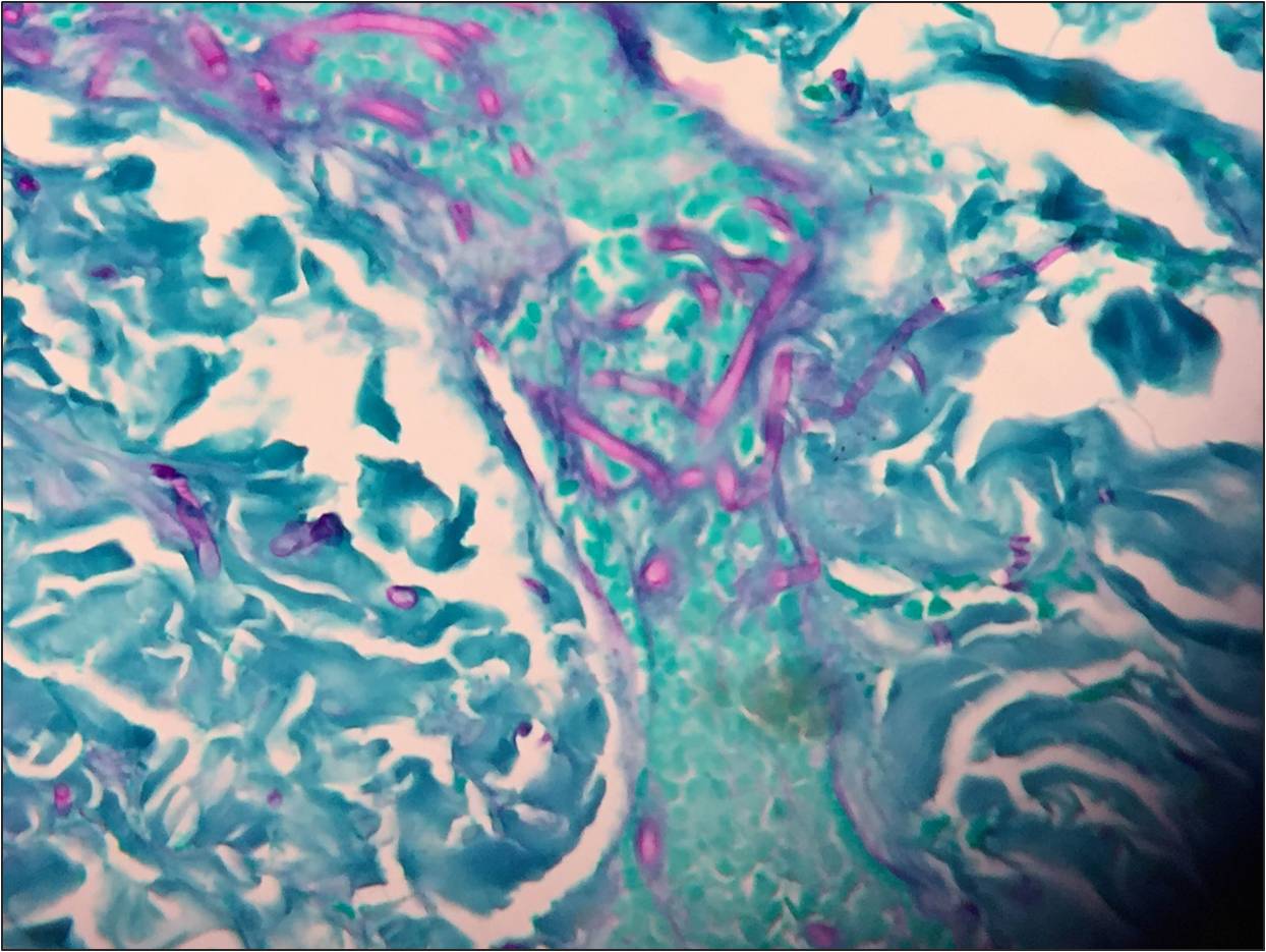
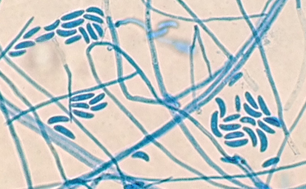
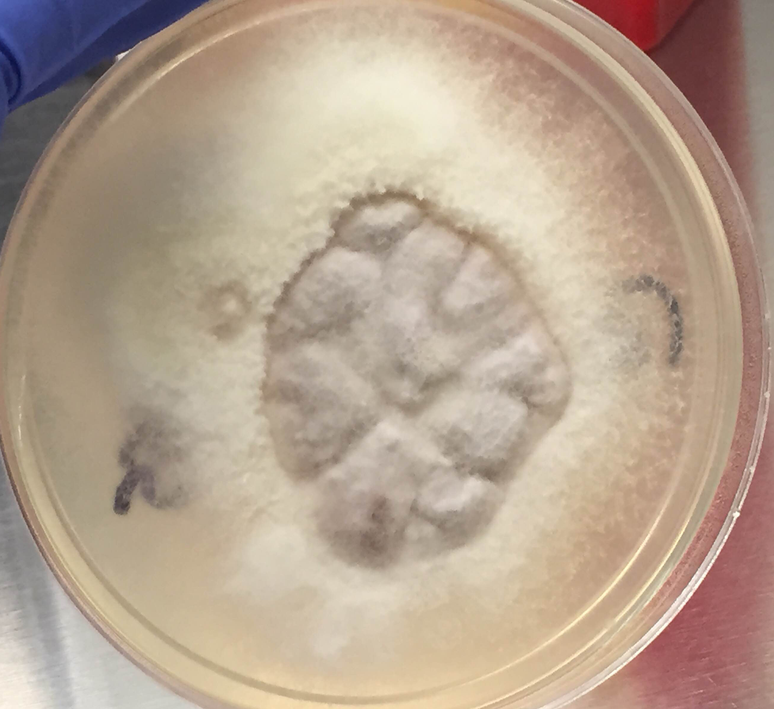
Routine blood culture, fungal culture from the endotracheal tube, and fungal culture from the skin lesion biopsy specimens all had fungal elements on KOH stain. Young growth of a whitish, fluffy mold was present on all cultures within two days. Histopathology on the punch biopsy of a skin lesion on the thigh showed septate hyphae within the dermis, epidermis, and invading the vasculature that was particularly apparent with GMS stain (Figure 1a and 1b). Within a few days, the fungal cultures showed septate hyphae with microconidia using lactophenol cotton blue tape preparation, and shortly thereafter the mold developed into macroconidia with multiple septations taking on canoe-like forms (Figure 2). The white, cotton-like colonies developed a pink tinge (Figure 3). These characteristics allowed for the identification of the growth as Fusarium sp.

Septate hyphae on GMS stained section of the skin punch biopsy.
Microscopic identification of Fusarium by lactophenol cotton blue stain.
Colony of Fusarium growing on inhibitory mold agar (IMA).

Fusarium is an opportunistic hyaline mold with infection most commonly seen in immunocompromised hosts. It can cause keratitis through contamination of contact lenses, penetration due to trauma, or use of immunosuppressive steroid ophthalmic solution. It is increasingly becoming the cause of disseminated infection in neutropenic hosts with a broader spectrum of disease, which includes: skin lesions, fungemia, rhinocerebral involvement and pneumonia. In these cases, without an immune system to fight the infection, mortality is high. Inhalation of airborne conidia, ingestion from water sources or access through mucosal membranes are all potential points of entry.

The colony growth on plated fungal media is rapid, usually maturing within four days. On microscopic examination, Fusarium hyphae are septate, approximately 3-6 microns wide with acute angle branching. Microconidia are small, oval-shaped, and no larger than 4 x 8 microns in size. These can look like Acremonium sp. Macroconidia are canoe- or sickle-shaped with the largest dimension being about 80 microns in length, exhibiting 3-5 septatations.


Jodi Music, MD, is an AP/CP resident at UT Southwestern Medical Center.

-Erin McElvania TeKippe, Ph.D., D(ABMM), is the Director of Clinical Microbiology at Children’s Medical Center in Dallas Texas and an Assistant Professor of Pathology and Pediatrics at University of Texas Southwestern Medical Center.

A “Primer” on DNA and the Consequences of Mutation

We can’t talk about Molecular Diagnostics without possibly talking about DNA, and with DNA, comes mutation. Much of the work I do revolves around searching for specific variants in a patient’s DNA sequence. We all have mutations in our DNA, but does that mean we all are affected negatively? Absolutely not. Spontaneous mutations occur during normal process such as DNA replication and repair within our cells. They also can arise from exposure to ionizing radiation, UV exposure, and chemical agents. Some mutations are passed down through reproductive cells. Mutations can be categorized as harmless or sometimes hurtful giving rise to gene defects, copy number variants, metabolic deficiencies, and cancer, while others result in positive effects.

Where do mutations occur?

Somatic mutations occur in cells that aren’t reproductive in nature. These mutations for the most part do not have a blatant effect on the organism because our normal body cells are able to counteract the mutated cells. However, sometimes mutations in somatic cells can affect division of cells, which has the potential to result in forms of cancer. On the contrary, germ-line mutations occur in reproductive cells and have the possibility of being passed down through generations, resulting in the presence of the mutation in all of the organism’s cells.

Understanding DNA

A T G C bases, along with a sugar, and a phosphate group combine to form a polymer of nucleotides. This polymer backbone of alternating sugar and phosphates is what we call DNA (Deoxyribonucleic Acid). DNA is transcribed to RNA (Ribonucleic Acid) which instead of Thymine (T) contains a Uracil (U). Finally, RNA is translated to protein.

Pyrimidines       Purines
Single-ringed organic bases Double-ringed organic bases
Thymine (T)

Cytosine (C)

Uracil (U)

Adenine (A)

Guanine (G)

Hydrogen Bonds Between Complimentary Strands of DNA
Adenine bonds Thymine   A = T(U)

Guanine bonds Cytosine   G ≡ C

Two Hydrogen Bonds

Three Hydrogen Bonds

mut1
Nucleic acid→ triplet codon (amino acids)→ polypeptides→ proteins (approx 50 amino acids in length)

Amino acids are the triplet codons that make up proteins. It is extremely important to become familiar with the codon table. You don’t need to memorize it, however you should at a minimum know what triplets code for START and STOP codons. You will quickly see that there are multiple codes for a single amino acid. This becomes important when considering the effects of mutation:

mut2

Like I mentioned earlier, many of the tests I perform involve screening for variants in DNA sequence. I accomplish this through different methods, which I will touch on in future blog posts. We could easily discuss mutations for hours and hours, but for now, it’s probably most important to gain a quick (and simple) understanding of the types of mutations and what the resulting effects can be. For the sake of demonstration, let us consider the following normal (wild type) sequence pattern:

NOTE: Uracil replaces Thymine because of transcription from DNA to single stranded messenger RNA (mRNA)

Transition Mutations occur when mispairing results in a purine being replaced with a different purine, or a pyrimidine replaced with a different pyrimidine:

A→G

C→T

Transversion Mutations occur when a purine is replaced with a pyrimidine or vice versa:

A→T

C→G

Missense Mutations have the ability to result in a change in the amino acid. This happens when the codon triplet is altered, and the amino acid sequence of the encoded protein is changed:

mut3

Sometimes, Missense Mutations are silent:

Notice how the structure of the gene product is unchanged. This happens because all amino acids can be encoded by more than one triplet codon.

Frame shift mutations can result from either a single base insertion or a single base deletion.

Insertion.
Insertion.
Deletion.
Deletion.

Nonsense mutations are exactly what they are called… Nonsense! Here, a base substitution changes an amino acid into a stop codon. Nonsense mutations result in abnormal termination of translation and the protein product is shortened:

mut7

Chromosomal Abnormalities

Sometimes changes in the actual chromosomal structure or number take place in the cells. These changes can result in deletions, duplications, inversions, and translocations of chromosomes. Chromosomal deletions and insertions are simply the loss or gain of chromosomal material. Inversions result from the removal, flipping, and then reconnection of the chromosomal material within the same chromosome.

Translocations are more complicated and involve switching of genetic material between two chromosomes. A reciprocal translocation occurs when parts from two different chromosomes exchange. We call a reciprocal translocation “balanced” when there isn’t a gain or loss of genetic material between the chromosomes (both chromosomes remain fully functional). An “unbalanced” reciprocal translocation affects an offspring’s phenotype due to extra or missing genes.

This information I have touched on is very general, however, it doesn’t mean it is not of importance. These basic concepts are the fundamentals needed to begin understanding mutation as they will provide us with the framework for recognizing the needs and importance of molecular diagnostic testing!


Test your Molecular Knowledge

  1. What amino acids do the following codons code for:
    1. GAU
    2. AUG
    3. UAG
    4. CAG
    5. UGA
  2. Classify the following mutations:
    1. T→A
    2. T→C
    3. AGA→UGA
    4. AGA→AAA
    5. AGA→CGA

Answers

1 a) Aspartic Acid b) Methionine (start) c) Stop codon d) Glutamine e) Stop codon. 2 a)Transversion b) Transition c) Nonsense d) Missense e) Silent

L Noll Image_small

-LeAnne Noll, BS, MB(ASCP)CM is a molecular technologist at Children’s Hospital of Wisconsin and was recognized as one of ASCP’s Top Five from the 40 Under Forty Program in 2015.

Microbiology Case Study: 76 Year Old Female with Upper Back Pain

Case History:
A 76 year old female presents with a two year history of worsening upper back pain. Imaging revealed compression fractures of the first three thoracic vertebrae (T1-T3). Fine needle aspiration and a core biopsy of the T3 vertebral body were examined in surgical pathology. There was acute and chronic granulomatous inflammation with fungal organisms observed on histologic examination. Surgery for decompression and fusion of C5-T6 vertebrae was performed and tissue was sent for fungal culture.

Potato flake agar shows a tan-brown fungus.
Mycosel agar shows beige-white fungal growth.
Scotch tape prep shows septate hyphae with unbranched conidiophores and single, terminal, "lollipop" conidia.
Scotch tape prep shows septate hyphae with unbranched conidiophores and single, terminal, “lollipop” conidia.
Silver stain of involved bone with fungal organisms exhibiting broad-based budding.

Laboratory Identification:
The workup revealed a thermally dimorphic fungus with a mold form growing in the laboratory at 25°C and a yeast form present in the surgical pathology specimen. The mold form is moderately slow growing and has septate hyphae with small, round, terminal conidia often described as “lollipops.” The yeast form is large (8-15 microns) with broad based buds and double contoured cell walls. The immune system reacts to the presence of the fungus by forming granulomas and leads to acute and chronic inflammation within the involved tissue. The organisms can occasionally be seen within giant cells in histologic sections. The silver stain, as seen above, highlights the organisms.

Discussion:
The fungus described above exhibits the features of Blastomyces dermatitidis. This organism resides in soil and decaying plant matter and is endemic to eastern North America including the Mississippi and Ohio River Valleys as well as areas surrounding the Great Lakes and St. Lawrence River. The most common primary sites of involvement for Blastomyces are cutaneous and pulmonary. Following a primary infection, the disease can progress to disseminated blastomycosis which involves other sites such as bone.

The primary site of infection in this case is unknown. There was no history of cutaneous ulcers and chest imaging was unremarkable. The patient did have a remote history of bloody sputum production which she had attributed to “dental difficulties” that she was experiencing and has since resolved. This may have been evidence of a primary pulmonary infection preceding the vertebral involvement; however it is difficult to say with certainty.

The classic double contoured cell walls are not evident on the silver stain of the surgical pathology specimen in this case. This may be due to the fact that the bone required decalcification before histologic sections could be taken. The decalcification process may have caused an artifactual loss of the double contour. Despite the fact that this classic finding was not seen, the macroscopic and microscopic morphology is most consistent with Blastomyces.

The patient is being treated with long-term itraconazole and is currently doing well.

-Britni Bryant, MD is a 2nd year anatomic and clinical pathology resident at the University of Vermont Medical Center.

 Wojewoda-small

-Christi Wojewoda, MD, is the Director of Clinical Microbiology at the University of Vermont Medical Center and an Assistant Professor at the University of Vermont.