The folks at Nebraska Medical are using an integrated approach to treat patients infected with Ebola virus. You can read about it exclusively on the Lab Medicine website.
Month: October 2014
Reminiscing Tampa
ASCP 2014 at Tampa provided the perfect getaway for a New Yorker forced to wear fleece early October. The same attire seemed to be mocking me the moment I stepped out of the Tampa International Airport on Wednesday night. It was a pleasant surprise and I gleefully tucked it right into my suitcase.
At the hotel, I took a quick glance at the lecture schedule. Having already missed the first day, I was eager to extract the best out of the next two days. I was thrilled to see an array of topics specially aimed at residents. Also, many lectures focusing on novel or state-of-the-art techniques, including molecular methods, virtual microscopy, digital pathology, informatics, etc. It seemed to me like “The future beckons!!” Being a hard-core morphologist, it was a tough call for me, as I would have to forego a host of other good lectures. But I decided to focus on the resident review courses and ancillary techniques.
Keeping with my agenda, I set the ball rolling on day two by attending the lecture on “Automating Anatomic Pathology.” It was an eye opener for me, dealing with the scope and future of automation in anatomic pathology lab. “Anatomic Pathologist’s Role in Patient Safety” was the next. Dr. Silverman cited studies revealing that soft tissue lesions, with an error rate of 20-30%, led the list of organ specific error rates. He deliberated on the importance of second opinions in error reduction. He aptly concluded his lecture with the remark, “the pathologist is the Final Quality Assurance Officer or ‘the buck stops here.’” It was a huge wake up call for me.
I moved on to my first lecture on Molecular Pathology, “Welcome to the Beginning: Molecular Pathology for the General Pathologist and Molecular Pathologist.” It was just the right one for me and helped me firm up basic concepts. In the evening I attended “Molecular Diagnostic Methods in Oncology: an update on practical aspects.” Dr. Larissa Furtado and Dr. Yue Wang from University of Chicago were simply brilliant in elucidating the role of molecular techniques in oncologic practice. The prior morning session, helped me understand the deliberations in this talk much better.
I made it a point to attend most of the Resident Review courses. Though my Board Exams are two years away, I took it as a perfect platform to acquaint myself with the “hot” topics. I spent almost the entirety of day three attending the courses. A packed audience was testimony to these sessions’ popularity. Most of the speakers were brilliant. The case based presentations followed by an interactive voting format helped keep us all fully involved. However, the lab administration and last day hematology section could have been better.
In between, I found some time to listen to one of my all time favorites: Dr. Goldblum’s trademark lecture on soft tissue pathology. He quipped in his inimitable style “Don’t hunt for lipoblasts to diagnose a liposarcoma” and warned us of the vast plethora of “pseudolipoblasts” lurking around. Rather, he stressed the importance of analyzing the entire histology in the correct clinical context.
Let’s wander into the poster sessions! We had a total of twelve posters from our program itself, probably the largest representation from a single center. I had four posters and one of them was selected as a finalist in the Best Resident Poster section. It was an entirely new experience for me. However, I did some homework to prepare myself for the judging session. The judges on both the days were very pleasant and spent a significant amount of time discussing the work with me. It was disappointing not to get the award, though I knew the competition was tough.
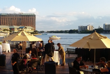
The evening Mixology Lab was the perfect concluding session in the backdrop of the setting sun across the scenic Hillsborough river. There was delicious food and wine as Dr. Baloch announced the various poster award winners. It was special for me for another reason, as my very good friend Shree Sharma was one of the “top 5 under 40” award winners.
It would be so improper if there were all work and no play. Friday evening provided the perfect opportunity to explore the city. I went out with friends to the Ybor City, taking the streetcars, which surprisingly provided 50% discount to conference attendees. Ybor City was such a happening place, full of fun. While strolling along the 7th avenue, we took pictures with people celebrating Gasparilla festival, dressed as pirates. A glass of sangria at the historic Columbia Restaurant provided the perfect toast to end the day.
My trip was not to end here as I had already registered for the TRIG Genomic Pathology Workshop for Saturday. This was my first exposure to such a session in molecular technology. We were divided into small groups. In a case based approach, the workshop deliberated on teaching principles related to the development of genomic assays and result interpretation. There were four cases pertaining to single gene testing, prognostic gene panel testing, how to design a cancer gene panel and whole genome sequencing, respectively. Both Richard Haspel and Andrew Beck were simply brilliant and they took special care to approach each group separately and clarify their doubts. It was a highly stimulating experience for me and I learned a whole new facet of pathology. The utilization of online genomic pathology tools for result interpretation appealed to me. It also gave me the opportunity to work with fellow residents from other programs in a very close and interactive manner. Though the warm sun outside beckoned, I believe this was the perfect finale for three full days of extensive learning activities.
A trip to Tampa would be incomplete without a visit to the Florida Aquarium. I took a relaxed tour of the aquarium after the workshop, visiting the lovely marine friends. When I boarded the flight back to New York on Sunday afternoon, I felt very content. It was also reassuring to see that ASCP indeed cares about resident education and needs. The meeting opened my eyes towards the new horizons in pathology and how many options lie before us for shaping our careers.

-Rifat Mannan, MD is a second year Pathology resident at Mt Sinai St.-Luke’s Roosevelt Hospital Center, New York.
Laboratory and Hospital Ebola Response
Laboratories are currently scrambling to define and put into place procedures for dealing with processing and testing of samples from highly infectious patients. The CDC has guidelines for healthcare workers and for laboratories specifically (http://www.cdc.gov/vhf/ebola/hcp/index.html). They also are very willing to help. Because Dallas had actual cases of Ebola, our hospital in Dallas mounted a hospital-wide response, in which the CDC and Texas State and County Health Departments were involved early on and throughout. This blog post describes the plans we instituted.
It quickly became clear that we did not want to transport infectious material through the hospital if we could avoid it, keeping everything infectious isolated in a single area. The hospital cleared an ICU wing which contained two negative pressure rooms, and the laboratory used an ICU room two doors away to create a mini-lab. The entire ICU wing was closed off as an isolation zone. No samples will leave the isolation zone unless they are headed for the CDC or State lab, and those will be couriered directly from the isolation zone.
All testing that can be, will be done on the I-stat in the patient room, including electrolytes, BUN, creatinine, ionized calcium and blood gases. A meeting was held with the ICU physicians who will be treating patients, to ask what testing they could foresee requiring other than those available on the I-stat. Their final list included platelets, CBC and coag tests, and originally also asked for ammonia and liver function tests. The only test we could not provide for them was ammonia. We couldn’t find a way to perform ammonia on a whole blood sample and had decided not to centrifuge any samples due to the possible risks of aerosolizing the sample and additional risks associated with aliquotting samples.
For the coag tests, we chose to use the I-stat PT/INR. Knowing that PT/INR on the I-stat is not FDA approved for anything other than Coumadin monitoring, we performed a full CLIA validation of the PT/INR in order to be able to use it for Ebola patients. Using the I-stat this way causes the PT/INR to become a high-complexity test, therefore only those individuals with appropriate licensure, training and competency will be performing the test at bedside.
Testing other than what is available on the I-stat will be done in the mini-lab set up in the nearby ICU room. It will be performed by lab personnel in full PPE, including PAPR (powered air purifying respirators), 3 layers of gloves, etc, all within the isolation zone. Lab testing in the mini-lab will occur once a day, with a possibility of twice a day. We purchased an Abaxis Piccolo for performing the liver enzymes and a Sysmex pocH-100i for the CBC and platelets. Both these analyzers will be run in the mini-lab room. The piccolo will be run inside a biosafety cabinet (BSC) which was put in the room because the piccolo is not a closed system. Sample pipetting into the piccolo carousel will occur in the BSC.
As far as blood utilization, the plan is to perform a one time, ABO only, blood typing on admission of a patient. A blood bank technologist in full PPE will perform the ABO only blood type manually in the BSC in the mini-lab. This ABO only typing has also been validated on samples allowed to settle rather than being centrifuged. The plan is for any patients to receive type O-negative blood if transfusions are required. However if they should require type-specific blood products for any reason (i.e. shortage of O-negative), it was felt that performing the blood type early before viral titers are really high would be better than waiting.
To work in the isolation wing, personnel must don full isolation PPE, including PAPR, etc, with a multi-step system in place for both donning and doffing the equipment. A buddy system is used throughout, with training on all procedures being continuous. The lab personnel who have volunteered to staff the mini-lab have undergone the PPE training. All of this perhaps excessive care is being taken in order to protect all other patients, as well as all healthcare team members, both lab and non-lab. Although Ebola may never reach our hospital, we live in a world where global travel makes if very likely that we will see patients with this or other highly infectious diseases appear in our facilities. It’s important to be as prepared as possible.

-Patti Jones PhD, DABCC, FACB, is the Clinical Director of the Chemistry and Metabolic Disease Laboratories at Children’s Medical Center in Dallas, TX and a Professor of Pathology at University of Texas Southwestern Medical Center in Dallas.
What is your top concern about the Ebola Virus
A Bayfront Convention – ASCP 2014 in Tampa, FL
From October 8-10th this year, ASCP members met at the Tampa Bay Convention Center. The convention center overlooks the picturesque calm waters where the Hillsborough River drains into Tampa Bay, waters which are alight at night with city lights and reflections from neon-lit bridges. Opposite the convention center stands the imposing figure of Tampa General Hospital, the metropolitan area hospital at which the University of South Florida residents undergo portions of their training.
Inside, the atmosphere was quiet and relaxing on the first day. Pathologists, cytotechnologists, laboratory professionals, residents, fellows, and others mulled about, some sipping coffee and catching up on news, others hurrying to get to one of the many available lectures or seminars.
Some lectures were star-studded, others from lesser-known speakers, but they were outstanding overall in subject and quality. Dr. Richard DeMay’s lecture on cytopathology was a real treat; he interjected humor and humility into his lecture, a remarkable feature for someone with an internationally renowned series of books under his belt. It was fascinating to watch him speak, with his keen blue eyes and wavy brown hair, with a single shock of white at the front. His demeanor was poised but colloquial, brilliant but accessible. I had the pleasure of shaking his hand after and thanking him for his contributions to the field, but others were more prescient; attendees lined up afterward to get their books signed and have photos taken.
Some of the more popular lectures had standing room only, although arriving 10-15 minutes prior to the start guaranteed a seat. Pathologists – old and young – stood up against walls or sat on the floor, fumbling with beverages and notepads, to hear about Head and Neck Surgical Pathology and Medical Liver Pathology. Yet other lectures had to be missed; I regret not being able to attend what I heard was a high quality lecture given by Steven Marionneaux, MS, MT(ASCP) on the topic of platelet counts and their impact on transfusion protocols.
The resident review courses, designed for pathology residents for the purpose of board review, were well done also. They were narrower in focus than many of the other lectures, but cut into the meat of their subjects. For the fourth-year residents who attended, no doubt the reviews served as a free complement to the Osler Review courses, which began on the Sunday in Tampa following the convention.
By Thursday, the posters and exhibits were up, and the exhibit hall (Science Connections Central) was bustling with activity. Presenters from all over the country (and some international) with varied backgrounds were there, with posters on everything from laboratory media for HPV testing to the utility of peripheral blood examinations of myelodysplastic syndromes.
The exhibits were the standard fare, with laboratory hardware vendors, molecular testing services, and booksellers all present. My favorite, after meandering for some time, was the Pathology Outlines booth with Dr. Nat Pernick. He was gracious enough to share his impetus for founding his company, which was to eliminate the need to carry books when he went from site to site doing PRN work in the Northeast, He was also gracious enough to give me an autograph. I had learned my lesson from the previous day.
After rounds of lectures, and a boisterous Lab Management University graduation ceremony, ASCP 2014 began to wind down. The Friday lectures grew more sparsely attended throughout the day, but many stayed for the ending awards ceremony.
On Friday evening, at the cusp of dusk, drinks and hors d’ouvres were served, and sharply dressed laboratory professionals watched as ASCP President Dr. Steven Kroft thanked everyone for coming, and the poster awards were handed out. The international award recipient gave an excellent improvisational speech, telling the assembly that he was honored to be studying in the United States, and that he looked forward to becoming “stronger together,” a nod to the ASCP’s newly minted motto. Yet my favorite award recipient was Dr. Kun Jiang of Moffitt Cancer Center, one of my attending physicians and in my considered opinion one of the most talented pathologists in the country. With his characteristic humility, he gave no speech and hurried off the stage too quickly to be photographed, but we were glad to see recognition of his hard work and talent. He was the recipient of much hand-shaking and back-slapping when he returned to his table.
Dusk came over the bay, but the convention was not yet over. Residents were invited to a classy meet-and-greet reception at Jackson’s Bistro, an upscale restaurant just a short walk away. Dr. Kroft appeared again to remind the residents that we are the future of pathology, and to inspire us to embrace the legacy we were being left with. Dr. Rebecca Johnson was there also, and it was interesting talking to her. I learned that the pathology board exams are not scaled with a Gaussian distribution, with the necessity of a certain number of exam failures, but are structured using a standards-based approach. This ensures that minimal criteria are met, and failure is not essential to the examination model. So, theoretically, everyone can pass on the first time. That knowledge was perhaps as inspirational as Dr. Kroft’s parting words.
The music popped on and residents mingled with residents, students, attendings, and a few others who showed up. It was a lively and convivial atmosphere with swimming lights, laughter, and good times. Smiling faces abounded as a room full of stressed and overworked people took at least one night out of the year to live a little. They also exchanged stories and news, cards and numbers. It was one of those moments of being caught up in l’esprit de temps, not as part of a country or a movement, but as part of a select group of people who have dedicated their lives to the accurate diagnosis of disease. We are a truly unique group in these modern times, caught between the legendary accomplishments of our forebears and a growing world of scientific modernity. I looked over the water for a moment, over the orange and white dots and the neon streaks, and I wondered, what will our future be?
-Michael Markow, MD is a third-year resident at the University of South Florida, Tampa, FL
Name That Cytogenetic Abnormality
A 36-year-old male presents with recurrent epistaxis and fatigue of several days’ duration. Physical examination reveals numerous ecchymoses scattered over his limbs and trunk. A CBC shows the following:
- Hgb 9.2 g/dL (normal = 13.5 – 17.5 g/dL)
- WBC 31×109/L (normal = 4.5 – 11 x 109/L)
- Platelet count 23 x 109/L (normal = 150 – 450 x 109/L)
Review of the blood smear shows numerous hypergranulated immature myeloid cells. Rare cells like the cell below are also present.

What cytogenetic abnormality is most likely present in the abnormal cells?
- inv(16)
- t(8;21)
- t(14;18)
- t(15;17)
- t(11;14)
The answer is D, t(15;17). This is a case of acute promyelocytic leukemia (or AML-M3 in the old FAB classification). The key to the diagnosis is the cell in the image above, which is an immature myeloid cell containing innumerable Auer rods. This cell is called a faggot cell because the Auer rods resemble a bundle of sticks (or faggot). Faggot cells are specific for acute promyelocytic leukemia; they are not seen in any other hematologic malignancy.
Other clues to the diagnosis which are not entirely specific for acute promyelocytic leukemia include the anemia and thrombocytopenia (which point towards bone marrow failure), and the leukocytosis (which presumably is comprised mostly of the hypergranular myeloid cells noted on the blood smear).
Acute promyelocytic leukemia (APL) is a type of acute leukemia in which the predominant cell type is the promyelocyte. The malignant promyelocytes in APL have a distinctive appearance which is different from that of normal promyelocytes. In most cases, the malignant promyelocytes in APL contain innumerable small azurophilic granules – but in rare cases, the promyelocytes are hypogranular.
The characteristic morphologic finding in APL is the faggot cell, as shown above. When you see faggot cells, you can make the diagnosis of APL based on morphology alone, without waiting for molecular or cytogenetic studies (which will show the characteristic t(15;17) of APL – but which take some time to perform).
Making an immediate, morphologic diagnosis is critical in cases of APL, because patients with APL cannot be given routine acute myeloid leukemia chemotherapeutic agents. The granules in the malignant promyelocytes contain substances which quickly activate the coagulation system. Traditional chemotherapeutic agents cause cell lysis and release of the procoagulant substances, which puts the patient at high risk for disseminated intravascular coagulation (DIC).
Patients with APL are given a drug called all-trans retinoic acid (ATRA) that overcomes the maturation block caused by the translocation between chromosomes 15 and 17. Following ATRA therapy, the malignant promyelocytes mature into segmented neutrophils, and the risk of DIC diminishes.
The other cytogenetic translocations in this question are seen in different disorders: inv(16) is seen in some cases of acute myelomonocytic leukemia (AML-M4); t(8;21) is seen in some cases of acute myeloblastic leukemia with maturation (AML-M2); t(14;18) is seen in follicular lymphoma; and t(11;14) is seen in mantle cell lymphoma.

-Kristine Krafts, MD, is an Assistant Professor of Pathology at the University of Minnesota School of Medicine and School of Dentistry and the founder of the educational website Pathology Student.
Poll Friday
ASCP Annual and Resident Council Meetings from the Big Guava
I just spent most of this past week at the ASCP Annual Meeting in Tampa. Even though many of us had just met, every night we socialized over food and drinks (and for some, over a hockey game because the arena was just across the street from the convention center). Inevitably, our conversations would touch on our training, boards, fellowships, and the job market…slightly different journeys to similar destinations.
This past January, I served as the resident on the Annual Meeting Steering Committee Education Working Group. At that time, which was freezing in Chicago, I was glad to be in warm Tampa (during Gasparilla, their quasi-Mardi Gras-like pirate festival). Since I worked half a day and flew in late, I had missed the tour of the convention center and USF’s Center for Advanced Medical Learning and Simulation (CAMLS). But I was there representing the resident voice when we finalized and scheduled all the educational sessions that attendees enjoyed this past week at the Annual Meeting. Since I had also helped with making sure that the marketing was more resident-focused, I was glad to see many residents in attendance. It’s always nice to see the final product of the fruits of one’s labors so attending this past week meant a lot to me.
I usually don’t visit too many posters at conferences because I’m usually presenting a poster. But this time as a member of the AMSC EWG, I served as a poster judge and was able to speak with many of the poster presenters, even international ones from Spain and France! It was surreal to be on the other side and asking questions and thinking thoughts that judges probably once thought of me. Some even came up and asked for feedback after the judging was over and I hope I helped with my comments.
I also was able to be a resident attendee as well. I attended the Thyroid Ultrasound FNA CAMLS and performed ultrasound-guided FNAs of silicone slabs filled with “olives” as nodules. And I found that it’s much harder that I previously realized. But I was able to use my newly learned skill when I performed a breast FNA this week. Most of the talks I attended focused on hematopathology and molecular pathology topics. I also attended Dr. DeMay’s ‘basics of cytology’ session which was jam packed and even asked him to autograph my copy of “baby DeMay” after his talk (gosh, I’m such a groupie) which I had with me since I’m on cytology now. Others took selfies and pictures with the cytopathology rock star.
The Mixology Lab where the poster and oral presentation as well as the 40 under 40 winners were announced was a great hit – good food, free drinks, and a fun time where attending physicians and trainees mingled next to the azure, calm Hillsborough River. And the fun didn’t end there as we closed the conference with a Resident Reception at the sushi bar across the river that was attended trainees, attending physicians, lab professionals, and friends/spouses of attendees. I even saw a Conga line composed of attending physicians, resident council members, and fellow trainees!
After the meeting, I stayed for the ASCP resident council meeting. It always inspires me to see those committed to organized medicine (or any cause) at work. Everyone was passionate, not afraid to speak up, and brought different skills and experiences to the table. ASCP is always looking for new leaders. But I realize that it’s not always easy to find opportunities to become involved with so I’ll try to advertise those I hear about here on this blog. Feel free to email me to pass along your name within the organization. I promise that getting involved with organized medicine is always rewarding and you will develop leadership skills that will help for when you are a pathologist without even realizing it.
Fellow readers, for the next few weeks, I’ll be taking a break and you’ll be hearing from other trainees about their experiences at the Annual Meeting and with ASCP.
-Betty Chung, DO, MPH, MA is a third year resident physician at Rutgers – Robert Wood Johnson University Hospital in New Brunswick, NJ.
CDC Press Release–Passenger Notification
CDC and Frontier Airlines Announce Passenger Notification Underway
On the morning of Oct. 14, the second healthcare worker reported to the hospital with a low-grade fever and was isolated. The Centers for Disease Control and Prevention confirms that the second healthcare worker who tested positive last night for Ebola traveled by air Oct. 13, the day before she reported symptoms.
Because of the proximity in time between the evening flight and first report of illness the following morning, CDC is reaching out to passengers who flew on Frontier Airlines flight 1143 Cleveland to Dallas/Fort Worth Oct. 13.
CDC is asking all 132 passengers on Frontier Airlines flight 1143 Cleveland to Dallas/Fort Worth on October 13 (the flight route was Cleveland to Dallas Fort Worth and landed at 8:16 p.m. CT) to call 1 800-CDC INFO (1 800 232-4636). After 1 p.m. ET, public health professionals will begin interviewing passengers about the flight, answering their questions, and arranging follow up. Individuals who are determined to be at any potential risk will be actively monitored.
The healthcare worker exhibited no signs or symptoms of illness while on flight 1143, according to the crew. Frontier is working closely with CDC to identify and notify passengers who may have traveled on flight 1143 on Oct. 13. Passengers who may have traveled on flight 1143 should contact CDC at 1 800-CDC INFO (1 800 232-4636).
Frontier Airlines Statement
“At approximately 1:00 a.m. MT on October 15, Frontier was notified by the CDC that a customer traveling on Frontier Airlines flight 1143 Cleveland to Dallas/Fort Worth on Oct. 13 has since tested positive for the Ebola virus. The flight landed in Dallas/Fort Worth at 8:16 p.m. local and remained overnight at the airport having completed its flying for the day at which point the aircraft received a thorough cleaning per our normal procedures which is consistent with CDC guidelines prior to returning to service the next day. It was also cleaned again in Cleveland last night. Previously the customer had traveled from Dallas Fort Worth to Cleveland on Frontier flight 1142 on October 10.
Customer exhibited no symptoms or sign of illness while on flight 1143, according to the crew. Frontier responded immediately upon notification from the CDC by removing the aircraft from service and is working closely with CDC to identify and contact customers who may traveled on flight 1143.
Customers who may have traveled on either flight should contact CDC at 1 800 CDC-INFO.
The safety and security of our customers and employees is our primary concern. Frontier will continue to work closely with CDC and other governmental agencies to ensure proper protocols and procedures are being followed.”
Internationally Safe
With the serious and concerning news about international contagious disease, it’s always appropriate to remind ourselves of safety, both personal and protective. What laboratory professional has not donned the gown, the mask, the gloves…in an effort to protect ourselves, and also protect the patients we serve? We all have…but we all have also occasionally been cavalier about it.
In these times of viruses and antibiotic-resistant strains of microbes—and who knows what iterations of the above are in the “evolutionary muck” of the future—we stand in the cautionary shadow of the devastation they can cause. The invention of the microscope only served to give us a view of our un-seeable enemies, and they are countless.
I travel extensively, internationally and within the USA, and the risks of contagion are all around. It helps to keep yourself personally prepared by encouraging a robust immune system, eating/sleeping and hydrating well, and staying as healthy as is possible—but as we all know that is not always enough. It will also serve us well, as laboratory professionals, to both practice and teach personal protection in compromised situations. When at work, it’s obvious…but when in someone else’s lab, or hospital, or clinic, or even railway station, we must be diligent and alert to the unseen dangers of contagious disease contamination. Laboratory scientists are trained to treat every single action, specimen, and encounter as if it were a threat to health and safety, and yet…do we?
Life is short, disease is inevitable, and safety precautions are a must…but also a choice. Choose wisely, and don’t compromise! If your hospital/laboratory/healthcare system is following PPE and international safety regulatory compliance, good for you and those around you. We are the most knowledgeable infectious control specialists on the planet, and we have the obligation to lead the way in international and personal safety.
And as I mentioned in my last blog, let’s roll up our lab coat sleeves—and put those gloves and masks on…we have a lot of work to do!
–Beverly Sumwalt, MA, DLM, CLS, MT(ASCP) is an ASCP Global Outreach Volunteer Consultant.
