Microbiology Case Study: An 89 Year Old with an Infected Wound

Case History

An 89-year-old gentleman presented with a cough, subjective fever, and potential wound infection on his forehead. He has a past medical history notable for hyperlipidemia, coronary artery disease, chronic congestive heart failure, past STEMI, history of gastric lymphoma, diabetes, Parkinson disease, and stage III chronic kidney disease. Two months prior to presentation, he fell down the steps of his apartment and suffered a laceration of his forehead that ultimately required surgical repair. He was discharged home with wound care instructions. He was doing relatively well up to 5 days prior to presentation when he started to develop a small, soft, erythematous lesion on his forehead with swelling and non-purulent appearing serous drainage at the site of repair. After it persisted, he presented to the emergency department where he underwent a small incision and drainage. A specimen was collected in a sterile syringe and sent to the microbiology laboratory for culture.

nocarabs1
Image 1. Gram stain from a fluid culture illustrating filamentous branching gram-positive bacilli (100x, oil immersion).
nocarabs2
Image 2. Modified Kinyoun stain from a fluid culture illustrating filamentous branching modified acid fast bacilli (100x, oil immersion).
nocarabs3
Image 3. Chocolate agar illustrating chalky white colonies.

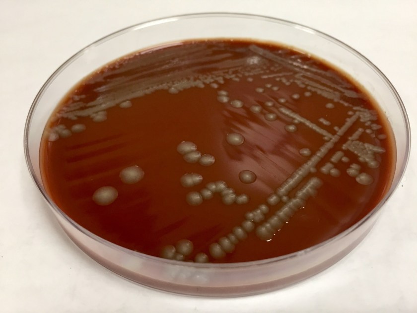
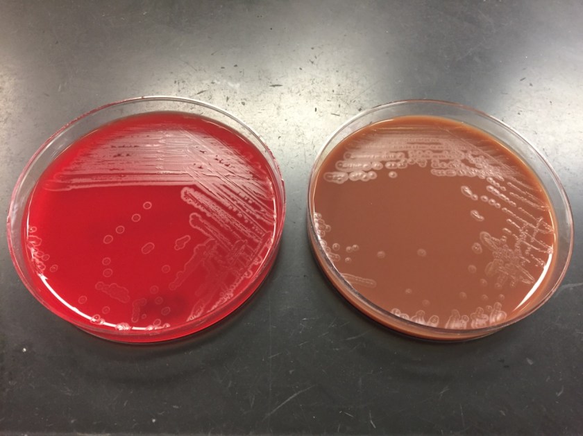
The organism was confirmed as Nocardia abscessus/asitica by a reference laboratory.

Discussion

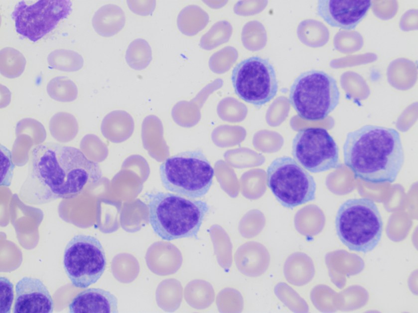
Nocardia is a genus of aerobic, weakly Gram-positive, catalase-positive, rod shaped bacteria that are modified acid-fast and form beaded branching filaments. They grow slowly on commonly used nonselective culture media, with colonies becoming evident in 3-5 days, and have the ability to grow in a wide temperature range. Colony morphology is variable, with colors ranging from white to orange and texture ranging from smooth to chalky. They are saprophytic organisms that are commonly found in soil, organic matter, and in the oropharynx as normal flora. Virulence factors for Nocardia include catalase, superoxide dismutase, and cord factor. Cord factor prevents lysosomal fusion with the phagosome, thus inhibiting phagocytosis by macrophages.

There are more than 80 species of Nocardia causing various forms of human disease, the most pathogenic of which are: N. asteroides, N.braziliensis, N.caviae, N.nova and N.abscesses. Symptoms can range from a localized lung or cutaneous infection to disseminated disease. Most infections involve the lung initially following inhalation of the organisms which commonly spread to extrapulmonary sites with the disease being more severe and likely to disseminate in immunosuppressed patients. In addition, the skin can be a site of primary infection through traumatic inoculation in immunocompetent patients.

Primary cutaneous infections occur in 5% of cases and manifest in one of the following ways: lymphocutaneous infection, mycetoma, superficial cellulitis, or localized abscesses. The most commonly involved sites for cutaneous infections are the extremities, though infection can affect any area, including the head and neck.

Treatment for primary cutaneous nocardiosis includes antimicrobial therapy and surgical irrigation and drainage when appropriate. Though antibiotic therapy is recommended, spontaneous resolution can occur without treatment. Trimethoprim-sulfamethoxazole is used most frequently for nocardiosis with the usual duration of therapy being 2-3 months in those cutaneous disease.

References

  1. Wilson JW. Nocardiosis: Updates and Clinical Overview. Mayo Clinic Proceedings. 2012;87(4):403-407. doi:10.1016/j.mayocp.2011.11.016.
  2. Vijay Kumar GS, Mahale RP, Rajeshwari KG, Rajani R, Shankaregowda R. Primary facial cutaneous nocardiosis in a HIV patient and review of cutaneous nocardiosis in India. Indian Journal of Sexually Transmitted Diseases. 2011;32(1):40-43. doi:10.4103/0253-7184.81254.
  3. Lee TG, Jin WJ, Jeong WS, et al. Primary Cutaneous Nocardiosis Caused by Nocardia takedensis. Annals of Dermatology. 2017;29(4):471-475. doi:10.5021/ad.2017.29.4.471.
  4. Outhred, A.C., Watts, M.R., Chen, S.CA. et al. Nocardia Infections of the Face and Neck. Curr Infect Dis Rep 2011 Apr;13(2):132-40. doi: 10.1007/s11908-011-0165-0.


-Clayton LaValley, MD is a 2nd year anatomic and clinical pathology resident at the University of Vermont Medical Center.

Wojewoda-small

-Christi Wojewoda, MD, is the Director of Clinical Microbiology at the University of Vermont Medical Center and an Associate Professor at the University of Vermont.


Lymphocytosis Can Be Anything

Case History

A 63 year old patient presented with a high white cell count of 108 K/uL and thrombocytopenia of 110 K/uL.

Peripheral smear examination revealed marked lymphocytosis with presence of numerous small to medium sized lymphoid cells with round to oval nuclei, clumped nuclear chromatin and variable amount of cytoplasm, some with cytoplasmic projections. As the features were consistent with a lymphoproliferative disorder peripheral blood was sent for flow cytometry.

lymph1

lymph2

Based on the morphology the differential diagnosis included B-cell lymphoproliferative disorders such as marginal zone lymphoma, hairy cell leukemia/variant, or less likely chronic lymphocytic leukemia and/or mantle cell lymphoma.

Flow cytometry revealed presence of clonal B-cells expressing CD19, CD20, Cd11c, CD103 and FMC-7. The cells were negative for CD5, CD10, and CD25.

The phenotype together with the morphology and CBC findings were diagnostic of hairy cell leukemia variant.

Discussion

Hairy cell leukemia variant ( HCL-v) is a B-cell lymphoproliferative disorder that resembles classic hairy cell leukemia but exhibits variant cytological and hematological features such as leukocytosis and also shows variant immunophenotype including absence of CD25, CD123 and/or annexin A1.

HCL-v is about one tenth as common as HCL (hairy cell leukemia) with an annual incidence of approximately 0.03 cases per 100,000 population. There is slight male preponderance. Patients with HCL-v typically present with leukocytosis with an average WBC of 30 K/ul and /or thrombocytopenia.

The 5 year survival rate is around 50-60%. Most patients require therapy which can range from splenectomy to combination chemotherapy with Rituximab.


Reference

  1. WHO classification of Tumors of Haematopoietic and Lymphoid Tissues; IARC 2017


Vajpayee,Neerja2014_small
-Neerja Vajpayee, MD, is the director of Clinical Pathology at Oneida Health Center in Oneida, New York and is actively involved in signing out surgical pathology and cytology cases in a community setting. Previously, she was on the faculty at SUNY Upstate for several years ( 2002-2016) where she was involved in diagnostic work and medical student/resident teaching.

Microbiology Case Study: A 74 Year Old Female with Right Knee Swelling

Case History

A 74 year old female presented to the ED with a chief complaint of fever, right knee swelling and pain for three days. Past medical history was significant for a right total knee arthroplasty approximately 5 months prior, with no significant complications. Physical exam revealed the patient to be febrile (103 degrees Fahrenheit), a swollen right knee that was warm to the touch and erythema surrounding the surgical incision site. Routine labs were obtained while in the ED which revealed a leukocytosis with an elevated ESR and CRP. Imaging was ordered and showed a large joint effusion of the right knee with intact hardware. Arthrocentesis was performed which returned 80 cc of cloudy yellow fluid with no crystals identified by light microscopy, a nucleated cell count of 169,200/cmm of which 97% were neutrophils.

Laboratory Identification

The primary gram stain was reported as polys and gram negative bacilli present. Cultures revealed a pure moderate growth on sheep blood and chocolate agar with no growth on the MacConkey agar. Colony morphology on the sheep blood agar was smooth, gray with no hemolysis appreciated. The key biochemical and physiologic characteristics of the isolate included: positivity for indole, nitrate reduction, catalase, ornithine decarboxylase, and fermentation of mannitol and sucrose; negativity for urea and maltose fermentation.  The isolate was identified by MALDI-TOF as Pasteurella multocida. Upon further questioning, the patient admitted to living with two indoor cats but denied any recent history of bites or scratches.

pastmulti1
Image 1. Chocolate agar with smooth gray colonies.

Discussion

Pasteurella multocida is a non-motile, oxidase positive, small -gram negative bacilli capable of fermenting glucose. This organism is part of the normal flora of the gastrointestinal tract and nasopharynx of wild and domestic animals. Humans who have extensive exposure to animals may be found to have Pasteurella multocida as part of their upper respiratory tract flora. With no significant virulence factors, this organism is often viewed as an opportunistic pathogen which requires mechanical disruption of anatomic barriers as occurs with bite and scratch wounds from cats and dogs. Though most infections are associated with bites or scratched from animals, infection can occur with non-bite exposure to animals. The typical disease caused by Pasteurella multocida is a focal soft tissue infection following a bite or scratch. However, chronic respiratory infections in patients with preexisting chronic lung disease and heavy animal exposure, and bacteremia with metastatic abscess formation have been documented.

Biochemical characteristics can be utilized in identifying the different Pasteurella species. The key biochemical and physiologic characteristics for Pasteurella multocida include: positivity for indole, nitrate reduction, catalase, ornithine decarboxylase, and fermentation of mannitol and sucrose; negativity for urea and maltose fermentation.

The vast majority of these organisms are susceptible to penicillin, thus susceptibility testing is generally unnecessary. Additionally, soft tissue infections caused by animal bites are frequently polymicrobial and warrant use of therapeutics with a broader spectrum. However, should the need arise to perform susceptibility testing, the Clinical and Laboratory Standards Institute (CLSI) does provide break points for Pasteurella multocida.

References

  1. Forbes BA, Sahm DF, Weissfeld AS. Bailey & Scott’s Diagnostic Microbiology. Mosby; 2007.
  2. Koneman EW. Koneman’s Color Atlas and Textbook of Diagnostic Microbiology. Lippincott Williams & Wilkins; 2006.


-Justin Rueckert, DO is a 2nd year anatomic and clinical pathology resident at the University of Vermont Medical Center.

Wojewoda-small

-Christi Wojewoda, MD, is the Director of Clinical Microbiology at the University of Vermont Medical Center and an Associate Professor at the University of Vermont.


Microbiology Case Study: A 57 Year Old Male with Fever, Backache, and Urinary Retention

Case History

A 57 year old male presented to the ED with the chief complaint of fever (103 degrees Fahrenheit), urinary retention, backache and headache. The patients past medical history is significant for penile cancer status post total penectomy with perineal urethrostomy and pelvic lymph node dissection approximately 12 months prior, recent urinary tract infection (2 weeks prior, treated with 7days of antibiotics), recent pace maker placement (2 months prior), Group B Streptococcus cellulitis of the left leg complicated by bacteremia (7 months prior). The patient requires use of in and out catheters for urination as a result of the penectomy and perineal urethrostomy; however he has had difficulty with catheterization recently secondary to urethral stenosis.

Physical examination revealed the right inner thigh to be erythematous, warm, and mildly tender, clinically consistent with cellulitis. The cellulitis appeared to be spreading along the medial aspect of the thigh and involving the lower leg. While in the ED, two sets of blood cultures were obtained and a urinalysis was significant for 1+ blood, 1+ nitrite, 1+ leukocyte esterase. He was subsequently treated with intravenous fluids and ceftriaxone and admitted to the hospital.

Laboratory Identification

Both sets of the blood cultures were positive (3/4 bottles), with the first bottle being positive after 11 hours of incubation. Gram smears of the bottles revealed the presence of gram positive cocci resembling Streptococcus. Per laboratory procedure, the positive bottles were analyzed utilizing the Luminex Verigene platform and resulted as Streptococcus species. The blood culture broth was subcultured to sheep blood agar and revealed a pure isolate of medium sized slightly opaque gray colonies with a large zone of beta hemolysis. The bacteria were found to be catalase negative and PYR negative. The organism was identified by a latex agglutination assay as Group C streptococci.

bsc1
Image 1. Sheep blood agar plate with beta hemolytic colonies.

Discussion

Group C streptococci designates Streptococcus species which react with Lancefield group C typing serum. Group C streptococci are comprised of several different Streptococcus species including S. dysgalactiae subspecies equisimilis, S. dysgalactiae subspecies dysgalactiae, S. equi subspecies equi, and S. equi subspecies zooepidemicus. The most commonly isolated species in human clinical specimens is S. dysgalactiae subspecies equisimilis. Group C streptococci are considered normal flora of human skin, nasopharynx, gastrointestinal tract and genital tract. The mode of transmission for these organisms includes endogenous isolates gaining access to sterile sites and person to person transmission. No definitive unique virulence factors have been identified to date; however similar virulence factors to those of S. pyogenes and S. agalactiae are likely. The diseases caused are similar to those caused by S. pyogenes and S. agalactiae including bacteremia, endocarditis, arthritis and skin and soft tissue infections. Disease generally occurs in patients in immunocompromised states or with multiple comorbidities. There have been documented cases of zoonotic infections with S. equi subspecies zooepidemicus in patients with farm animal exposure, however not all infections are associated with animal exposure. S. equi subspecies zooepidemicus is a causative agent of bovine mastitis and has been documented as the etiologic agent in several outbreaks attributed to ingestion of inadequately pasteurized dairy products. Group C streptococci are susceptible beta-lactam antibiotics, and penicillin is considered the drug of choice for treatment.

References

  1. Forbes BA, Sahm DF, Weissfeld AS. Bailey & Scott’s Diagnostic Microbiology. Mosby; 2007.
  2. Koneman EW. Koneman’s Color Atlas and Textbook of Diagnostic Microbiology. Lippincott Williams & Wilkins; 2006.
  3. Pelkonen S, Lindahl SB, Suomala P, et al. Transmission of Streptococcus equi subspecies zooepidemicus infection from horses to humans. Emerging Infect Dis. 2013;19(7):1041-8.


-Justin Rueckert, DO is a 2nd year anatomic and clinical pathology resident at the University of Vermont Medical Center.

Wojewoda-small

-Christi Wojewoda, MD, is the Director of Clinical Microbiology at the University of Vermont Medical Center and an Associate Professor at the University of Vermont.

Microbiology Case Study: A 62 Year Old Man with a Lung Mass

A 62 year old male without a significant past medical or smoking history was referred to pulmonology for an abnormal chest CT.  Three months prior to presentation, the patient had developed a cough after doing some home remodelling that involved sanding drywall.  The cough became severe and blood-tinged, including some clots, so the patient sought medical attention.  The patient denied any other symptoms and reported feeling well overall.  Physical exam findings were all within normal limits.  A chest X-ray showed a round lesion in the left lower lobe.  Follow-up chest X-rays showed that the lesion had decreased in prominence but had not resolved.  Subsequently, a chest CT was performed that showed a 2.8cm mass-like focal area of consolidation in the left lower lobe without associated lymphadenopathy.  Because malignancy could not be excluded, the patient underwent bronchoscopy with biopsies obtained for cytopathologic evaluation as well as mycobacterial and fungal cultures.

blastoderm1
Image 1: Cytologic preparation (alcohol-fixed, Papanicolaou-stained) of lung, left lower lobe, 2.8cm mass, fine needle aspiration.

The cytologic preparation of fluid from a fine needle aspiration (Image 1) shows granulomatous inflammation with patchy necrosis.  Typically, a mixed inflammatory reaction is observed, with neutrophils, granulomas, epithelioid histiocytes, and foreign body giant cells.  Examination reveals several round-to-oval yeast cells, measuring 9-13μm in diameter.  Single broad-based (4-5 μm wide) buds and thick, double contoured, refractile cell walls are also characteristic of the yeast forms visualized here, leading to a rapid presumptive diagnosis.

blastoderm2
Image 2: Scotch Tape touch preparation of one white colony growing on potato flake agar (25°C) after 10 days of incubation.

Growth of the fungus on various culture media is more sensitive than direct examination and yields a definitive diagnosis.  On potato flake agar incubated at room temperature (25°C), one white colony that was tan on the reverse began growing at 8 days.  Typically, colonies appear in 1-4 weeks and range from white (initially) to brown (with age).  Microscopic examination of a Scotch Tape touch prepared at 10 days (Image 2) demonstrates the mold form of this dimorphic fungus has delicate, septate hyphae with right-angle conidiophores that bear single, terminal conidia (resembling lollipops).  A DNA probe is used to confirm the identification of Blastomyces dermatitidis.

Discussion

As described above, Blastomyces dermatitidis is a thermally dimorphic fungus.  In the environment, the mold form of B. dermatitidis is found in wet soil, particularly when enriched by animal droppings and decaying organic matter (1).  When a susceptible host (healthy or immunocompromised) disrupts wet earth that contains B. dermatitidis, infectious conidia are inhaled into the lungs.  Adult men are more likely to have blastomycosis, likely because they partake in outdoor activities (ex. hunting, fishing) that are associated with environmental exposure to airborne conidia.

Symptoms of blastomycosis are variable, ranging from asymptomatic or transient flu-like to severe pulmonary involvement.  Patients may present with symptoms of acute pneumonia (fevers, chills, cough, hemoptysis, and dyspnea) that can be indistinguishable from viral or bacterial causes.  Other patients, with chronic pneumonia, have systemic symptoms (weight loss, low-grade fevers, night sweats, productive cough, and chest pain) that overlap with pulmonary tuberculosis, histoplasmosis, or bronchogenic malignancy.  In addition to the primary pulmonary infection, approximately half of patients develop extrapulmonary symptoms from hematogenous dissemination to almost any organ; most commonly to skin, bones, male genitourinary, and the central nervous system.

Regardless of symptoms, a majority of patients with blastomycosis will have chest X-ray findings, alveolar infiltrates or a mass lesion involving any location that are non-specific and may mimic malignancy.  The mortality rate is 0% in healthy hosts and up to 30% in immunocompromised people, frequently due to disseminated disease.  There are no guidelines for susceptibility testing of dimorphic fungi.  The preferred treatment of mild to moderate pulmonary blastomycosis is itraconazole for 6-12 months.  Conversely, amphotericin B is used in moderately severe disease to treat chronic pulmonary symptoms, disseminated blastomycosis, CNS involvement, immunocompromised or pregnant patients.

Reference

  1. Saccente M, Woods GL. Clinical and laboratory update on blastomycosis. Clin Microbiol Rev. 2010;23(2):367-81.


-Adina Bodolan, MD is a 1st year anatomic and clinical pathology resident at the University of Vermont Medical Center.

Wojewoda-small

-Christi Wojewoda, MD, is the Director of Clinical Microbiology at the University of Vermont Medical Center and an Associate Professor at the University of Vermont.

Microbiology Case Study: A 19 Year Old Male with Fever and Chills

Case History

A 19 year old African American male presented to the emergency department (ED) with complaints of fevers, chills, nausea, vomiting, a “head-splitting” headache and abdominal pain. He reported that his fevers and chills had increased in severity, reaching a max of 104°F, and had 7-8 episodes of non-bloody emesis on the day of presentation. Travel history was significant for a recent return from a two year visit to his home country of Ghana. He did state he was bitten by mosquitos there about a week before his symptoms began. In the ED, vital signs showed a fever of 102.6°F, increased respirations (36 per minute) and a normal pulse and blood pressure (98 beats per minute and 120/65, respectively). Initial CBC showed a hemoglobin of 13.2 g/dL and a hematocrit of 37.3%. Platelet count was decreased (31,000 TH/cm2). A malaria screen was ordered to look for the presence of blood parasites.

Laboratory Identification

plasfal1
Image 1. The rapid detection test for malaria antigens showed a strong positive band for Plasmodium falciparum (T1) and a weak positive band for common malarial antigens (T2).   
plasfal2
Image 2. Giemsa blood smear revealed multiple intracellular trophozoites (ring forms) (100x oil immersion).

The BinaxNOW rapid malaria screening test was positive for both P. falciparum and common malarial antigens, making a possible mixed infection unable to be ruled out (Image 1). The thin blood smear revealed numerous trophozoites with multiple ring forms in one red blood cell and appliqué forms, findings characteristic of P. falciparum (Image 2). No advanced forms, including schizonts and gametocytes, were identified. The high level of parasitemia (approximately 5.5%) also supported the diagnosis of P. falciparum.

Discussion

Malaria is a disease infecting humans through the bite of the female Anopheles mosquito and affects many worldwide, particularly in the tropic and subtropic regions of Africa and Southeast Asia. According to the Centers for Disease Control and Prevention (CDC), an estimated 212 million cases of malaria occurred in 2015 with 429,000 deaths attributed to malaria. In the United States, the majority of cases are diagnosed in travelers and immigrants returning from endemic areas. Rapid diagnosis of malaria, especially in the most aggressive Plasmodium falciparum species, is of utmost importance in order to provide prompt treatment to the patient to minimize morbidity and mortality. Clinical findings can be non-specific, especially early in the disease course, and it is important to ask about travel and exposure history. In the case of P. falciparum, cyclic tertian fevers, chills, headache, nausea, vomiting and muscle aches are common.

Definitive diagnosis is achieved by examination of thick and thin blood smears in the clinical laboratory. These two Giemsa stained smears are prepared in order to recognize the Plasmodium organisms (thick smear) and identify the particular species causing infection (thin smear). This approach remains the gold standard for laboratory confirmation of malaria. In the case of P. falciparum, the most common microscopic findings include visualizing early intracellular trophozoites (two chromatin dots connected by a thin cytoplasm). Multiple rings in a single red cell and appliqué forms (trophozoites at the edge of the red cell) are common as well. The crescent shaped gametocyte is also a diagnostic form of P. falciparum, with schizonts being rare in peripheral blood smears.

Immunochromatographic testing is gaining popularity due to their ability to rapidly (10-15 minutes) detect malaria antigens. This makes them a useful alternative to microscopy where resources do not allow for adequate microscopic examination or trained staff is unavailable around the clock. Although these tests are useful in some clinical settings, cost, accuracy and overall performance need to be considered prior to implementation.

Following identification, another important aspect of the laboratory diagnosis is determining the level of parasitemia, as this aids in the classification of disease severity and how anti-malarial drugs should be administered and in what setting. This determined by the following equation using the thin smear: (number of infected RBCs/total number of RBCs) x 100. At least 500 RBCs should be counted, but in the case of lower levels of parasitemia upwards of 2,000 cells is recommended for the most accurate percentage. Other important points include that gametocytes should not be included in the count and red cells infected with more than one trophozoite should be counted as one infected cell.

In the case of our patient, his parasitemia level of approximately 5.5% classified him as a severe malaria infection and he was transferred to the intensive care unit for close monitoring and treatment with IV quinidine, as this drug is associated with hypoglycemia and QT prolongation. After 24 hours, his parasitemia level was 2.6% but due to significant prolongation of the QT interval on EKG from 407 ms to 560 ms, he was switched to oral atovaquone-proguanil (malarone) after consultation with experts at the CDC.  Parasitemia level was 0.4% after an additional 24 hours. The patient was discharged home after completion of therapy and was well at follow up outpatient visit.

ET

-Eric Tillotson, MD, is a second year Anatomic and Clinical Pathology resident at the University of Mississippi Medical Center.

Stempak

-Lisa Stempak, MD, is an Assistant Professor of Pathology at the University of Mississippi Medical Center in Jackson, MS. She is certified by the American Board of Pathology in Anatomic and Clinical Pathology as well as Medical Microbiology. She is the director of the Microbiology and Serology Laboratories. Her interests include infectious disease histology, process and quality improvement and resident education.

Microbiology Case Study: A 65 Year Old Man with Pneumonia

Case History

A 65 year old male with a history of systolic heart failure secondary to non-ischemic (alcohol-induced) dilated cardiomyopathy underwent cardiac transplantation on 10/11/2016. He was hospitalized between 3/1/17 and 4/15/17 for neutropenia and CMV viremia. Two days after discharge, he presented to the hospital with a gradual-onset of left-sided sharp chest pain described as “soreness” over his rib cage and exacerbated by breathing. Associated symptoms included fever, malaise, and fatigue. In the emergency department, vital signs included: BP 144/75 mmHg, T 40.2°C, RR 24/min, HR 101 bpm, SpO2 97% on room air. A CBC revealed a normal white blood cell count and a chest X-ray demonstrated a lingular opacity. The immunocompromised patient was admitted for sepsis secondary to presumed pneumonia following recent hospitalization. He was treated empirically for hospital-acquired pneumonia with vancomycin and piperacillin-tazobactam. After a urine antigen test detected the presence of Legionella pneumophila serogroup 1, antibiotic treatment was changed to levofloxacin and an induced sputum culture was obtained for Legionella surveillance.

legion1
Image 1. Sputum culture on BCYE agar with PAV shows Legionella pneumophila colonies that are circular with smooth edges, grey-white, and glistening in addition to few usual oropharyngeal flora.

Discussion

Legionnaires’ disease, caused by Legionella bacteria, is a cause of 1-9% of both community-acquired and hospital-acquired pneumonias. Symptoms of fever, chills, cough, and chest pain are similar to other causes of pneumonia; however multiple organ systems may be involved, producing additional symptoms including gastrointestinal (diarrhea, nausea, and vomiting) and central nervous system (headache and confusion) findings. Legionella was first discovered after a 1976 outbreak of pneumonia among Pennsylvania State American Legion members who attended a convention at a Philadelphia hotel that had infected water in the air conditioning system; it is reported that 29 out of 182 infected people died. At present, the mortality rate of Legionnaires’ disease ranges from less than 10% in treated community-acquired cases to approximately 30% for hospital-acquired cases.

The genus Legionella contains greater than 60 species of which approximately 20 are human pathogens. Legionella pneumophila (consisting of serogroups 1-16) is the most common cause of Legionnaires’ disease and, in particular, L. pneumophila serogroup 1 causes 70-90% of cases. The organisms are ubiquitous in nature, particularly in warm freshwater environments including lakes and streams, where they infect and multiply within single-celled host organisms. Of pathogenic concern, they can be present in high numbers in human-made complex water systems (such as cooling towers, whirlpool spas, humidifiers, and decorative fountains). After environmental aerosols are inhaled or contaminated water is aspirated into the lungs, alveolar macrophages are infected by the obligate intracellular bacteria. Host risk factors for developing Legionnaires’ disease include organ transplantation, immunocompromised state, immunosuppresion, age greater than 60 years, chronic lung disease, and smoking.

In the microbiology laboratory, Legionella are mesophilic (20-45 °C) obligate aerobes. The small, thin gram negative rods react poorly with Gram stains and are not usually stained in direct clinical samples. The patient’s Gram smear revealed moderate neutrophils, few squamous epithelial cells, and mixed gram positive and gram negative organisms present. Sensitivity for detecting the biochemically inert and fastidious bacteria is increased with culture on buffered charcoal yeast extract (BCYE) agar. For sputum samples that are likely contaminated with usual oropharyngeal flora, BCYE agar with polymyxin B, anisomycin, and vancomycin (PAV) media are used. After 3-5 days of incubation, Legionella colonies appear convex, circular, 3-4 mm in diameter, grey-white to blue-green, and glistening. This identification was confirmed by MALDI-TOF MS. Laboratory in vitro susceptibility studies are not recommended on individual isolates, as they do not correlate with clinical responses. Monotherapy with a fluoroquinolone (Levofloxacin) or macrolide (Azithromycin) is active against Legionella.


-Adina Bodolan, MD is a 1st year anatomic and clinical pathology resident at the University of Vermont Medical Center.

Wojewoda-small

-Christi Wojewoda, MD, is the Director of Clinical Microbiology at the University of Vermont Medical Center and an Associate Professor at the University of Vermont.

Microbiology Case Study: A 24 Year Old Refugee with Eye Irritation

Case History

A twenty-four year-old male Kenyan refugee had been in the United States for about a month when he received a mandatory health screen for infectious diseases. He had no complaints and stated that overall, he was generally healthy. Physical exam was significant only for bilateral red conjunctiva. He stated at times his eyes get irritated and have since birth. As part of routine work-up, an ova and parasite stool exam was ordered. Organisms were detected as seen in Image 1.

giardia1
Image 1. Trichrome stained slides of patient’s stool sample.


Discussion

The patient’s stool examination showed Giardia cysts. Two nuclei are visible in the figure above with centrally located karyosomes. Also visible are the intracytoplasmic fibrils, seen as a darker purple area.

Giardia is a flagellated protozoan that causes giardiasis, a diarrheal illness. It is the most commonly diagnosed intestinal parasitic disease in the United States. It is known as Giardia intestinalis, Giardia lamblia, or Giardia duodenalis. The most common mode of transmission is drinking water contaminated with feces from infected mammals (1).

Symptoms vary and can last 1 week to years if untreated (2). Typical symptoms of giardia are “greasy, foul-smelling, frothy stools that float.” Interestingly, less common symptoms can be itchy skin, hives, eye and joint swelling (3). Retinal arteritis and iridocyclitis has been noted as well (4). It is possible that this patient’s eye irritation is due to a chronic giardiasis infection. Common treatment is usually with an antibiotic/antiparasitic drug like metronidazole (Flagyl).

Diagnosis of Giardia can be made by demonstrating the pear shaped trophozoites and/or ovoid cysts in feces. A key identifier for this parasite is the presence of the two to four nuclei with a central karyosome and intracytoplasmic fibrils that make the parasite look like a face under the microscope. However, because Giardia is excreted intermittently, it is recommended to sample three stool specimens on separate days (5). Due to problems in concentrating the organism for identification on a trichrome stain, a fecal immunoassay is available that is more sensitive and specific (5).

References

  1. https://www.cdc.gov/parasites/giardia/index.html
  2. Robertson LJ, Hanevik K, Escobedo AA, Mørch K, Langeland N. Giardiasis–why do the symptoms sometimes never stop?. Trends Parasitol. 2010;26(2):75-82.
  3. https://www.cdc.gov/parasites/giardia/illness.html#seven
  4. Wolfe MS. Giardiasis.[PDF – 8 pages] Clin Microbiol Rev. 1992;5(1):93-100
  5. https://www.cdc.gov/parasites/giardia/diagnosis.html


-Angela Theiss is a pathology resident at the University of Vermont Medical Center.

Wojewoda-small

-Christi Wojewoda, MD, is the Director of Clinical Microbiology at the University of Vermont Medical Center and an Associate Professor at the University of Vermont.

Microbiology Case Study: A 10 Year Old Boy with Right Knee Pain

Case History  

A 10 year old Caucasian male presented to the pediatric emergency department due to significant pain in his right knee with exposed hardware. His past medical history was noteworthy for Perthe’s Disease, a condition leading to avascular necrosis of the femoral head. In addition, he is affected by an autosomal dominant congenital disorder and has had ischemic strokes in the past. Recently, he had surgery performed on multiple joints to correct abnormalities and they were complicated by dehiscence & infection with methicillin sensitive Staphylococcus aureus. On current admission, his mother reported he has been afebrile and was consistently taking cephalexin to treat the above infection. He was taken to the operating room for incision & drainage and hardware removal of the knee. Bacterial cultures were collected at the time of surgery and he was started on IV clindamycin.

Laboratory Identification

eikcor1
 Image 1. Clear, spreading colonies that showed “pitting” of the agar after 48 hours incubation at 37°C in 5% CO2.  
eikcor2
Image 2. Gram stain directly from the colony showed slender gram negative rods (100x oil immersion).

On direct Gram stain, there was no organisms seen and rare white blood cells. Initially, there was no growth at 24 hours, but two morphologies were observed on the second day. The first organism was identified as the S. aureus that was previously isolated from this site. The second organism was clear with spreading colonies that pitted the agar (Image 1). A distinct bleach like odor was observed. There was no growth on MacConkey agar even though the Gram stain showed gram negative rods, making this organism most likely to be classified as fastidious (Image 2). Benchtop biochemical tests were negative for catalase and positive for oxidase. MALDI-TOF mass spectrometry identified the isolate as Eikenella corrodens. 

Discussion

Eikenella corrodens is a fastidious Gram negative rod that is a member of the HACEK family. It is considered normal flora in the oral cavity and possibly the gastrointestinal tract of humans.  Infection results from these endogenous sources and can be the result of poor oral hygiene, mucositis or dental procedures. E. corrodens causes juvenile and adult periodontitis and is commonly implicated in bacteremia and infective endocarditis, particularly in IV drug users who lick needles prior to injection. Infections of the abdomen, bones/joints and brain are less common.

In the laboratory, E. corrodens is slow growing and is usually present as clear, spreading colonies after 48 hours incubation at 37°C in 5% CO2 on blood and chocolate agars. A unique feature of the organism is that it pits or corrodes the agar, lending to its species name. Also, E. corrodens produces a bleachy smell due to the production of hypochlorite. It does not grow on MacConkey agar despite the fact it is a Gram negative rod. Biochemical tests are negative for catalase, positive for oxidase and negative for indole. Automated instruments and MALDI-TOF mass spectrometry are both able to identify E. corrodens with confidence.

Susceptibility guidelines can be found in the 3rd edition of the CLSI M45 document. In general, E. corrodens is susceptible to penicillin, board spectrum cephalosporins, carbapenems, azithromycin and fluoroquinolones. Resistance to narrow spectrum cephalosporins, macrolides and clindamycin has been documented. In general, susceptibility testing should be performed on E. corrodens when it is isolated from a normally sterile site or is identified in pure culture. Beta lactamase testing is recommended routinely on E. corrodens, and if positive, the isolate is resistant to penicillin, ampicillin and amoxicillin. In the case of bite wounds caused by E. corrodens, susceptibility testing may not be necessary if it is treated with amoxicillin-clavulanate acid (Augmentin) due to a high probability of susceptibility to this antibiotic.

In the case of our patient, he responded to the antibiotic therapy used to treat his S. aureus and E. corrodens infections and healed well. He was placed on long term oral antibiotic therapy until additional hardware is able to be removed at a future date.
RA

-Rim Alkawas, MD, is a first year Anatomic and Clinical Pathology resident at the University of Mississippi Medical Center. 

Stempak

-Lisa Stempak, MD, is an Assistant Professor of Pathology at the University of Mississippi Medical Center in Jackson, MS. She is certified by the American Board of Pathology in Anatomic and Clinical Pathology as well as Medical Microbiology. She is the director of the Microbiology and Serology Laboratories. Her interests include infectious disease histology, process and quality improvement and resident education.

Pseudohyperkalemia in Patients with Severe Leukocytosis

It has been reported many times that falsely elevated potassium can be seen in patients with severe leukocytosis from chronic lymphocytic leukemia (CLL). Early recognition of factitious hyperkalemia is very important to prevent inappropriate and potentially hazardous treatment. One case we observed in our institution, again, emphasized the importance and urgency to recognize these instances.

In this case, patient was a 58 year old male with recently diagnosed CLL. His potassium level rose from normal levels at admission to 8.9 mmol/L on repeated blood draws. Patient was asymptomatic with good strength on physical exam, and had no abnormalities on EKG or telemetry. Insulin/glucose and calcium gluconate was administered to correct potassium level and to prevent cardiac effect of hyperkalemia. The hyperkalemia result was initially thought to be due to emerging tumor lysis syndrome and was not brought to our attention until another specimen obtained had a potassium level greater than the measurable range (10.0 mmol/L). Specimens were not hemolyzed and white blood cell count was as high as 455K/µL.

Given his history of CLL, we suspected pseudohyperkalemia, an entity that has been attributed to the combination of the fragility of the leukemic lymphocytes and the mechanical stress on the cells during specimen transportation and centrifugation. Our clinical team was notified immediately, and a whole blood specimen was collected and hand carried to the laboratory. Without centrifuging, the whole blood specimen was analyzed on a blood gas analyzer and showed a potassium level of 4.2 mmol/L!!! Potassium-lowering treatment was discontinued.

Artifactually elevated potassium level is commonly seen due to red blood cell hemolysis, but not well appreciated is its occasional occurrence in patients with extreme leukocytosis from CLL. It is important for laboratorians to recognize this pattern and to notify our clinical teams so that patients are not inappropriately treated.


Xin-small

-Xin Yi, PhD, DABCC, FACB, is a board-certified clinical chemist, currently serving as the Co-director of Clinical Chemistry at Houston Methodist Hospital in Houston, TX and an Assistant Professor of Clinical Pathology and Laboratory Medicine at Weill Cornell Medical College.