Hematology Case Study: Brrrr, It’s Cold Outside!

A CBC was received on a 70-year-old surgical inpatient at our facility and was run in automated mode on our Sysmex XN analyzer. On the first run, the analyzer gave flags for RBC agglutination and MCHC >37.5. These flags require evaluation of the high MCHC with investigation of a cold agglutinin, lipemia or icterus. A smear review for RBC agglutination was also indicated. The sample was incubated at 37°C for 30 minutes and the CBC was repeated. Results are shown below in Table 1.

Table 1. CBC results before warming at 37°C and after 15 minutes incubation.

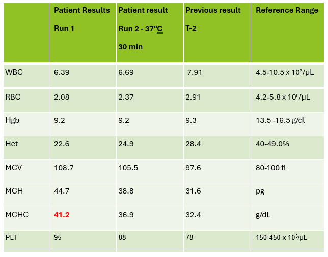
On Run 2, the MCHC is now below 37.5, so there was no operator alert to incubate further or to investigate the high MCHC. This result was validated with the comment “37°C results, possible cold agglutinin.” However, there was still an RBC agglutination flag, a high MCV, and the hemoglobin and hematocrit still don’t look great, i.e. they don’t follow ‘the rules of 3’. We know that these rules are only valid for normal samples, but if we look at the previous results from 2 days ago, the Hgb 9.3 and Hct 28.4 did follow the ‘rules of 3’ and additionally, today’s results look like the patient’s hemoglobin has not changed but the hematocrit has dropped. The RBC did go up on the second run, but it is considerably lower than 2 days ago. The indicies are also inconsistent with the previous sample. “Hmmm…What could cause this?” Instead of validating, this is what you should be asking yourself.

Cold agglutinins are known for causing pre-analytical and analytical spurious results with CBCs. With cold agglutinins, IgM antibodies bind to the RBCs after exposure to cold, causing RBC agglutination which leads to a classic pattern of false results. There is an increased MCV because the RBCs are clumped and sticking to one another, making the analyzer think these larger clumps are individual RBCs. This, in turn, will make the RBC count appear deceased because large clumps of RBCs going through the RBC aperture are counted as one RBC. The hematocrit is lowered because the volume of a clump is less than those cells individually. Hemoglobin concentration, on the other hand, is not affected by cold agglutinins, but because the hematocrit and RBC are falsely lowered, this makes the MCH and MCHC spuriously markedly increased. Cold agglutinins may be subtle, like this one, but some have extremely high MCHC and MCV, and extremely low RBC and Hct. Clues that you have a cold agglutinin are not only the high MCHC, but also flags from your analyzer such as “RBC agglutination”. The hematocrit will likely seem too low for the hemoglobin, and you may even see a hemoglobin that is higher than the hematocrit (yes it happens!) and RBC values so low as to be incompatible with life.

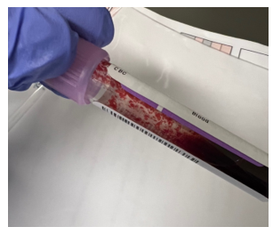
The first thing is to do if you get these spurious results is to compare the parameters and instrument flags and decide if the results are consistent with a cold agglutinin. Other factors that may cause a high MCHC include lipemia, icterus, low sodium, abnormal proteins, hemolyzed samples and samples from patients with hemoglobinopathies. The patterns of result with these samples will show a high MCHC, but with a normal or low MCV, and you won’t usually see an RBC agglutination flag. A sample with cold agglutinins may also appear grainy or clumpy to the naked eye. With a suspected cold agglutinin, warming the sample for 15-30 minutes will allow the RBCs to disperse and improve the results. However, these results must be reviewed, and if there are still instrument flags and/or if there is still clumping, the results may not yet be corrected or ‘correct’. Be sure to review a smear from the tube before warming and after. The first smear confirms the presence of the cold agglutinins and clumping, and the second smear should confirm the resolution of the clumping after warming.

This sample did have the characteristic grainy appearance of a cold agglutinin, and the post warming results did look a bit better, but the RBC agglutination flag was still present, and a review of the smear showed that the sample still had RBC clumping. After warming for another 30 min, the sample was quickly mixed and placed back on the analyzer. The results of this 3rd run are shown below, in Table 2.

Figure 1. EDTA tube with RBC agglutination
Table 2. CBC results on 3 runs. Run 3, after 60 min of incubation.

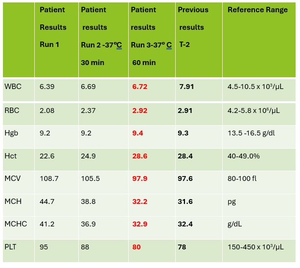
Notice that hemoglobin has not changed as it is not affected by the cold agglutinins, but after warming for 60 minutes, the RBC, hematocrit and indicies all now look consistent with the previous sample drawn 2 days ago, and a review of the smear showed no RBC agglutination. These results from the 3rd run are ready to validate.

Cold agglutinins are IgM autoantibodies that react best at 4°C but may also react at room temperature. They are generally not clinically significant and may be found in many healthy individuals. These natural cold autoantibodies occur at low titers, less than 1:64, and have no activity at higher temperatures. However, because they react at room temperature, they are notorious as a pre-analytical and analytical factor that causes spurious CBC results. They can also cause difficulties in Blood Banking during ABO/Rh typing and antibody detection.

Cold agglutinins have various clinical manifestations. Benign cold agglutinins generally do not cause hemolytic anemia and need no treatment. Most benign cold autoantibodies have anti-I specificity, are polyclonal, low titer, and do not react above 30°C. Cold agglutinins associated with Mycoplasma pneumoniae, and infectious mononucleosis are usually clinically insignificant. In cases where they do cause hemolytic anemia, the antibodies are polyclonal IgM with normal κ and λ light chains. The anemia is acute and generally spontaneously resolves in several weeks without treatment.

Though most cold agglutinins are benign and do not cause RBC destruction, when they do, they can cause hemolytic anemia that varies in severity from mild to life-threatening. This chronic cold agglutinin disease (CAD) is now known to be a form of autoimmune hemolytic anemia caused by a bone marrow lymphoproliferative disorder. Chronic CAD is a cold-autoantibody autoimmune hemolytic anemia (cAIHA) that is caused by an autoantibody produced by the clonal B cell lymphocytes. This antibody is usually monoclonal IgM with κ light chains and “I” or “i” specificity. These pathological cold agglutinins are high titer and usually react at 28°C to 32°C, and even up to 37°C. The highest temperature at which the antibodies continue to be activated is called the thermal amplitude. Because these can act at higher thermal amplitude, they may lead to CAD. In CAD the IgM autoantibodies bind to red cell antigens at 30-32°C, typically in the cooler extremities. IgM’s structure, a large immunoglobin pentamer, makes it an effective activator of the classical complement system. As the blood circulates to the central parts of the body, the RBCs warm up and the IgM antibodies dissociate from the RBC membranes, but the complement activation will continue, leading to RBC hemolysis and a cAIHA.

Chronic CAD occurs most often in adults over 50, is more common in women, and produces anemia with varying severity. Patients may be seasonally affected. In the winter, the temperature of blood may fall below 30°C in the extremities, activating the cold agglutinins. Patients may experience acrocyanosis of the hands, feet, ears, and nose with exposure to cold. They may also experience other cold related symptoms such as numbness and Raynaud’s. Patients with chronic CAD and mild anemia are therefore monitored with a ‘wait and see’ plan and advised to avoid cold temperatures. In patients with more severe anemia, it is found that targeting the underlying lymphoproliferative disorder provides the best treatment. Rituximab has been used to achieve partial remission. Therapeutic plasma exchange is also used in severe cases to rapidly remove cold agglutinins.

I have been thinking about cold agglutinins recently because of the number I have seen come into our lab this winter. As I am writing this, watching the temperature outside drop in anticipation of more snow coming in tonight, cold agglutinins came to my mind again. I used to live in the cold northern Northeast, but after moving further south, we see fewer cold agglutinins in hematology than I used to see. This winter we have had some cold spells, and, interestingly, I’ve seen more cold agglutinins. That led me to ask myself if cold agglutinin disease is really more common when patients are exposed to cold temperatures. I remember learning myself, and telling my students that that the treatment for mild to moderate CAD was to advise patients to move to someplace warmer. It has been assumed for many years that CAD worsens in colder climates or seasons. Interestingly, there have been a number of studies done since the 1950’s that examined the relationship of cold temperatures and CAD. The studies used hemoglobin, bilirubin and LDH for monitoring. Early case reports had findings that supported the theory of more anemia and higher LDH in the winter. (Dacie, Lyckholm)One recent article in 2022 found a 4-fold difference in the incidence of CAD between cold (Norway) and warm (Italy) climates. (Berentsen). However, atthe same time another study found that there was no statistically significant seasonal variation in hemoglobin, but that LDH levels were higher in winter. It concluded that these conditions should be monitored through all seasons because of the risk of hemolysis and thrombotic episodes. It was also demonstrated that though there may not be obvious statistical difference in CAD between cold and warm months, that there is a large variability of disease severity across patients and even with an individual patient. (Roth).

In conclusion, when working in any department of the laboratory, quality results are important. Results on the patient chart that vary considerably from day to day because sometimes a cold agglutinin has been effectively resolved in lab testing and other days results after 15-30 minutes of warming are just reported without a good review of the smear and the parameters, are confusing, and could affect patient care. If one tech reports the results after 30 min incubation with values that are still spurious, and the next tech resolves the agglutination with further warming, the lab will be reporting out inconsistent results. A patient who actually has stable CBC results may have deltas and what appear to be erratic results. Cold agglutinins do take time to resolve, but with over 80% of samples autoverifying with the use of auto verification, we have time to work on these problem samples. If something doesn’t look or feel right about a sample, look at all the parameters, check the instrument flags and operator alerts, check the previous results and investigate any changes. It is important to review results carefully, because we want to report out the best results possible.

References

S Berentsen, W Barcellini, S D’Sa, U Randen, THA Tvedt, B Fattizzo, E Haukås, M Kell…

Blood, The Journal of the American Society of Hematology, 2020•ashpublications.org

Climent F, Cid J, Sureda A. Cold Agglutinin Disease: A Distinct Clonal B-Cell Lymphoproliferative Disorder of the Bone Marrow. Hemato. 2022; 3(1):163-173. https://doi.org/10.3390/hemato3010014

Nikousefat Z, Javdani M, Hashemnia M, Haratyan A, Jalili A. Cold Agglutinin Disease; A Laboratory Challenge. Iran Red Crescent Med J. 2015 Oct 17;17(10):e18954. doi: 10.5812/ircmj.18954. PMID: 26566452; PMCID: PMC4636857.

Patriquin, C.J. and Pavenski, K. (2022), O, wind, if winter comes … will symptoms be far behind?. Transfusion, 62: 2-10. https://doi.org/10.1111/trf.16765

Rodak, Bernadette F., et al. Hematology: Clinical Principles and Applications. 5th ed. St. Louis, Mo., Elsevier Saunders, 2016

Röth A, Fryzek J, Jiang X, Reichert H, Patel P, Su J, et al. Complement-mediated hemolysis persists year round in patients with cold agglutinin disease. Transfusion. 2022; 62: 51–59. https://doi.org/10.1111/trf.16745

Socha-small

-Becky Socha, MS, MLS(ASCP)CMBBCM graduated from Merrimack College in N. Andover, Massachusetts with a BS in Medical Technology and completed her MS in Clinical Laboratory Sciences at the University of Massachusetts, Lowell. She has worked as a Medical Technologist for over 40 years and has taught as an adjunct faculty member at Merrimack College, UMass Lowell and Stevenson University for over 20 years.  She has worked in all areas of the clinical laboratory, but has a special interest in Hematology and Blood Banking. She currently works at Mercy Medical Center in Baltimore, Md. When she’s not busy being a mad scientist, she can be found outside riding her bicycle.

Lessons Learned During a Cytology Staffing Shortage

No one wants to be short-staffed. Cytology programs across the country are either closing or shifting to a Master’s-only degree (to finally reflect our expanded scope of responsibilities), and during the pandemic, it seems there was a mass exodus of retiring baby-boomer cytologists. We’re in a crisis to say the least. As a prominent clinical rotation site, we have no shortage of cytology students. So much that whenever we’ve had an open position in the past, we were confident an eligible student would be able to fill our void. Over the past year, we’ve noticed that students had already secured jobs before they came to us for their clinical rotation. It wasn’t a problem until we realized that we were no longer immune to the nationwide staffing shortage. After one of our cytologists tragically passed away in November of 2022, we made do by working overtime. Before we were able to fill the empty position fifteen months later, another cytologist left to teach. Finally securing an amazing candidate with experience, we knew that we had students rotating through during the winter months, and things started looking up. We encouraged our first student to apply after being blown away by her already-fine-tuned her locator skills and hired her to start as soon as she graduates this summer. We were feeling assured that by the end of the summer (and my supervisor’s retirement), we would be fully staffed, fully trained, and ready to take on the world again. And then, another cytologist let us know she was moving to New England and her last day was five weeks from now. And just last week, another cytologist put in her notice. We’re down 4 cytologists in 15 months, the latter 3 within just 2 months. It’s the highest turnover our department has ever experienced, and our optimism was crushed. Fortunately, we do have more students rotating through this summer, but with 3 positions to fill before August, we’re treading water like our lives depend on it.

Sound familiar? I’m certain that the pervasive staffing shortage is plaguing medical laboratories all over the country. But how do we not let this impact our services? How do we continue to provide the same level of exemplary care while preventing burnout in our team? I’d like to share some lessons learned during our shortage from both a management and cytologist perspective, and I’m eager to hear if you have any you’d like to share from your own experiences.

Lesson #1 – Analyze Service Impact & Develop a Contingency Plan

Will the staffing shortage negatively impact turnaround time? With CLIA’s maximum screening limit of 100 slides in no less than an 8-hour day, a reduction in cytologists shifts the burden of the workload which can risk exceeding limits. Factoring in non-screening activities, such as performing Rapid Onsite Evaluation (ROSE) for FNA procedures, sending out tests for ancillary studies (ThyroSeq, Afirma, HPV, etc.), accessioning, scheduling, slide filing, cleaning biopsy carts, compiling statistics for QA, assisting in the cytoprep lab, maintaining continuing education, etc., it’s far too easy to exceed those limits. We pride ourselves on a one-day turnaround time. Our clinicians and patients expect it, and we refuse to sacrifice that feat.  The most significant concern is the rising number of scheduled FNA procedures and not enough people to safely attend them all. We examined productivity and available time for FNAs given the number of cytologists present and daily case/slide workload. First, we looked at the number of slides that need to be screened for the day and divided it by the max mandated screening rate. Then, we counted the number of cases that need to be accessioned and the time involved. This process includes reconciling clinical history and histologic correlation, resolving the plethora of pre-analytical errors (please show us a perfect system for order entry). Considering the time spent on all other activities beyond accessioning and screening including assisting in the cytoprep laboratory, and what remains is the number of hours available for biopsies. We compare this to what has actually been scheduled for the day. Quite often, we are available for much less than what is requested and we must reallocate our resources. Postponing or reallocating out our prep assist duties, filing, and cart cleaning is an option with the cytoprep technicians also working overtime. If and when the prep techs are caught up on their work, they are able to clean carts for us. As for filing slides and paperwork, try to utilize your hospital’s resources, such as volunteers, who are incredibly valuable. Try to also share or reallocate statistics or other QA activities to reduce the burden on one employee while still maintaining operations. You could hire a temporary administrative assistant with a background in medical terminology to assist with accessioning as another option. The worst case scenario would be asking clinicians to “self-collect” FNAs in a balanced salt solution and sending it up to the lab to be processed. Our clinicians value our ROSE services, especially to confirm viability and to ensure we have sufficient material for ancillary studies such as molecular, IHC, and flow cytometry, and not being present would be an ethical dilemma for us all. To help mitigate this, we worked with the schedulers and clinicians across various departments to level out the biopsy schedule, and we postponed or reallocated non-screening activities to be able to handle the FNA workload to the best of our staffing level.

Lesson #2- Go LEAN

Now is a great time to go LEAN, if you haven’t already. And if you think you have, do it again. Analyze your lab for forms of waste. Are there non-value-added activities that are interfering with daily operations? Is your workflow optimized? How much of your cytologist’s time is spent waiting on biopsies? Waiting to call the cytologist to the procedure after the clinician has scrubbed in and marked the targeted lesion could save the cytologist 10-45 minutes of time. By reducing excess and unproductive biopsy wait time, the cytologist can be more productive within the laboratory. You could also reduce motion waste by having one cytologist attend multiple biopsies in the same department within a short time frame. For example, if an ultrasound-guided biopsy is scheduled for 10:15 AM and a CT-scan biopsy is scheduled for 11:00 AM, the same cytologist could attend both without having to return to the lab just to be called back down to radiology. Reducing excess employee movement between departments can also reduce potential care delays by having the cytologist present, moving with the nurses and proceduralist. Similar to the previous lesson on developing a contingency plan with reallocation of resources, how much of the cytologist’s talent is wasted on miscellaneous tasks that outside of the scope of high complexity testing, such as filing, scheduling, and cart cleaning? These are tasks that could be easily assigned to an administrative assistant or prep tech. And lastly, is the lab “over-prepping?” Many hospital laboratories only produce one liquid-based preparation (such as a ThinPrep slide) for morphology and a cell block for ancillary studies. If you are also making cytospins and smears or other additional preparations that offer a higher level of quality than is actually required to make the diagnosis, it could be considered waste. To reduce supply costs and time spent both prepping and interpreting excess material, monitor the laboratory for overproduction and overprocessing waste. This is especially helpful in reducing turnaround time and freeing up existing resources for other tasks.

Lesson #3 – Promote Mental Health & Self-Care

I especially thank my supervisor for this lesson because he and our cytopathology director have always maintained the family-comes-first and quality-of-life philosophies. Recognize that you and your cytologists are humans and not automated machines. Working in a short-staffed state with an abundance of overtime for more than a year can quickly manifest in burnout. You have to protect the gems that you still have. One thing I learned from my supervisor is to continuously seek feedback. How can we prevent burnout and protect both our mental and physical well-being? The main concern was quality of life, which was flourishing when we worked 4-10’s. While the overtime is not mandatory, we had to switch back to a 5-day work week to compensate for the staffing shortage. With that said, the remaining cytologists feel a sense of duty to our patients and therefore have extended their days to 9- to 10-hour days 5 days per week just to cover basic laboratory operations. We anticipate that once our March-start cytologist is fully trained to handle biopsies which run afterhours and our June-start cytologist is fully trained on accessioning, we can return to the 4-10 workweek. But for now, we maintain morale by knowing that the future is bright and we have 3 exceptionally strong senior techs remaining who are fully prepared to train any new hires. While management responsibilities have also shifted during a staffing shortage, a good leader must sharpen their intuition and emotional intelligence, checking in with their employees who are under extreme stress. Too often the manager forgets to check in with themselves while weathering a storm.  Remember the airplane oxygen mask metaphor – you must care for yourself before you attempt to help others.Make sure your employees know that too. Patients and their specimens need us, but we cannot provide exceptional services unless we take care of ourselves first.

Lesson #4 – Communicate Intentions & Goals Early & Often

Communication- It’s a two-way street. Please, for everyone involved, consider giving MORE than the minimum required notice. For our department where we clearly do much more than just screening slides and attending FNAs, you must leave enough time to train others on how to handle the processes you own, such as send-out tests or statistics. It is crucial to give the remaining cytologists sufficient time to learn these processes and be able to ask questions while you’re still onsite. Communicating your intent to resign earlier than the minimum required time also enables management to shift their duties and either actively recruit or simply consider prospective candidates to help close the gap. Please also understand that indicating your intent to leave a laboratory does not mean that management will give up on you during your remaining tenure. If anything, leadership will ensure that you are able to accomplish any residual goals within the organization and help you prepare for the next stop on your journey. This principle applies to the entire duration of your career within the laboratory. At the beginning of your tenure, be open and honest about your short-term and long-term goals both career-wise and outside of the workplace so that management can help you customize a plan to achieve those aspirations. Should your goals or intentions change, be transparent. Pivoting is not a form of weakness. While it isn’t easy to brave a storm, especially as the effects of the shortage are exponentially more evident, it’s not only okay to seek help, but strongly encouraged. If you feel overwhelmed or on the verge of burnout, lean on your team members, communicate your concerns to your manager, and take time to ground yourself. Sometimes leaving a laboratory only reduces familiar burnout, and by starting over elsewhere, the unfamiliar may turn out to be more stressful, yet sometimes that new challenge is exactly what you needed. Just keep in mind that the storm will not last forever, and the laboratory sun will shine again.

-Taryn Waraksa-Deutsch, MS, SCT(ASCP)CM, CT(IAC), has worked as a cytotechnologist at Fox Chase Cancer Center, in Philadelphia, Pennsylvania, since earning her master’s degree from Thomas Jefferson University in 2014. She is an ASCP board-certified Specialist in Cytotechnology with an additional certification by the International Academy of Cytology (IAC). She is also a 2020 ASCP 40 Under Forty Honoree.

Shish Kebabs, Pears and Grapes: All “Clue(s)” to Diagnosing Microbial Infections on Pap Smears 

What is the test?

In the 1920s, the Greek physician Georgios Papanikolaou developed a method of cervical cancer screening, now reliably used and colloquially known as a “Pap smear”. During a Pap smear, a healthcare provider swabs cells from the cervix for further analysis by the lab​1​, including cytopathologic examination. Regular utility of Pap smears in women aged 21 to 65 has decreased the incidence and mortality of cervical cancer by at least 80% in the United States alone​2​. 95% of cervical cancer cases worldwide are caused by persistent human papilloma virus (HPV) infection of the cervix​3​. However, other sexually transmitted infections (STIs) and common non-STIs with associated morphological changes can also be identified on Pap smears. 

Bacteria

Bacterial vaginosis (BV) is the most common cause of abnormal discharge in young women, characteristically with a “fishy” ammonia-like odor. Although not strictly considered an STI, this infection is associated with severe rare complications including pelvic inflammatory disease (PID), infertility and premature labor. The vagina typically have an abundance of Lactobacillus spp., which is a gram positive bacilli (Figure 1). However, if the normal vaginal flora is disrupted with high abundance of gram negative bacteria such as Gardnerella vaginalis or Mobiluncus, then BV may occur. On pap smears, ‘clue cells’ which are squamous epithelial cells with coccobacilli, appear as dark purple staining and cloudy appearance suggest BV. In cervicovaginal samples typically found in women using intrauterine device usage, clumps of filamentous bacteria suggestive of Actinomyces species may also be visible. For cases pertaining to bacterial etiologies, staining of the rods or filaments usually warrant suspicion of infection. Other the other hand although Chlamydia trachomatis is the most common bacterial STI in the United States, with up to 4 million new cases every year, diagnosis on pap smears is not the definitive diagnosis since the cytopathology is non-specific. The interpretations include visualization of inflammatory exudates and inflammatory cells. Oftentimes, further testing is needed. 

Figure 1. Lactobacilli spp. bacilli seen as part of normal flora (red arrow, top left and top middle). Example of a clue cell (red arrow): intermediate squamous cell coated with grape-like short coccobacilli (Gardnerella vaginalis) with a shift in normal flora (top right). What appears to be a clue cell (red arrow) at first glance is actually an intermediate squamous cell covered with normal flora upon closer inspection (bottom left). Follicular cervicitis due to Chlamydia infection (bottom right). Note lymphohistiocytic aggregates with polymorphous lymphocytes and histiocytes (red arrow). Tangible body macrophages may also be identified (ThinPrep, 40X).

Parasite

Trichomonas vaginalis is the most prevalent non-viral STI in the United States, strongly associated with an increased risk of HIV infection among women​4​. Majority of the patients may exhibit symptoms such as burning, itching and vaginal discharge. T. vaginalis is a parasitic flagellated protozoan although in some cases, a flagella may be found. Typically, it is of a pear-shape and staining of a nucleus may be visualized (Figure 2).

Figure 2. Pear-shaped round-oval Trichomonas spp. (red arrows) ranging in size from 15-30 µm present singly and in groups, known as “Trich parties”. Note pale vesicular eccentrically located nucleus and eosinophilic cytoplasmic granules. Reactive cytological changes (black arrow) such as nuclear enlargement and perinuclear halos are also identified. (ThinPrep, 40X. Case courtesy of Dr. Edina Paal, VA Medical Center, Washington DC).

Fungus

Yeast infections (vulvovaginal candidiasis) due to Candida infection occur in 75% of women at some point in their lives. Classic clinical presentation includes itching, erythema, and thick white “cottage-cheese” discharge. There is also an increased association with HIV infection, diabetes, and any cause of immunosuppression (e.g. transplant, chemotherapy, steroids). Morphologically, budding yeast forms (conidia, small and oval measuring 3-6 µm) and pseudohyphae (long filamentous spores) are found. The combination of pseudohyphae and yeast forms are referred to as “sticks and stones” and frequently, squamous cells lined up along the pseudohyphae are found referred to as having “shish kebab” appearance (Figure 3)5. Note that no true septation is found.

On pap smears, sometimes there could be superficial mucosal infections that may appear associated with enlarged hyperchromatic nuclei with halos, which can be confused with low grade squamous intraepithelial lesions.

Figure 3. Shish kebab: entangled streaming intermediate and superficial squamous cells along Candida spp. pseudohyphae (red arrow, top). Partially treated Candida infection. Note poorly formed granular pseudohyphae (red arrow) with significant treatment effect (red arrow, bottom) (ThinPrep, 40X).

Viruses

HPV with its associated risk of cervical cancer remains the most crucial microorganism to detect on Pap smears. Given HPV’s association to cancer development, it is crucial to examine the samples for cervical lesions and their associated pathologies. HPVs in the low-risk category typically is associated with low-grade squamous intraepithelial lesions and HPVs in the high-risk category is associated with high-grade squamous intraepithelial lesions and invasive squamous cell carcinoma.  Other viral etiologies seen in PAP smears include Herpes simplex virus (HSV) and cytomegalovirus (CMV). Infections with HSV can be asymptomatic but the most common symptom is the development of vesiculopustular or small ulcerative lesions on the genitalia. The classic cytopathological findings of HSV infection are the 3 “Ms”: multinucleation, nuclear molding, and margination of chromatin. CMV infections are rare, usually asymptomatic and can be transient. On the pap smears, the infected cells may appear with intranuclear inclusions surrounded by a halo.

Conclusions

While Pap smears are routinely performed on women and can provide a presumptive diagnosis, current developments in molecular technologies (e.g. Nucleic acid amplification tests (NAATs)) is transforming the field. There are several FDA-approved molecular platforms available for clinical diagnostic labs to test for HPV and can even genotype the strain to determine risk levels. Crucially, the remaining sample from liquid-based Pap tests​6​ can also be submitted for both NAAT testing and HPV-DNA testing, facilitating a quicker turnaround time and obviating the need for additional patient sampling.  Most recently, a PCR-based test is now on the market (Cepheid, Sunnyvale, CA) that can diagnose all three etiologies, BV, Candidiasis, and Trichomoniasis, within 60 minutes from a single specimen.  In summary, accurate examination of the Pap smear often incidentally provides the first step in the diagnosis and further work-up of all these infectious diseases.

References 

​​1. Pap Smear: MedlinePlus Medical Test [Internet]. [cited 2024 Feb 4]. Available from: https://medlineplus.gov/lab-tests/pap-smear/ 

​2. PDQ® Screening and Prevention Editorial Board. Cervical Cancer Screening (PDQ®): Health Professional Version. National Cancer Institute [Internet]. 2022 [cited 2024 Feb 4];1–26. Available from: https://www.cancer.gov/types/cervical/hp/cervical-screening-pdq 

​3. Lei J, Ploner A, Elfström KM, Wang J, Roth A, Fang F, et al. HPV Vaccination and the Risk of Invasive Cervical Cancer. New England Journal of Medicine. 2020 Oct 1;383(14):1340–8.  

​4. Davis A, Dasgupta A, Goddard-Eckrich D, El-Bassel N. Trichomonas vaginalis and Human Immunodeficiency Virus Coinfection Among Women Under Community Supervision: A Call for Expanded T. vaginalis Screening. Sex Transm Dis [Internet]. 2016 Sep 15 [cited 2024 Feb 5];43(10):617–22. Available from: https://pubmed.ncbi.nlm.nih.gov/27631355/ 

​5. Kamal Meherbano M.  The Pap smear in inflammation and repair. Cytojournal [Internet]. 2022 Apr 30; 19:29. Available from: doi: 10.25259/CMAS_03_08_2021

​6. Hawthorne CM, Farber PJ, Bibbo M. Chlamydia/gonorrhea combo and HR HPV DNA testing in liquid-based pap. Diagn Cytopathol [Internet]. 2005 Sep [cited 2024 Feb 5];33(3):177–80. Available from: https://pubmed.ncbi.nlm.nih.gov/16078250/ 

-Zoon Tariq is a pathology resident at George Washington University. Her interests include surgical pathology and cytopathology.

-Rebecca Yee, PhD, D(ABMM), M(ASCP)CM is the Chief of Microbiology, Director of Clinical Microbiology and Molecular Microbiology Laboratory at the George Washington University Hospital. Her interests include bacteriology, antimicrobial resistance, and development of infectious disease diagnostics.

Homicide by Unspecified Means

Let’s imagine you are a forensic pathologist, called by investigators to the basement of an abandoned house where a building inspector found human remains. Upon your arrival, you identify a human skeleton, still partially encased in trash bags. The plastic trash bags have melted over the exposed surfaces, and there are charred cans of lighter fluid lying on top of the body. After a full examination of the body at your morgue, however, you cannot find any remaining signs of injury. One need not be an expert to recognize that the circumstances in which this body was found are extremely concerning, regardless of the absence of injuries at autopsy. This is when the diagnosis of “homicide by unspecified means” (“HUM”) enters consideration.

As we have previously discussed (see Undetermined, Undetermined – Lablogatory), in situations of extensive soft tissue loss or incomplete remains, we may not be able to identify the cause of death. It is unfortunately not uncommon for bodies of homicide victims to be concealed, delaying their discovery and allowing decomposition to progress. Out of a desire to hide the victim’s identity or to conceal the crime itself, attempts may be made at dismembering or destroying the body, which can further hinder efforts to identify injuries. Still, there are situations which are easily recognized as suspicious – yet we need to be careful to not rush to conclusions. There are less malignant explanations for some strange circumstances – for example, a person who accidentally overdoses may be moved to a different location, to divert law enforcement attention from a specific house or person. A hiker in an isolated location may suffer a natural cardiac event but not be found before decomposition and animal predation have occurred.

There are five criteria required to meet the diagnosis of “homicide by unspecified means,” a term which was originally coined by the late Dr. Joe Davis in Miami, Florida. These criteria were designed to ensure that all possible alternatives are thoroughly considered before arriving at the diagnosis. The criteria, as delineated in the original 2010 article, are:

  1. Objectively suspicious circumstances of death. This would include evidence that the body was deliberately concealed (in trash bags, luggage, or a shallow grave, for example), attempts were made to destroy the body (e.g. with fire, or bleach), or that the victim was restrained. There may be evidence at the scene (or in the victim’s home) of significant blood loss, or there may still be non-lethal injuries (for example, a shallow laceration, or numerous bruises) identifiable on the body.
  2. No anatomic cause of death. Meeting this criterion requires the completion of a full autopsy, despite the potential lack of tissue or complete remains. Sometimes evidence of lethal trauma is still identifiable – for example, gunshot wounds or knife marks on bone – and in this situation, the better “cause of death” is the specific type of injury. In other situations, there may be significant cardiovascular disease found at autopsy; the presence of a competing, natural cause of death must be carefully weighed with the other evidence.
  3. No toxicological cause of death. This essentially means we have excluded an overdose as a possible cause of death. At times, this is a difficult standard to meet – in the example of skeletal remains, no material may be present to test. Other remains may be so decomposed that obtaining quantitative results (rather than qualitative) is impossible. Even if results are ‘positive’ for drugs of abuse, there is strong evidence that intoxicated individuals are at increased risk for interpersonal violence, and it would be a public disservice to automatically ascribe these deaths to overdose. This criteria, much like criteria #2, needs to be considered carefully in the context of all other findings.
  4. No environmental, circumstantial, or historical causes of death. One always needs to consider the scene investigation and surrounding environment. Take the example above of a hiker in an isolated location – a death from hypothermia can have no or minimal findings at autopsy, let alone in the context of decomposition. Deaths due to drowning, epileptic seizures, or transient exposure to a toxic agent (like carbon monoxide) are similarly difficult to identify in these circumstances. If any of these possibilities cannot be confidently excluded, the diagnosis of “HUM” should not be made.
  5. A more specific cause of death cannot be suggested by the data set. Essentially a reminder to review the totality of the evidence. Ruling a death as a ‘homicide’ sets off a chain of events which could result in a person being permanently incarcerated or executed. It is not a ruling to be made lightly.

Not every jurisdiction uses “HUM” as a term. Some will rule the cause of death as purely ‘undetermined’ and the manner of death ‘homicide’. In either case, the point is the same – there are clear indicators of homicidal violence, yet we cannot determine the specific type – and these criteria are still helpful to make sure alternative manners of death are thoroughly considered.

References:

  1. Matshes EW, Lew EO. Homicide by unspecified means. Am J Forensic Med Pathol. 2010 Jun;31(2):174-7. doi: 10.1097/PAF.0b013e3181df62da. PMID: 20436340.

Krywanczyk A, Gilson T. Homicide by Unspecified Means: Cleveland 2008 to 2019. Am J Forensic Med Pathol. 2021 Sep 1;42(3):211-215. doi: 10.1097/PAF.0000000000000657. PMID: 33491950.

-Alison Krywanczyk, MD, FASCP, is currently a Deputy Medical Examiner at the Cuyahoga County Medical Examiner’s Office.

Microbiology Case Study: Traveler’s Diarrhea in a 59 Year Old Patient

Case History

A 59-year-old man presented to the Emergency Room with bright red blood per rectum, associated with nausea, vomiting, abdominal cramping, and persistent watery diarrhea. Several days earlier, he had returned from a three-week trip to the Dominican Republic. On physical examination, he was afebrile. His abdomen was soft and not tender or distended.

A stool sample was sent to the Microbiology Lab for PCR testing, and both Vibrio and Vibrio cholera targets were detected. The stool was then plated for culture confirmation. Hemolytic colonies grew on the blood agar plate (Figure 1), and yellow (original medium color green) colonies grew on thiosulfate citrate bile sucrose selective agar due to sucrose fermentation (Figure 2). Gram stain from these colonies showed gram negative, curved, comma-shaped rods (Figure 3) and MALDI-ToF identification revealed Vibrio albensis. The specimen was sent to the Department of Public Health for confirmation, which reported Vibrio cholerae O1 serovar Ogawa with O1 antigen typing. 

Figure 1. Growth on blood agar
Figure 2. TCBS-sucrose fermentation
Figure 3. Gram negative, curved, comma-shaped rods

Discussion

Vibrio albensis is a gram negative, halophilic bacterium belonging to the Vibrionaceae family. V. albensis is a recently identified species within the Vibrio genus and is believed to be a member of the Vibrio cholera complex. Although primarily considered non-pathogenic, V. albensis has been associated with rare cases of human infections, particularly in individuals with compromised immune systems.1 While previously reported studies indicated V. albensis as a non-O1, non-O130 serogroup of V. cholerae,2the Department of Public Health confirmed our patient’s isolate as O1 serovar Ogawa by O1 antigen typing. V. albensis is an emerging pathogen with limited information regarding its clinical significance and optimal management. The infections are predominantly associated with contaminated seawater or seafood exposure. The primary transmission mode is through open wounds or ingesting raw or undercooked seafood.3

Clinical presentation of this organism can be similar to V. cholerae, as diffuse watery diarrhea, the hallmark of cholera, or asymptomatic. Other case reports presented bacteremia, septicemia, and urinary tract infections.4,5

Laboratory diagnosis of Vibrio cholerae albensis infection involves isolating and identifying the bacterium from stool samples. This can be achieved using selective culture media, such as thiosulfate-citrate-bile salts-sucrose (TCBS) agar, which allows for the growth of V. cholerae and its variants.6

Antimicrobial resistance is a growing concern in the management of cholera. Studies have reported varying resistance levels to commonly used antibiotics in V. cholerae, including V. cholerae albensis. It is essential to monitor antimicrobial susceptibility patterns to guide appropriate treatment strategies.7 Further research is needed to better understand the epidemiology, clinical manifestations, and optimal treatment strategies for V. albensis infections as members of V. cholerae complex are being identified/recognized with more advanced diagnostic tools.

References

  1. Baker-Austin C, et al. (2016). Vibrio albensis sp isolated from a mesophilic bacterial culture, abalone (Haliotis spp.), and seawater. International Journal of Systematic and Evolutionary Microbiology, 66(1), 187-192.
  2. Ahmed AOE, Ali GA, Hassen SS, Goravey W. Vibrio albensis bacteremia: A case report and systematic review. IDCases. 2022 Jun 30;29:e01551. doi: 10.1016/j.idcr.2022.e01551. PMID: 35845827; PMCID: PMC9283503.

3. Sharma P, et al. (2018). Vibrio albensis: An Emerging Pathogen Causing Necrotizing Fasciitis. Journal of Clinical Microbiology, 56(3), e01454-17.

4. Araj GF, Taleb R, El Beayni NK, Goksu E. Vibrio albensis: An unusual urinary tract infection in a healthy male. J Infect Public Health. 2019 Sep-Oct;12(5):712-713. doi: 10.1016/j.jiph.2019.03.018. Epub 2019 Apr 10. PMID: 30981654.

5. Sack RB, et al. (2004). Cholera. The Lancet, 363(9404), 223–233.

6. Centers for Disease Control and Prevention (CDC). (2021). Laboratory Methods for the Diagnosis of Vibrio cholerae. Retrieved from https://www.cdc.gov/cholera/laboratory.html

7. Ceccarelli, M., et al. “Editorial–Differences and similarities between Severe Acute Respiratory Syndrome (SARS)-CoronaVirus (CoV) and SARS-CoV-2. Would a rose by another name smell as sweet.” European review for medical and pharmacological sciences 24.5 (2020): 2781-2783.

-Eros Qama, MD, is a 2nd year AP/CP pathology resident in the Department of Pathology at Montefiore Medical Center in Bronx, NY

-Phyu Thwe, Ph.D, D(ABMM), MLS(ASCP)CM is Associate Director of Infectious Disease Testing Laboratory at Montefiore Medical Center, Bronx, NY. She completed her medical and public health microbiology fellowship in University of Texas Medical Branch (UTMB), Galveston, TX. Her interests includes appropriate test utilization, diagnostic stewardship, development of molecular infectious disease testing, and extrapulmonary tuberculosis.

The 3 Hazards of Hazardous Waste Management

Managing chemical (hazardous) waste in the laboratory is easily one of the most complicated areas of  safety to understand. The regulations are set forth by the Environmental Protection Agency (EPA) and enforced by them or representatives of their state branches. For laboratories that are housed in hospitals or other large facilities, hazardous waste is often removed and handled through other departments like environmental services or maintenance. In the eyes of the EPA, the waste generated by the lab is the responsibility of the lab until it gets to its final disposal location. If other departments manage lab waste, the lab should routinely make sure it is being handled appropriately.

One of the most common areas where hazards occur in waste management regards storage. There are two types of chemical waste storage areas that can be designated in a facility, a Satellite Accumulation Area (SAA) and a Central Accumulation Area (CAA). Chemical waste is initially stored in a SAA which must be within the line of vision from where the waste is generated. Labs may store up to 55 gallons of waste in a SAA, and the EPA does not permit moving waste from one SAA to another. If the waste stored at the SAA is flammable, it should be kept inside of a flammable storage cabinet, but that cabinet would also need to be visible from the point of waste generation.

A second common issue surrounding hazardous waste is container labeling. In most US states, it is required that all containers of chemical waste display the words “hazardous waste.” The label must also show a description of the waste (i.e. stain waste, xylene waste, etc.). Finally, there must be some sort of hazard warning on the label. That warning may be in the form of a pictogram, a NFPA or HMIS warning label. If waste is poured into an empty reagent container, no elements from the original product label may be used, even if the waste is the same as the original reagent. Cross out the original label and place a new complete hazardous waste label on the container.

Containers placed in a SAA should not have an accumulation start date on the container. Facilities are allowed to store waste on site for specific periods of time based on the generator status selected as part of the EPA registration process. However, that storage time limit does not start until the waste is moved to the Central Accumulation Area. All waste in the CAA must be labeled with an accumulation start date.

The third hazard that crops up often in laboratories surrounds recordkeeping. Chemical waste vendors will come to the facility and they may remove waste containers from the CAA or any SAA. When they remove waste, they present a waste manifest which must be signed by a facility representative. Whoever signs that initial manifest must have a specific hazardous waste training that is required by the Department of Transportation (DOT). It is easy for a lab to monitor their own staff training, but if a different department signs waste manifests for lab waste, you need to check that those signing have the required training documentation.

Keep initial waste manifests in a file. The facility should receive final waste manifests within 45 days, and those final copies should be matched up with the initial paperwork so the lab can be sure all waste has been delivered to its final destination point. If the manifest records are kept in other areas of the facility, a lab representative should make routine checks to ensure records are kept up to date.    

As you can see, it is fairly easy to make an error when managing hazardous waste for your facility. The regulations are complicated, and we only scratched the surface of them in this blog. Perform waste audits regularly, and include all storage areas and departments in the facility that may handle your waste. Reach out to the EPA or a state branch representative and feel free to ask questions. Managing hazardous chemical waste can be tricky, but it can be done so that the lab follows all regulations and laboratory staff can remain safe.

Dan Scungio, MT(ASCP), SLS, CQA (ASQ) has over 25 years experience as a certified medical technologist. Today he is the Laboratory Safety Officer for Sentara Healthcare, a system of seven hospitals and over 20 laboratories and draw sites in the Tidewater area of Virginia. He is also known as Dan the Lab Safety Man, a lab safety consultant, educator, and trainer.

Forensic Pathology and Heritable Cardiovascular Disease: Room for Growth

In most areas of the United States, the sudden and unexpected death of a previously healthy person falls under the jurisdiction of a forensic pathologist. Forensic pathologists are therefore often in a position to diagnose a multitude of potentially heritable diseases including cardiomyopathies, channelopathies, and aortopathies. Importantly, these conditions can remain clinically undetected until lethal complications occur—for example, a cardiac arrhythmia or an aortic dissection—and the first chance for diagnosis may come at autopsy.  While finding a genetic diagnosis at this stage obviously comes too late for our patient, it affords families the chance to seek diagnosis and proactive treatment. If we identify a causative mutation, cascade testing of family members can identify those who are at risk and those. This information can also give peace of mind to family members who do not carry the gene and allow them to safely forgo follow-up screening.

Despite the potential benefits of postmortem genetic testing, there are many obstacles which have prevented routine implementation. One of the main barriers is cost. Insurance companies do not reimburse for postmortem genetic tests, and while the affordability has improved, a single panel (testing multiple genes at once) typically costs hundreds of dollars. If expanded testing (whole exome or whole genome) is needed, the cost is even greater.  Medical Examiner and Coroner offices are funded by local or state governments and have limited resources which are typically strained by high rates of homicides and overdoses. On such tight budgets, the occasional genetic test may be pursued – one or two a year, perhaps – but more regular testing is out of reach. Some families are willing and able to pay for genetic testing, but many cannot afford the cost.

Additional challenges can arise when interpreting the results of genetic testing, which are more complex than a simple ‘positive’ or ‘negative’ result. The classification of cardiovascular gene variants as either benign (normally found in the population) or pathogenic (causative of disease) is more challenging than the classification of cancer-related genes, and many fall in the indeterminate category of ‘VUS’ – a variant of uncertain significance.  Typically, in clinical settings when the patient is still alive, a genetic counselor helps the family interpret the findings. With a “VUS”, they may help coordinate further testing of family members to identify whether the variant shows an association with the disease phenotype; they can also be a point of contact if genetic variants are reclassified as more data becomes available. Having the input of a clinician is imperative because family members need phenotypic screening as well – by CT or MRI scans, echocardiogram, electrocardiogram, and/or other modalities – as the yield of postmortem genetic testing is far from 100%, even for disorders that show clear autosomal dominant inheritance patterns.

Some medical examiner offices have successfully developed regional systems to ensure that decedent’s families receive coordinated care in the event a heritable cardiovascular disease is suspected. These systems are multidisciplinary and include the forensic pathologists, genetic counselors, cardiologists, and specialists in connective tissue disease, and are often informally built networks of clinicians with a common goal. While this is an excellent way to care for families, the majority of the United States lacks such coverage because of regional variation in death investigation and access to medical specialists. Even in the face of these limitations, forensic pathologists can have a huge impact by simply recognizing the potential for genetic disease and saving a sample for possible testing. Genetic testing is expensive and complex, but a phone call to a next-of-kin is cheap and straightforward. Freezing a sodium-EDTA tube of whole blood is also relatively inexpensive, yet preserves an otherwise irretrievable sample. Notifying families of their potential risk for disease, and encouraging them to seek medical diagnosis and treatment, provides a forensic pathologist the opportunity to potentially save lives—a rarity in our field.

-Alison Krywanczyk, MD, FASCP, is currently a Deputy Medical Examiner at the Cuyahoga County Medical Examiner’s Office.

Microbiology Case Study: The Importance of Process Workflows and Stains in Positive Blood Culture Bottles with No Organisms Seen

Case History

An 83 year old female with history of type 2 diabetes presented to the emergency room (ER) with two month history of dysuria and diarrhea. Upon admission, Clostridiodes difficile (C. difficile ) GDH/TOX with reflex to nucleic acid amplification test (NAAT), urine culture, and 2 sets of blood culture specimens were collected for testing. C. difficile test was positive for GDH and Toxin A/B production. Urine culture was positive with 50,000 – 99,000 colony forming units (CFU)/mL of Klebsiella pneumoniae. Patient was administered ceftriaxone, metronidazole and vancomycin and discharged three days post admission. On day of discharge, blood cultures were documented as “no growth at 3 days”. The patient visited her primary care physician the day after discharge. Two days later, the primary care office was notified of a corrected report. The patient’s blood culture collected on day of admission was being corrected from no growth to a positive with growth of Fusobacterium nucleatum. The patient was contacted to check on her status and medication compliance was verified. Patient continued to demonstrate symptom improvement during follow up at physician’s office 10 days post discharge.

What happened here?

The blood culture was collected at an acute care hospital. The system microbiology laboratory policy for a first blood culture positive that is read as no organisms seen (NOS) from an acute care hospital laboratory, is to inoculate appropriate media and prepare three smears. Gram stain is to be performed on one of the smears and if organisms are not seen, a stained and unstained smear, along with inoculated media, are to be sent to the core microbiology laboratory (core lab). The bottle is then to be reloaded on the blood culture instrument.

The policy for a second positive alert from the same NOS bottle is to inoculate appropriate media and prepare 3 cytospin smears. Gram stain is to be performed on one of the cyto-smears and if organisms are still not seen, a stained and unstained smear are to be sent to the core lab along with the positive bottle and inoculated media. The core lab is to use the unstained cytospin smear to make and read an acridine orange (AO) stain.

In this patient’s case, the anaerobic blood culture bottle from one set flagged as positive on day 4 of incubation. The gram stain was read as NOS and inoculated media and smears were sent to the core lab. The bottle was reloaded on the instrument and the bottle alerted as positive a second time that day. The cytospin gram stain was read as NOS and inoculated media, smears, and blood culture bottle were sent to the core lab. At the core lab, a new unspun smear was made from the bottle. AO was performed on this smear and read as NOS.

In our laboratory, if a gram stain result is not entered for a blood culture, it will auto verify as a negative blood culture every 24 hours with a final negative result at 5 days. Both blood culture sets on this patient resulted as no growth at 5 days.

At the 48 hour (6 days post-collection) culture read, BAP, CHOC, and MAC demonstrated no growth, but the CDC ANA had growth. The isolate was setup for MALDI identification and resulted as 99.9% Fusobacterium nucleatum.

Per protocol, a review of smears was performed on this discrepant smear-culture. The unstained cyto-smear sent by the acute care laboratory had not been stained with AO and read per protocol. Rather, as was mentioned above a Gram stain was made on a new unspun smear and read as NOS. Reviewing this AO stain during the investigation revealed long thin fluorescing rods with tapered ends (Fig 1)

The cyto-smear that had been sent after the second positive alert, but did not get stained and read was also stained with AO and long thin fluorescing rods with tapered ends were observed (Fig 2).

At this point in the investigation, the unspun gram stain from the 1st positive alert that was sent over from the acute care hospital laboratory was also reviewed. Knowing that the bottle was positive for F. nucleatum the technologist shared that she eventually saw rare long thin gram-negative rods with pointed ends that matched what was seen in the AO (Fig 3). As part of the investigation, safranin was added for an additional 10 minutes revealing the organism more clearly (Fig 4).

Figure 1. Acridine orange of unspun slide.
Figure 2. Acridine orange of cytosmear
Figure 3. Gram stain of unspun smear.
Figure 4. Gram stain of unspun smear with 10 minutes of safranin

Discussion

Fusobacterium is an obligate anaerobic gram-negative rod that gram stains as a light staining thin rod with pointed ends. Fusobacterium are found in oropharyngeal flora and are commonly seen in oral biofilms. It is a primary pathogen seen in peri-implantitis, root canal infections, dentoalveolar abscesses and spreading odontogenic infections. It can also be a pathogen seen in abscesses in various parts of the body and seen in the blood stream.

Due to the staining characteristics of Fusobacterium species, they often blend into the background of gram stains from positive blood cultures. As a result, the miss in gram stain and delay in culture growth combined with the late detection of Fusobacterium on blood culture instrumentation for these fastidious organisms can result in a false negative report that is only caught after the organism grows from the original NOS bottle.

Safranin is a secondary stain or counterstain, utilized in the Gram stain, that will stain the colorless gram negative bacteria pink or red. Legionella, Brucella melitensis, Legionella and Campylobacter species are all reported to be enhanced with safranin left on for 2 minutes and it is recommended if anaerobes are suspected and not seen to leave on for 3-5 minutes or use basic fuchsin in order to enhance the morphology of these organisms. For this reason, some laboratories routinely use basic fuchsin as the counterstain.

Acridine orange (AO) is a fluorochromatic dye which binds to nucleic acids of microorganisms and human cells. Acridine orange is a fluorochrome stain that binds to the nucleic acid of cells and bacteria.  RNA and single‐stranded DNA will appear orange and double‐stranded DNA found in human cells, with the exception of red blood cells, will appear yellow or yellow‐green, when visualized under UV light. Therefore, bacteria and fungi stain bright orange while epithelial, white blood cells, and background debris will appear pale green to yellow. To name a few, some common applications for AO include routine screening of normally sterile body fluids and rule‐out of Gram‐positive microorganisms versus crystal violet stain precipitate. Of important note, while not able to differentiate the actual organism, it does work for detecting Mycoplasma and Ureaplasma.

Conclusion

Blind subculturing of NOS gram stains from positive blood cultures, longer staining with safranin, and AO stains are beneficial to be added to the micro lab armamentarium. They are especially beneficial when added into the protocol for processing sterile body site specimens in which organisms, that stain lightly and blend in the background, may be suspected. For further review, Special Media or Stains for Fastidious and Infrequently Encountered Organisms can be found in the Clinical Microbiology Procedures Handbook 5th edition.

References

Kononen E, Nagy E, Conrads G. 2023. Bacteroides, Porphyromonas, Prevotella, Fusobacterium, and Other Anaerobic Gram-Negative Rods. Manual of Clinical Microbiology 13th edition. ASM Press, Washington, DC.

Tille PM.  Bailey & Scott Diagnostics Microbiology, 14th ed., St. Louise, Mosby, Inc., 2017.

Dallas SD and Harrington A. 2023. 3.2 Staining Procedures. Clinical Microbiology Procedures Handbook 5th edition. ASM Press, Washington, DC.

-Jennifer Tedrick MLS(ASCP), Technical Specialist

-Maureen Bythrow, M(ASCP), Microbiology Manager

-Frances Valencia-Shelton, PhD, D(ABMM), SM(ASCP)CM is the Clinical Infectious Diagnostics Director for the Baptist Health System in Jacksonville, FL. She is actively engaged in the Jacksonville Area Microbiology Society and the American Society for Microbiology. Her interests include defining and utilizing clinical best-practice for testing and reporting. She is equally interested in learning with and educating others in the field of clinical microbiology.

Microbiology Case Study: A 62 Year Old Lung Transplant Recipient with Shortness of Breath

Presenting History 

A 62 year old male, former smoker, with status post double lung transplant three months prior, presented to the lung transplant clinic for a follow-up appointment in July complaining of shortness of breath, which had worsened over the past 3 weeks and prompted the need for O2 again with minimal daily activities. He denies any chest pain, fevers, headaches, dizziness, N/V/D. He was admitted for further management of possible organ rejection and worsening respiratory function tests.  

The patient was started on IV solumedrol followed by a prednisone taper. A chest CT (non-contrast) showed patent bronchial anastomoses and stable bilateral small right greater than left loculated pleural effusions. Respiratory pathogen panel results were negative, and Cryptococcus Antigen and titer were negative. He then underwent bronchoscopy and biopsy, showing no signs of rejection. BAL was sent to microbiology for cultures. Fungal culture grew 3 days after incubation (Fig 1), and the Lactophenol cotton blue (LCB) prep shows septate hyphae with long and short conidiophores in small groups, which was identified as Scedosporium spp.

Figure 1: (left to right) Sabouraud agar, Blood agar, Chocolate Agar
Figure 2: Lactophenol cotton blue (left) low magnification and (right) high magnification

Discussion

Scedosporium apiospermum is an environmental mold increasingly reported as an opportunist organism due to the increasing use of corticosteroids, immunosuppressants, antineoplastics, and indiscriminate use of broad-spectrum antibiotics.1 The organism can cause various diseases, including colonization in cystic fibrosis, neurological infection associated with near-drowning incidents, and disseminated disease in immunocompromised individuals.2,3

Laboratory diagnosis of a Scedosporium infection is primarily based on the histopathologic exam from a direct specimen or microscopic examination of lactophenol cotton blue prep of fungal culture growth combined with the clinical or radiographic findings suggesting infection. Since the microconidia of Scedosporium could resemble Blastomyces spp, care should be taken to rule out the dimorphic mold. Scedosporium grows well and faster than Blastomyces on routine mycological media such as Sabouraud’s glucose agar, blood agar, and chocolate agar. Patient’s travel/demographic history is particularly important since Blastomyces is commonly found in Ohio and Mississippi River Valley regions and endemic in Southcentral and Southeastern US whereas Scedosporium is ubiquitous.4 

Scedosporium growth is also observed on the media with a high concentration of cycloheximide5 which is inhibitory for clinical Aspergillus species. A competing fungal flora of rapidly growing Aspergillus and Candida species is frequently present. Isolation using benomyl agar6 or cycloheximide-containing agar is then recommended. Culture of sputum or bronchoalveolar lavage (BAL) or secretions from the trachea or external ears, particularly in CF patients, may be hampered by their mucoid consistency. 

Typically, fungal identification is achieved primarily via microscopic examination in clinical microbiology laboratories. At the same time, more laboratories have adopted matrix-assisted laser-desorption-ionization Time-of-Flight (MALDI-ToF) for more accurate and rapid identification. Microscopic examination from a fungal culture requires a significantly longer time for mold sporulation. With MALDI-ToF, identification can be achieved rapidly as soon as sufficient growth for protein extraction. Nucleic-acid-based identification methods, such as DNA polymerase chain reaction (PCR) combined with ITS (Internal transcribed spacer) or 28s rRNA, can also be used for identification directly from clinical samples or the mold grown on culture.7 Histopathologic examination is helpful for determining the presence of invasive mold infection, but it is not possible to establish definitive identification without culture because various hyaline molds have a similar appearance. For this reason, culture is still an essential part of the diagnostic evaluation. Culture is also vital for testing in vitro susceptibility since Scedosporium spp can be resistant to multiple antifungal agents.7

References

[1] Khan A, El-Charabaty E, El-Sayegh S. Fungal infections in renal transplant patients. J Clin Med Res. 2015;7:371–8.

[2]  K.J. Cortez, E. Roilides, F. Quiroz-Telles, J. Meletiadis, C. Antachopoulos, T. Knudsen, et al. Infections caused by Scedosporium spp Clin Microbiol Rev, 21 (1) (2008), pp. 157-197

 [3] W.J. Steinbach, J.R. Perfect Scedosporium species infections and treatments J Chemother, 15 (2003), pp. 16-27

[4] Kim MK, Smedberg JR, Boyce RM, Miller MB. The Brief Case: “Great Pretender”-Disseminated Blastomycosis in Western North Carolina. J Clin Microbiol. 2021 Nov 18;59(12):e0304920. doi: 10.1128/JCM.03049-20. Epub 2021 Nov 18. PMID: 34792387; PMCID: PMC8601235.

[5] Rippon JW. , Medical Mycology. The Pathogenic Fungi and the Pathogenic Actinomycetes3rd edn, 1998PhiladelphiaSaunders

[6] Summerbell RC. The benomyl test as a fundamental diagnostic method for medical mycology, J Clin Microbiol, 1993, vol. 31 (pg. 572-577)

[7] De Pauw B, Walsh TJ, Donnelly JP, Stevens DA, Edwards JE, Calandra T, Pappas, et al. European Organization for Research and Treatment of Cancer/Invasive Fungal Infections Cooperative Group; National Institute of Allergy and Infectious Diseases Mycoses Study Group (EORTC/MSG) Consensus Group. Revised definitions of invasive fungal disease from the European Organization for Research and Treatment of Cancer/Invasive Fungal Infections Cooperative Group and the National Institute of Allergy and Infectious Diseases Mycoses Study Group (EORTC/MSG) Consensus Group. Clin Infect Dis. 2008 Jun 15;46(12):1813-21. doi: 10.1086/588660. PMID: 18462102; PMCID: PMC2671227.

-Abdon Lopez Torres, M.D., is a second year AP/CP resident of Pathology Department at Montefiore Medical Center, Bronx, NY. He completed his medical degree in Saint George’s University in Grenada. He’s interested in pursuing a surgical pathology fellowship after completing his residency.

-Phyu Thwe, Ph.D, D(ABMM), MLS(ASCP)CM is Associate Director of Infectious Disease Testing Laboratory at Montefiore Medical Center, Bronx, NY. She completed her medical and public health microbiology fellowship in University of Texas Medical Branch (UTMB), Galveston, TX. Her interests includes appropriate test utilization, diagnostic stewardship, development of molecular infectious disease testing, and extrapulmonary tuberculosis.

Letting Safety Slip

On a recent trip to my parents’ house for Thanksgiving, the second-row seat in my wife’s crossover vehicle broke. My wife and I, along with our two daughters were excited to set out for a long holiday weekend, the first in many years. We took that vehicle with three rows of seating so that when we visited my folks, everyone can fit in one car. While on vacation, we returned from a park, and my father exited the second row, followed by my daughter. The lever was flipped in order to fold the seat forward.  When the lever was activated again to fold the seat back into its normal position, I noticed the pop-up indicator on the seat did not retract (the indicator lets you know when the seat is locked into place and safe for passengers). After tinkering with the lever, I discovered that the bottom right side of the seat was not locking completely into the floorboard. I immediately thought about the trip home. Our oldest child gets car sick when she rides in the last row, and our youngest’s car seat occupies the other second row seat. Should I take the risk and let my child ride in the semi-broken seat? After all, three out of the four sides were locked in place, and she would only be in danger if we got into an accident. I just had to make sure we drove extra carefully, and nothing would go wrong. The alternative was dealing with a carsick child- a very unpleasant option.

 I share this story because I have seen lab staff having to make similar decisions and potentially compromising their safety. I wonder how many of you reading this blog have one piece of broken equipment in your lab that you continue to use. Maybe it is not all the way broken. Perhaps it is just a centrifuge with a broken latch or lock. It might be a drawer with a missing handle, and the drawer falls off the track when you open it all the way. There are worse scenarios. Right now there is someone working in a lab where the biological safety cabinet sash doesn’t go down all the way, and all the chairs have at least one rip in the leather. I know lab chairs are not cheap, and the company that comes out to fit the BSC costs a pretty penny, but how much do you think do you think it would cost if something catastrophic occurred because these issues were not addressed?

Sometimes we don’t think too much about broken equipment until something bad happens. Why would someone continue to use a broken centrifuge? Would you get on a rollercoaster if it were broken? Would you put your child in a seat that was not fully locked into place? I hope not. I sometimes hear managers say they are looking into fixing the issue, or they are waiting to get a quote, but they are still using the broken equipment. We should never be complacent when it comes to safety. Accidents will happen, fires will occur, and people will get injured while working in the lab. We put safeguards in place to reduce these occurrences, but when we choose to work with broken equipment, we negate all of those efforts. If you notice a piece of broken equipment, you need to take it out of service immediately and let your supervisor or manager know. Managers may not be aware of everything that happens in the department, and they depend upon staff to keep them in the loop when equipment gets damaged. Do not encourage working in an unsafe environment.

We made the executive decision to let our daughter ride in the far back row on the trip home. It was raining and we knew there would be a great deal of traffic. My child’s life was on the line, so of course I chose to do the right and safe thing. Did we have to make a few extra stops? We sure did, about three extra stops were included because she felt nauseated.  We were actually about 15 minutes from home before she got sick. I knew it would happen; it was just a matter of time. I didn’t mind this time because it beat the alternative of having something happen to her if we were involved in an accident. In life we have to assume the worst will happen so we can make decisions that protect those we care about. It made the trip a little longer, a little messier, but for safety’s sake we have to be willing to take the long road, work a little harder, and maybe even be inconvenienced at times. Lab life isn’t always easy either, but it is worth the effort to protect those in our department. We should always take on the work to make sure the patients, our coworkers, ourselves, and even our loved ones are always as safe as possible.

-Jason P. Nagy, PhD, MLS(ASCP)CM is a Lab Safety Coordinator for Sentara Healthcare, a hospital system with laboratories throughout Virginia and North Carolina. He is an experienced Technical Specialist with a background in biotechnology, molecular biology, clinical labs, and most recently, a focus in laboratory safety.