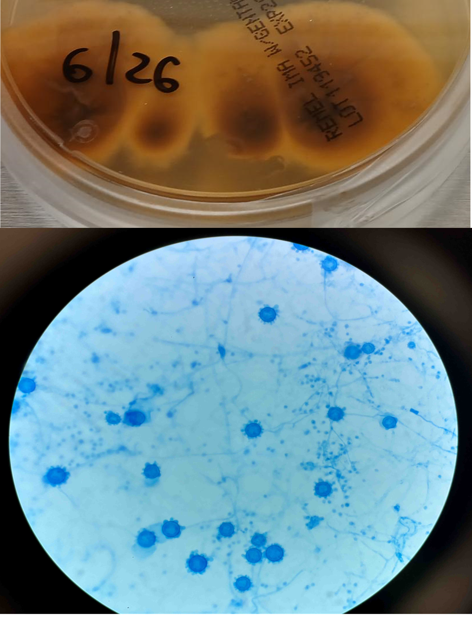
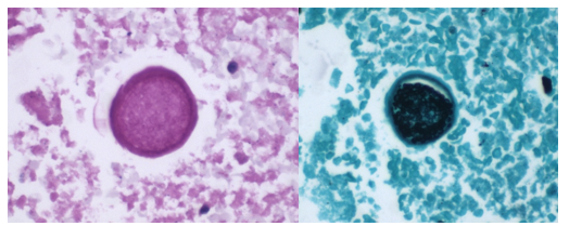
Body identification is one of the core responsibilities of a forensic pathologist yet is also probably the most common one to be overlooked. Most of the deceased people who come to our office are visually identifiable, and the identity may already have been confirmed if they were transported to the hospital. In some situations, though, the body may be disfigured by fire, decomposition, injury, or there may only be partial remains recovered. In incidents with multiple fatalities, we need to be sure the correct remains are returned to the correct family. Particularly when foul play is involved, there may be intentional attempts to conceal the decedent’s identity or disguise them as another person.
There are two different levels of identification: “positive” identification (the gold standard) and “presumptive” identification.
The three generally accepted forms of “positive” identification are DNA comparison, fingerprint comparison, and radiograph comparison. While television shows have DNA “matches” coming back in the time it takes for a commercial break, DNA identification can pose challenges. A pre-existing specimen from the decedent or close family members is needed for comparison, which means you need to already have some suspicion of who they are. If they’ve been previously arrested or charged with a felony (the laws vary slightly by state), their DNA may have been uploaded to the Combined DNA Index System (CODIS), and a match may potentially be obtained blindly by uploading the decedent’s sample. By comparison, one can collect fingerprints from a decedent and submit them to the Automated Fingerprint Identification System (AFIS) for relatively rapid identification. Many people have been fingerprinted in their lifetime, whether for relatively minor arrests, employment, or background checks. However, there are still limitations. The hands (or at least fingertips) need to be intact, with printable skin. For mummified remains, the tissue can be rehydrated by soaking in sodium carbonate or sodium hydroxide to obtain legible prints. “Degloved” remains, where the skin has sloughed from the hands due to decomposition, can be fingerprinted by inserting one’s gloved hand into the sloughed skin.
Radiographs, like DNA, are limited by the need to have a pre-existing sample from the decedent (meaning you need to know who they might be). Radiographs are invaluable when trying to identify someone with no usable fingerprints, or no fingerprints on file. A variety of locations can be used for comparison, including the dentition, frontal sinuses, vertebral processes, healed fractures, or orthopedic implants. Serial numbers on implanted devices can also be traced back to the decedent, although not all implantable devices have such markings.
“Presumptive” identifications are based on many other common sense factors including context, visual identification, tattoos, belongings, and clothing. Depending on the context of the case, a presumptive identification may suffice. For a decomposed body in a secure apartment occupied by a single, elderly person the neighbors haven’t seen in days, monogrammed dentures within the mouth may be sufficient. But in a fire with three charred female victims, aged 20-25, it’s much more important to confirm the identifications by a positive method. As I mentioned earlier any situation involving foul play may provide motivation for to conceal a victim’s identity, and so all homicide victims must be positively identified. It’s often taken for granted that the tag on our patient’s toe is accurate, but we need to approach our autopsies with the same level of diligence a laboratorian has when evaluating the label on a blood tube. Knowing who your patient is, and who your sample comes from, is the first critical step for any pathologist.
The mummified remains of a young adult were found in an abandoned house; while the fingertips were initially too dessicated to yield fingerprints, rehydration revealed excellent ridge details. Fingerprints were then uploaded to AFIS, and the decedent was identified within an hour.For skeletal remains with intact teeth, dental radiographs of the remains can be used for identification; however, for edentulous patients, a different strategy must be used.
-Alison Krywanczyk, MD, FASCP, is currently a Deputy Medical Examiner at the Cuyahoga County Medical Examiner’s Office.
A 47 year old male presented to the emergency department with a two-week history of progressively worsening left-sided flank and abdominal pain with radiation to his back. He also endorsed associated nausea, vomiting, dizziness, lack of appetite, and a 1-month history of intermittent fever, chills, night sweats, cough, and chest pain. Patient history was also notable for travel throughout the Amazon basin in Peru and Colombia, then to Panama where he felt ill while hiking in the rainforest. He also had sustained injuries to his left flank and head while traveling.
Given his symptomology, further evaluation and management were undertaken. CBC showed a drop in hemoglobin from 10.3 g/dL to 8.5 g/dL, and imaging revealed splenomegaly with a caudally located subcapsular hematoma (Figure 1). Small hemoperitoneum on the pelvis with small non-loculated fluid along the bilateral paracolic gutters was also observed. The patient received two units of packed red blood cells to stabilize his hemoglobin levels.
Laboratory Diagnosis
Malaria smears were ordered given his history travel and evidence of anemia. Plasmodium vivax was identified on thin smear at a parasitemia of 0.1%. The Infectious Disease service was consulted and the patient received IV artesunate followed by oral artemether/lumefantrine and was also started on primaquine for hypnozoite eradication as he was not G6PD deficient. The malaria-associated splenic rupture was well managed with conservative treatment, and no surgical intervention was needed.
Figure 1. Computer Tomography scan of the abdomen reveals a caudally located subcapsular hematoma (white circle).Figure 2. Peripheral blood smears (Giemsa stain, 1000x magnification, oil immersion) from the presented patient. A) An enlarged red blood cell with an irregular membrane containing a ring form (black arrowhead) and a mature trophozoite (white arrowhead) of P. vivax. B) An enlarged red blood cell with dot-like staining and a ring form, consistent with P. vivax (black arrowhead). C) P. vivax gametocyte (black arrowhead) and mature trophozoite (white arrowhead). Infected cells are enlarged and the mature trophozoite expands to nearly fill the red blood cell.
Discussion
Malaria is a severe and sometimes fatal tropical disease caused by Plasmodium sp.The World Health Organization (WHO) estimates 300-500 million humans suffer from malaria each year, of whom >1 million die.1 Five species of Plasmodium cause human malaria: P. falciparum, P. malariae, P. vivax, P. ovale, and P. knowlesi. Patients with malaria initially present with a combination of non-specific symptoms such as fever, chills, headache, and general malaise. Moreover, it can cause various complications, including severe anemia, neurologic defects, nephrotic syndrome, hepatosplenomegaly, and even splenic rupture.
Splenic rupture is an infrequent but life-threatening complication of malaria. Due to a paucity of reported cases, the incidence of pathological splenic rupture in natural vector-transmitted malaria is poorly defined. Additionally, cases of splenic rupture in the setting of malaria are likely to be underreported due to a lack of diagnostic methods, especially in endemic regions.2 One case series determined splenic rupture during malaria was fatal 22% of the collected cases, and it was likely to be the most frequent life-threatening issue of Plasmodium vivax infection reported worldwide.3 Similarly, P. vivax tends to cause more pronounced splenomegaly and a higher incidence of splenic rupture when compared to other Plasmodium species as noted in experimental and clinical studies.2.4 However, the underlying mechanism is incompletely established.
The cause of splenic rupture in malaria infections is thought to be a complication of the macroscopic consequences of different microscopic infiltrative phenomena. Specificities of clotting regulation in the spleen red pulp and acute spleen-specific pooling/unpooling of platelets may lead to increase in intrasplenic tension, making the spleen prone to rupture.3 Although malaria-related splenic rupture is essentially a ‘pathological rupture’ in the absence of overt trauma, minor trauma can also be a triggering event, which possibly contributed to the splenic rupture in this case.
Malaria patients with splenic rupture may present with abdominal pain, hypotension, tachycardia, nausea, and vomiting, which can be difficult to distinguish from signs and symptoms of malaria. Referred left shoulder pain (Kehr sign) caused by diaphragmatic irritation from peri-splenic effusion may also occur. The physical examination can show left upper quadrant or diffuse abdominal tenderness, abdominal rigidity or guarding, and splenomegaly. Radiological studies (i.e., ultrasound or CT scan) play a critical role in diagnosing malaria-associated splenic rupture, and the main findings may include splenic enlargement, splenic tear, subcapsular/intrasplenic hematoma, peri-splenic effusion, and hemoperitoneum. Non-radiological workup by contrast (e.g., decreased hemoglobin/hematocrit level) is generally non-specific.
Management of malaria-associated splenic rupture generally follows the same principles as traumatic injuries. Early diagnosis and appropriate management are essential for a favorable prognosis. It is suggested that conservative treatment can be considered in patients with stable hemodynamics. However, the risk of delayed hemorrhage still exists, which makes strict monitoring crucial. Some data suggest surgical intervention should be considered a first-line treatment if tears involve the splenic hilum.5 Although it may be challenging to decide on surgery versus conservative strategy in clinical practice, there are several definitive surgical indications including signs of peritonitis and unstable hemodynamic status despite aggressive resuscitation. Of note, splenectomy may increase the risk of severe infection caused by encapsulated bacteria and complicatedmalaria; therefore, spleen-preserving procedures should be attempted in selected patients, such as frequent travelers to malarious regions.6
References
Guerra, C.A., et al., The limits and intensity of Plasmodium falciparum transmission: implications for malaria control and elimination worldwide. PLoS Med, 2008. 5(2): p. e38.
Zingman, B.S. and B.L. Viner, Splenic complications in malaria: case report and review. Clin Infect Dis, 1993. 16(2): p. 223-32.
Imbert, P., C. Rapp, and PA Buffet, Pathological rupture of the spleen in malaria: analysis of 55 cases (1958-2008). Travel Med Infect Dis, 2009. 7(3): p. 147-59.
Schmidt, L.H., Plasmodium falciparum and Plasmodium vivax infections in the owl monkey (Aotus trivirgatus). I. The courses of untreated infections. Am J Trop Med Hyg, 1978. 27(4): p. 671-702.
Fuks, D., et al., Spontaneous splenic rupture during a febrile crisis of Plasmodium falciparum malaria. J Chir (Paris), 2005. 142(6): p. 403-5.
Jimenez, B.C., et al., Spontaneous splenic rupture due to Plasmodium vivax in a traveler: case report and review. J Travel Med, 2007. 14(3): p. 188-91.
-Bo Zhang, MD is an AP/CP resident at UT Southwestern Medical Center in Dallas, Texas. He is a first year resident interested in different aspects of surgical pathology.
-Andrew Clark, PhD, D(ABMM) is an Assistant Professor at UT Southwestern Medical Center in the Department of Pathology, and Associate Director of the Clements University Hospital microbiology laboratory. He completed a CPEP-accredited postdoctoral fellowship in Medical and Public Health Microbiology at National Institutes of Health, and is interested in antimicrobial susceptibility and anaerobe pathophysiology.
-Clare McCormick-Baw, MD, PhD is an Assistant Professor of Clinical Microbiology at UT Southwestern in Dallas, Texas. She has a passion for teaching about laboratory medicine in general and the best uses of the microbiology lab in particular.
Recently our lab was asked by physicians to start reporting band counts of ≧10% as critical values. While we have always reported bands when a manual differential is performed, we have also heard for years about other labs that have stopped reporting bands. Mayo Clinic stopped reporting bands in the 1990’s- other nationally and internationally known hospitals and community hospitals alike have followed suit. CAP proficiency surveys for cell ID do not separate segmented neutrophils and band neutrophils for cell IDs. Our hematology analyzer reports Immature granulocytes (IG) and we have learned about the benefits of IG over the band count. Thus, when our pathologists asked us to add a critical value for bandemia, we wondered if we were moving ‘backwards’.
Bandemia is defined as elevated band neutrophils in the peripheral blood. Neutrophils are produced to help fight infection. With infection, there is an increase in WBCs being released by the bone marrow into the peripheral blood. Bands are considered mature neutrophils and can fight infection, and in an effort to keep up with demand, some of these infection fighting cells which are released are bands. However, we are reminded that this is a nonspecific clinical finding. Bands can be elevated in many situations including inflammation, autoimmune disease, metabolic abnormalities, pregnancy, and treatment with granulocyte colony stimulating factors. Band counts of ≧10% have been used clinically as an indicator of serious bacterial illness. A finding of bandemia can help providers decide what steps need to be taken in evaluation of a patient, and in conjunction with clinical findings, can help in making a differential diagnosis.
The trouble with bands is that they are notoriously difficult to measure accurately and precisely. Bands are somewhat controversial and there are conflicting opinions on the utility of bands. Should bands be reported or included with neutrophils? A left shift reflects bone marrow response to bacterial infection, and this has been quantified as band count or immature granulocyte count. Is the IG a better parameter than the band count?
If your Hematology analyzer reports the absolute neutrophil count (ANC) with an automated differential, this includes all mature neutrophils. The theory that supports this is that neutrophilic bands and segmented neutrophils are both mature cells, and both fight infection. A true left shift would therefore be the presence of immature granulocytes (metamyelocytes, myelocytes and promyelocytes). Yet, we also now have analyzers that offer a 6-part differential that includes the immature granulocyte (IG) count. And we know that an automated differential is based on counting thousands of cells, and a manual differential is based on counting 100 cells. So, which is better? We can theorize that the IG is a better indicator of left shift because the automated diff count looks at more cells than a 100-cell manual diff.
Let’s say that we have a patient with a WBC of 20 x 103/μL. Left shift is defined as an absolute IG count >0.1 x 10³/μL. Automated diffs can count >30,000 cells depending on the WBC count. If the auto diff counts 32,000 cells and finds 1% IG, this means that the analyzer identified 320 IG. Absolute counts are calculated as % x the WBC, in this case 0.01 x 20,000=.2 x 103/μL. This meets the definition of a left shift. If we do a manual diff and count 100 cells and see 1 meta, the absolute IG would be .2 x 103/μL, again meeting the criteria for a left shift. However, if we did not see any metas or other IG on the manual diff, the absolute IG would be 0. Thus, performing a manual diff, the difference in seeing 1 cell or 0 would make the difference of reporting a suspected infection versus no suspicion of infection.
Table 1: Left shift? comparison of absolute IG when 1 or no IGs are seen on manual diff
With the advent of the IG count on automated differentials, labs have moved away from reporting bands. I have attended conferences and heard presentations about “banning” bands. A few years ago, I wrote a blog called “Beyond Bands: The Immature Granulocyte Count”, describing the benefits of using the IG count over bands in manual diffs. The above example would support “Ban the bands” arguments. Using the IG count from analyzers can take advantage of analyzing many more cells and give us statistically more precise values.
Results from auto diff can get to patients’ chart faster than a manual diff result, leading to faster treatment. To report bands, a manual differential must be done. We must wait for a slide to be made, dry, and a diff to be analyzed either under the microscope or on CellaVision. Bands are subjective, relying on technologist interpretation.
So, why are we suddenly being asked to make bandemia a critical value?
Physicians asked for this change and have cited cases where patients were seen in the emergency department (ED) and subsequently released, later to return, or experiencing negative outcomes. These patients had bands reported on differentials. Other area hospitals are reporting bands and have critical values for bandemia. Because patients often are seen at more than one area hospital, and doctors may have privileges at more than one of these, for consistency, this makes sense. But is also makes sense clinically.
Recent data has drawn renewed attention to bands as a reliable predictor of severity of patient condition. A number of research papers have been published that indicate that bands may indeed be important for patient care. A 2012 study investigated bandemia in patients with normal white blood cell counts. This cohort study found that patients with normal WBC counts with moderate (11%-18%) or high (>20%) band counts had increased odds of having positive blood cultures and in-hospital mortality. (Drees, 2012)
In 2019 a study showed that there was an “increasing risk for death with increasing bandemia, irrespective of leukocyte count. (Davis, 2019) A 2021 study done at Rhode Island Hospital showed a strong correlation between increasing percentages of bands on an initial emergency room CBC and the likelihood of significant positive blood cultures and in-hospital mortality. This was noted even at band levels below 10%. (Hseuh, 2021) S. Davis, MD, from the Department of Emergency Medicine, George Washington University School of Medicine and Health Sciences, in Washington, DC wrote that “While emergency physicians may find reassurance in a normal leukocyte count, the balance of evidence strongly suggests a more prudent approach would be to wait for the bands.” (S. Davis, 2021) In other words, wait for that manual differential. He stated that emergency room physicians get results from automated CBCs before the manual diff and do not see or are aware of any internal laboratory flags on these specimens. Physicians should be aware of reporting processes to avoid early discharge of otherwise well-appearing patients before band counts are reported. Last year, trends in bandemia and clinical trajectory among patients was reviewed in a retrospective chart review at George Washington University Hospital. They noted that “Bandemia clearance and trending, in conjunction with other existing clinical tools, may be of use as a marker of improvement in sepsis. Conversely, worsening bandemia may be predictive of a deteriorating clinical status and possibly a higher mortality.” They also noted that following trends of band levels in patients with sepsis or septic shock may help to predict a clinical course and overall prognosis. (Prasanna, 2022) Additionally, a band count greater than 10% is one of the American College of Chest Physicians/Society of Critical Care Medicine’s systemic inflammatory response syndrome (SIRS) criteria used to diagnose sepsis. (Chakraborty, 2022) These are just a few of the many articles that support a clinical utility of reporting the band count.
When we first learned that we would be reporting bandemia as a critical value, we realized that we would need to get everyone on the same page. We do most of our diffs on CellaVision, so, in theory, that should be easy, but, and a big but, is that bands are notoriously subjective. Different technologists may have been taught or have used their own definitions of bands. Variation can occur depending on slide quality, tech training, definition of bands used, and number of cells counted. So, how do we make this work? We need to be sure that all our technologists are reporting bands using the same criteria, so we are not reporting differing or confusing information to physicians. The concern lies in a physician making a significant clinical decision based on apparent changes in band counts that are not real but only reflect predictable statistical factors and unpredictable technologist variability.
In our laboratory, we approached the implementation of this new critical value as an educational opportunity. Differentials that had been performed on our CellaVision were reviewed, and it was apparent that bands were not being categorized consistently among techs. (See Figure1) We started with reviewing the definition of bands with all technologists and writing updated detailed procedures that includes these definitions. We use CAP’s definition of bands, which is the definition used in most textbooks and references for over 60 years. (See Table 1) Many examples of both bands and segmented neutrophils were added to our reference library on CellaVision. These included textbook perfect bands and some that may be more subjective. These reference cells can be used by techs to compare cells when making decisions as to in which category they belong. It was also stressed to techs that they need to look at cells carefully, and in a view that is large enough to see both detail and differentiation.
Figure1. Cells reported as segmented neutrophils on Differential. How many bands do you see?Table 2. CAP Definition of bands vs segmented neutrophilsFigure 2. Are these bands or segmented neutrophils?
Patient samples that had bands reported were located on CellaVision and multiple slides were made from these samples to be used as competency slides. In developing the differential evaluation tool, Rumke’s data showed that for a differential with a reported 12% bands, a second differential would have to have greater than 23% bands or fewer than 3% bands before the difference could be considered statistically significant. But this can be significant to our patients and patient care. In this example, diffs with <10% bands would not indicate bandemia, and diffs over 10% would initiate a critical bandemia call. And this could happen on the same slide depending on who did the diff, or on sequential samples on the same patient over a short period of time. These competency slides were assigned to techs to collect statistics on the mean and SD of the bands reported on each slide. Retraining and coaching will be provided as necessary. Follow up competency slides will be assigned, and statistics will be recalculated. The goal is to decrease variability in our band counts and to show that we have done so. This ongoing quality project has involved writing procedures, offering continuing education, assigning and reviewing competency slides, coaching technologists and reviewing slides with them and calculating statistics. Our goal is consistency and reporting meaningful results to our physicians.
As we saw in the studies cited above, % bands and trends are both important when evaluating clinical correlations. The chart below shows examples of how this might affect patient care. If a patient on presenting at the ED had a 19% band count, this would be called as a critical, and the patient would be further evaluated, and depending on clinical symptoms and medical history would likely have blood cultures drawn and be admitted to the hospital. If a second tech did the diff and reported 5% bands, the patient may be sent home without further evaluation. On subsequent CBCs, we could be giving confusing results to the physician if we are not consistent in our reporting. With multiple techs doing differentials on different shifts, it could look like this patient is getting better,
getting worse, or it could look like the patient is responding to therapy, and then the next day they had a setback. While we understand that bands will probably always be somewhat subjective, we need to narrow this down. By adhering to one definition, our goal is to report consistent and accurate results.
Table 3. Various results on differentials on the same patient over the course of 3 days, showing technologist dependent results.
ED physicians are looking for an early marker that can be used to identify septic patients as early as possible. Bandemia may be used as this marker. We therefore need to be as objective as possible when reporting bands. “Ultimately the band count is only one factor amongst several others which will be used in assessing the patient’s clinical state and in determining any subsequent medical management. Yet, identifying bands is important, and emphasizes the key role that our laboratory professionals play in identifying causes for concern” Dr Edgar Alonsozana, Mercy Medical Center, Baltimore, Md.
I welcome your comments about how your laboratories report bands and if bandemia is a critical value in your facilities!
References
P.Joanne Combleet, Clinical utility of the band count, Clinics in Laboratory Medicine, 2002;22:101-136
Al-Gwaiz, Layla A. and H H Babay. “The Diagnostic Value of Absolute Neutrophil Count, Band Count and Morphologic Changes of Neutrophils in Predicting Bacterial Infections.” Medical Principles and Practice 16 (2007)
S. Davis, R. Shesser, K. Authelet, A. Pourmand. “Bandemia” without leukocytosis: A potential Emergency Department diagnostic pitfall. The American Journal of Emergency Medicine,Volume 37, Issue 10, 2019
Harada T, Harada Y, Morinaga K, Hirosawa T, Shimizu T. Bandemia as an Early Predictive Marker of Bacteremia: A Retrospective Cohort Study. Int J Environ Res Public Health. 2022 Feb 17;19(4):2275.
Christine DeFranco DO and Terrance McGovern DO MPH St. Joseph’s Regional Medical Center, Paterson, NJ. Isolated Bandemia: What Should We Do with It? Critical Care, Oct. 2016,
Harmening, Denise. Clinical hematology and Fundamentals of Hemostasis, 4th ed. 1997
Prasanna N, DelPrete B, Ho G, et al. The utility of bandemia in prognostication and prediction of mortality in sepsis. Journal of the Intensive Care Society. 2022;0(0).
Takayuki Honda, Takeshi Uehara, Go Matsumoto, Shinpei Arai, Mitsutoshi Sugano, Neutrophil left shift and white blood cell count as markers of bacterial infection,Clinica Chimica Acta, Volume 457, 2016
Drees M, Kanapathippillai N, Zubrow MT. Bandemia with normal white blood cell counts associated with infection. Am J Med. 2012 Nov;125(11):1124.e9-1124.e15.
Leon Hsueh, Janine Molino, Leonard Mermel,
Elevated bands as a predictor of bloodstream infection and in-hospital mortality,The American Journal of Emergency Medicine,2021
-Becky Socha, MS, MLS(ASCP)CMBBCM graduated from Merrimack College in N. Andover, Massachusetts with a BS in Medical Technology and completed her MS in Clinical Laboratory Sciences at the University of Massachusetts, Lowell. She has worked as a Medical Technologist for over 40 years and has taught as an adjunct faculty member at Merrimack College, UMass Lowell and Stevenson University for over 20 years. She has worked in all areas of the clinical laboratory, but has a special interest in Hematology and Blood Banking. She currently works at Mercy Medical Center in Baltimore, Md. When she’s not busy being a mad scientist, she can be found outside riding her bicycle.
A middle-aged patient with a complex past medical history presented to the Emergency Department with the primary complaints of shortness of breath and cough. The patient had recently been admitted for community-acquired pneumonia and discharged with antibiotics 10 days ago. However, the patient continued to have a dry cough, a fever of 102 degrees F, and shortness of breath. On the new admission, they were noted to be hypotensive and labs showed elevated WBC (14.7), lactate of 7.3, proBNP of 852, and procalcitonin of 4.62. Bacterial blood cultures showed no growth. A chest x-ray showed worsening left lower lobe consolidation with extension to the left upper lobe, compared to the x-ray at the previous admission. A bronchoscopy was done and bronchoalveolar lavage (BAL) was submitted for Gram stain, aerobic bacterial culture, and fungal culture. The Gram stain and bacterial culture were negative. However, the fluorochrome stain showed many large budding yeast forms, most suggestive of Blastomyces dermatitidis. Lung tissue sample culture (Image 1), Gram stain (Image 2) and histopathology (Image 3) confirmed the identification as Blastomyces dermatitidis. The patient was expired before the results were released. Gross images of lungs are shown in image 4.
Image 1. Fungal growth on the Sabouraud dextrose plate observed after 2 days. Image 2. Gram stain of left lower lobe of lung tissue sample demonstrating budding yeast. Image 3. GMS and H&E stains of left lower lobe of lung sections demonstrating budding yeast. Image 4. Gross pictures of lungs showing left lower lobe hepatization and miliary pattern consolidation in the right lung.
Discussion
Blastomyces dermatitidis is a dimorphic fungus found in soil and decaying wood. Often found in areas close to a water source such as a lake, river, or stream.1 The fungus is endemic to parts of the United States, including parts of the Appalachian Mountains, the Great Lakes, and the Ohio and Mississippi River Valley.1 Human infection occurs when airborne conidia are inhaled. Blastomycosis (aka Gilchrist’s disease) may cause a broad range of clinical presentations. The most affected organs are the lungs and the skin.2 Osseous, genitourinary, central nervous system (CNS), and disseminated blastomycosis can also be seen, but at a lower frequency.2 Unlike opportunistic fungal pathogens such as H. capsulatum, C. immitis, and C. neoformans, B. dermatitidis no more likely to cause disease in immunocompromised population compared to the non-immunocompromised populatin.3 However, the disease is more severe, with higher morbidity and mortality, and more likely to be disseminated in immunocompromised patients.3
Although growth of fungi took 2 days in our case, Blastomyces is a slow growing fungi in that mycelial forms mature in 14-21 days. In suspicious cases, cultures should be hold up to 8 weeks. Furthermore, Blastomyces should be cultured as soon as possible since it does not survive well in samples. Morphologically, Blastomyces colony appears as a mold, which is white, prickly, and cottony at 25-30 °C However, at 35-37 °C, the colony appears as a yeast, which is tan, wrinkled, and waxy. Microscopically, at 37 °C, Blastomyces appear as round to oval cells with refractile and thick cell walls and measure 8 – 15 uM in diameter. Each yeast cell produces only one, broad-based (4-5 uM), bud. At 25-30 °C, septate hyphae form with short or long conidiophores. Round to pear-shaped conidia attach to the apex, resembling lollipops (Image 5).4
Image 5. Spherical, oval, or pyriform conidia rising from aerial hyphae, mold phase.
History of recent travel to the endemic areas or already living in there usually triggers the suspicion with clinical findings. There are different auxiliary diagnostic modalities for B. dermatitidis including culture, cytology smear, histopathology, urine antigen test, serum antigen test, serum antibody test, and molecular techniques. Serum antibody test exhibits high degree of cross-reactivity with other endemic mycoses.5 Serum and urine antigen testing has a sensitivity of 89 % and specificity of 79 % and it is useful in diagnosis, monitoring treatment, and detecting recurrence.5,6,7 Direct visualization of the distinctive yeast form with broad-based budding on a cytology smear of respiratory secretions or tissue samples using fluorochrome stain is considered as presumptive diagnosis and enough for initiation of antifungal therapy. However, a negative result in smear cannot exclude the diagnosis due to lack of sensitivity. Polymerase chain reaction (PCR) based assays have been developed to detect B. dermatitidis.8 However, although they are promising, utility has not been confirmed and there are no FDA approved assays.8
Only histopathology and culture can provide a definitive diagnosis. Although H&E may be enough in selected cases, periodic acid-Schiff (PAS) and Grocott’s methenamine silver (GMS) stains are useful to highlight the yeast form in tissue sections. In culture, seeing the characteristic morphologic and microscopic appearance is very useful for diagnosis. However, confirmatory testing with chemiluminescent DNA probe may still be necessary.9
Depending on the severity and site of the infection fluconazole, itraconazole, voriconazole, or amphotericin B can be used in treatment.10
References
Castillo CG, Kauffman CA, Miceli MH. Blastomycosis. Infect Dis Clin North Am. 2016;30(1):247-264.
Mazi PB, Rauseo AM, Spec A. Blastomycosis. Infect Dis Clin North Am. 2021;35(2):515-530.
McBride JA, Gauthier GM, Klein BS. Clinical Manifestations and Treatment of Blastomycosis. Clin Chest Med. 2017;38(3):435-449.
Maresca B, Kobayashi GS. Dimorphism in Histoplasma capsulatum and Blastomyces dermatitidis. Contrib Microbiol. 2000;5:201-216.
Wheat LJ. Antigen detection, serology, and molecular diagnosis of invasive mycoses in the immunocompromised host. Transpl Infect Dis. 2006;8(3):128-139.
Mongkolrattanothai K, Peev M, Wheat LJ, Marcinak J. Urine antigen detection of blastomycosis in pediatric patients. Pediatr Infect Dis J. 2006;25(11):1076-1078.
Tarr M, Marcinak J, Mongkolrattanothai K, et al. Blastomyces antigen detection for monitoring progression of blastomycosis in a pregnant adolescent. Infect Dis Obstet Gynecol. 2007;2007:89059.
Bariola JR, Hage CA, Durkin M, et al. Detection of Blastomyces dermatitidis antigen in patients with newly diagnosed blastomycosis. Diagn Microbiol Infect Dis. 2011;69(2):187-191.
Saccente M, Woods GL. Clinical and laboratory update on blastomycosis. Clin Microbiol Rev. 2010;23(2):367-381.
Chapman SW, Dismukes WE, Proia LA, et al. Clinical practice guidelines for the management of blastomycosis: 2008 update by the Infectious Diseases Society of America. Clin Infect Dis. 2008;46(12):1801-1812.
-Kadir Isidan, MS, MD is a pathology resident at University of Chicago (NorthShore). His academic interests include gastrointestinal pathology and cytopathology.
-Paige M.K. Larkin, PhD, D(ABMM), M(ASCP)CM is the Director of Molecular Microbiology and Associate Director of Clinical Microbiology at NorthShore University HealthSystem in Evanston, IL. Her interests include mycology, mycobacteriology, point-of-care testing, and molecular diagnostics, especially next generation sequencing.
When we enter the laboratory, we know of the dangers that can be encountered. Our training tells us there could be microbes and other potential pathogens in the samples we are about to analyze. We also learned how to protect ourselves; how our behavior while in the lab has consequences. We even know how to dress properly and what engineering controls we have at our disposal to keep us safe. We put on our personal protective equipment (PPE) before we start to work and remove it before leaving the lab. For some, these behaviors are automatic, actions that are done almost without even thinking. But is this the same for all who enter the lab? Do visitors who comes into the department know what they are really walking into or how to keep themselves safe in an environment that may be foreign to them? One common question asked by lab staff regarding visitors is “do they have to adhere to the lab safety policies and if so, why?”.
On a recent safety audit, I visited a lab that happened to be getting a new chemistry analyzer installed. I noticed the vendor team, which consisted of 5 individuals, were not wearing any PPE. There were backpacks, open water bottles, and cell phones sitting on the counters and floors. The new instrument was not hidden in a back corner of the lab far away from the daily work. It was close to the area where the lab process, spins, and runs patient samples. Members of the vendor team were lying on the floor and crawling around. How does that scene make you feel?
Vendors and service representatives are regular visitors in your lab. A laboratory can have a representative on site a dozen times before you even begin to use that piece of equipment. Once it is installed, you can bet you will see them multiple times for preventative maintenance and service calls. How does your lab welcome these guests? Do you let them in and have them get right to work? If they are there to repair an analyzer you are likely eager to have them get started, but do you ask them to wear a lab coat? Did they bring one of their own that was kept in their backpack? If so, do you think that coat is clean or was it used in a different lab, packed up, and brought to your lab? Vendor compliance is a safety issue for many labs because these visitors are not lab employees, yet they are in your department and may be putting themselves and your team at risk. Often vendors are seen with drinks in labs, using cell phones or touching instruments without gloves – behaviors lab folk are told not to follow. So why is it tolerated? It shouldn’t be, and you have the right to speak up and ask them to adhere to your lab policies.
What about other potential laboratory visitors? Do pathologists come in to look at a patient slide in Hematology? Do they just sit down at your bench and look at the slide without gloves or a lab coat? Is lab staff allowed to scan a smear without PPE? Probably not, and no one else should be allowed too either. The microscope has most likely been touched with dirty gloves, and no one else should touch the same scope without gloves. Even lab doorknobs are a consideration. Staff should wash hands before leaving the department. That means no one should use contaminated gloves to open the door.
Speaking up about these safety issues to lab visitors can feel uncomfortable. A conversation with a physician about safe practices in the lab can be daunting, but the cost of not speaking up can be high. Take the opportunity to show you care about visitors and want to keep them protected. Sometimes you know who is coming to the lab, and you feel confident they have been trained and will use the best safety practices. At other times, though, those guests may be unexpected and lacking in safety knowledge. Make sure to treat them with respect, give them the safety training and tools they need so they can leave both happy and healthy.
-Jason P. Nagy, PhD, MLS(ASCP)CM is a Lab Safety Coordinator for Sentara Healthcare, a hospital system with laboratories throughout Virginia and North Carolina. He is an experienced Technical Specialist with a background in biotechnology, molecular biology, clinical labs, and most recently, a focus in laboratory safety.
A 40-year-old woman with past medical history of polysubstance use disorder (cocaine, IVDU-heroin) presented with shortness of breath on exertion, weight loss, and weakness. Cardiac ultrasound showed aortic valve endocarditis with mild thickening of the aortic valve, vegetation and severe aortic regurgitation without stenosis. The patient was taken to the operating room for aortic valve replacement. A tissue biopsy was sent for surgical pathology workup and microbiology cultures. H&E staining of the right and left coronary cusp and noncoronary cusp and showed focal giant cell reaction, and acute inflamed granulation tissue consistent with vegetation/infective endocarditis. The Gram stain of the tissue specimen revealed gram positive cocci but no bacterial growth was seen. Fungal and mycobacterial cultures were also negative. Broad Range Bacterial PCR and Sequencing (BRBPS) of the heart valve detected Streptococcus mutans.
H&E staining of the heart valve revealed focal giant cell reaction (black arrow), and acute inflamed granulation tissue consistent with vegetation/infective endocarditis (left image). The Gram stain of the tissue specimen revealed gram positive cocci (right image). (H&E diagnosis credits: Dr. Irena Manukyan from George Washington University.)
Discussion
The first step of BRBPS is performing polymerase chain reaction (PCR) targeting the 16S ribosomal RNA (rRNA), a conserved gene across many bacterial species including mycobacteria. If PCR is negative, then sequencing is not performed but if PCR is positive, sequencing such as Sanger sequencing or Next generation sequencing will be followed. Subsequent bioinformatics analysis of the sequencing results will allow identification of the specific bacteria.1,2 In the patient here, the PCR for 16S rRNA was positive and sequencing revealed S. mutans.
S. mutans is part of the viridans-group of Streptococcus and can be identified as a gram positive cocci with growth that is alpha-hemolytic, optochin resistant, and bile insoluble. Viridans group Streptococcus invade the bloodstream following dental treatment or dental procedures and can be associated with high risk for infectious endocarditis in patients with underlying heart disorders. S. mutans first gained medical attention due to its role in dental caries.3S. mutans is considered part of the normal flora of the oropharynx, more specifically the dental plaque, and can form biofilms on the hard surface of the tooth. Bacterial strains that cause endocarditis have been shown to be able to bind to type I, III, and IV collagen, which are major components of the host cardiovascular tissues.4 The bacteria also have virulence factors that can cause strong adhesion to human endothelial cells. Additionally, aggregation and interaction with fibrinogen, platelets, and completement allow this bacteria to successfully cause cardiovascular diseases.5
In infective endocarditis, 20% of the cases are negative by conventional methods such microbiology cultures.6 Several studies have shown that in specimens from patients with endocarditis, 16s rRNA sequencing can correctly detect the organism that was grown in culture but also successfully detect organisms in specimens where there was no growth, allowing clinical teams to accurately treat patients, similar to our case here.7,8 For prosthetic heart valve tissue, the sensitivity of sequencing versus culture is 93% and 35%, respectively. A study describing periprosthetic joint infections using elbow joint fluids showed that sequencing was positive in 47 specimens but 8% of those were negative by culture.10 Additionally, 16s rRNA sequencing has been useful in identifying bacterial tick-borne organisms not typically grown in culture such as Borrelia, Anaplasmsa, Ehrlichia, and Rickettsia].11
The BRBPS test is validated for and ideally be tested on specimens from sterile sources (joint fluid, blood, heart valves, CSF) and ideal specimens are those where bacterial organisms can be visualized using microscopy. Limitations of BRBPS is that this test only detects bacterial organisms and will not detect viruses, fungi, and parasites. False-positive results are possible if the specimen is contaminated with patient flora. False-negative results may occur due to sequence variability affecting how the primers bind, presence of PCR inhibitors, or the quantity of nucleic acid material below the limit of detection. Clinical tests utilizing sequencing approaches may be costly at the moment in time; those who order sequencing tests need to understand the purpose of different sequencing tests so the most appropriate test is ordered. For example, if fungal pathogens are suspected, a sequencing test based on amplifying 28S rRNA or ribosomal ITS genes for the conserved genes found in fungal pathogens is appropriate.12 Such tests where you selectively enrich for specific targets such as 16s or ITS are ‘targeted NGS tests’ which have higher sensitivity for detection of microorganisms in sample types with large amounts of DNA. In comparison, metagenomics is a more agnostic approach and allows for detection of all nucleic acid in the specimen (including both host and microbial reads). While metagenomics can successfully detect pathogens involved in an infection, it can also detect the microbiome present in the same specimen. Hence, the one limitation is the background noise from human nucleic acid and the microflora [13-14. Another type of sequencing is whole genome sequencing where the microbial genome of a particular organism is sequenced and assembled. WGS aids in identification, typing, and determining the microbial susceptibility. WGS is helpful in identifying outbreaks or for epidemiological purposes, but the limitation is that a pure culture is needed. WGS requires a pure culture so this is a limitation for organisms that are non-viable in culture [13-14].
References
Rosey AL, Abachin E, Quesnes G, Cadilhac C, Pejin Z, Glorion C, Berche P, Ferroni A. Development of a broad range 16S rDNA real-time PCR for the diagnosis of septic arthritis in children. J Microbiol Methods. 2007 Jan;68(1):88-93. doi: 10.1016/j.mimet.2006.06.010. Epub 2006 Aug 14. PMID: 16904782.
Loesche WJ. 1986. Role of Streptococcus mutans in human dental decay. Microbiol Rev 50:353–380.
Nomura R, Naka S, Nemoto H, Inagaki S, Taniguchi K, Ooshima T, Nakano K. 2013. Potential involvement of collagen-binding proteins of Streptococcus mutans in infective endocarditis. Oral Dis 19:387–393. doi: 10.1111/odi.12016.
Otsugu, M., Nomoura R., Matayoshi, S., Teramoto, N., Nakanoa, K. Contribution of Streptococcus mutans Strains with Collagen-Binding Proteins in the Presence of Serum to the Pathogenesis of Infective Endocarditis. Infect Immun. 2017 Dec; 85(12): e00401-17.
Baddour LM at al; American Heart Association Committee on Rheumatic Fever, Endocarditis, and Kawasaki Disease of the Council on Cardiovascular Disease in the Young, Council on Clinical Cardiology, Council on Cardiovascular Surgery and Anesthesia, and Stroke Council. Infective Endocarditis in Adults: Diagnosis, Antimicrobial Therapy, and Management of Complications: A Scientific Statement for Healthcare Professionals from the American Heart Association. Circulation. 2015 Oct 13;132(15)
Peeters, B., Herijgers, P., Beuselinck, K., Verhaegen, J., Peetermans, W.E., Herregods, M.-C., Desmet, S., Lagrou, K. Addd diagnostic value and impact on antimicrobial therapy of 16s rRNA PCR and amplicon sequencing on resected heart valves in infective endocarditis: a prospective cohort study. Clinical Microbiology and Infection. 2017 Nov; 23(11): 888.e1-888.e5
Premru, M.M, Zupanc, T.L., Klokocovnik, T., Sabljic, E.R., Cerar, T. Broad-Range 16s rRNA PCR on Heart Valves in Infective Endocarditis. J Heart Valve Dis. 2016 Mar; 25(2):221-22
Miller, R. J.H., Chow, B., Pillai, D., Church, D. Development and evaluation of a novel fast broad-range 16S ribosomal DNA PCR and sequencing assay for diagnosis of bacterial infective endocarditis: multi-year experience in a large Canadian healthcare zone and a literature review. BMC Infect Dis. 2016 April 12:16:146
Flurin, L.; Wolf, M.J.; Greenwood-Quaintance, K.E.; Sanchez-Sotelo, J.; Patel, R. Targeted next generation sequencing for elbow periprosthetic joint infection diagnosis. Diagn. Microbiol. Infect. Dis. 2021, 101, 115448.
Kingry, L.; Sheldon, S.; Oatman, S.; Pritt, B.; Anacker, M.; Bjork, J.; Neitzel, D.; Strain, A.; Berry, J.; Sloan, L.; et al. Targeted Metagenomics for Clinical Detection and Discovery of Bacterial Tick-Borne Pathogens. J. Clin. Microbiol. 2020, 58, e00147-20.
Yeo, S.F.; Wong, B. Current status of nonculture methods for diagnosis of invasive fungal infections. Clin. Microbiol. Rev. 2002, 15, 465–484.
Mitchell SL, Simner PJ. Next-Generation Sequencing in Clinical Microbiology: Are We There Yet? Clin Lab Med. 2019 Sep;39(3):405-418
Hilt, E.E., Ferrieri, P. Next Generation and Other Sequencing Technologies in Diagnostic Microbiology and Infectious Diseases. Genes (Basel). 2022 Aug 31;13(9):1566. doi: 10.3390/genes13091566.
–Rami Abdulbaki, MD is a Pathology Resident (PGY-4) at The George Washington University Hospital. His academic interest includes hematopathology and molecular pathology.
-Maikel Benitez Barzaga, MD is a Pathology Resident (PGY-2) at The George Washington University Hospital. His academic interest include hematology, microbiology, molecular and surgical pathology.
–Rebecca Yee, PhD, D(ABMM), M(ASCP)CM is the Chief of Microbiology, Director of Clinical Microbiology and Molecular Microbiology Laboratory at the George Washington University Hospital. Her interests include bacteriology, antimicrobial resistance, and development of infectious disease diagnostics.
Emergency services were called to a fire in a small apartment building, in which the structure was completely engulfed. Most of the occupants had been evacuated – however, once the fire was extinguished, the charred remains of an adult woman were found in the debris.
At the autopsy of severely fire-damaged human remains, two key questions must be answered: 1) who is the decedent?, and 2) were they alive when the fire started?
Question #1 is particularly relevant in this case, as many people lived in the building. Presumptive identification based on the tenant list may seem reasonable at first, but this victim could represent a visitor, contractor, or subletter. When facial identification isn’t possible, radiographic identification can be done with dental x-rays or x-rays of other bones which may have unique features from healed trauma or degeneration. Additional methods of positive identification could include fingerprints (if still intact), or DNA comparison to first degree relatives.
Question #2 is of importance because fire can be used in an attempt to disguise the identity of a victim of violent crime and destroy evidence. Cutaneous evidence of trauma may be disguised by burns, so full body x-rays are taken of every fire-damaged body. X-rays can also reveal retained bullets, knife tips, or fractures unlikely to have been caused by the fire.
When deciding if a fire victim was alive when the fire started, we first examine the upper and lower airways for soot. Most fire victims do not die from cutaneous burns, but from smoke inhalation – including carbon monoxide (CO) toxicity, which is often apparent by cherry red discoloration of the blood and viscera. Postmortem carboxyhemoglobin measurements in house fire victims are typically greater than 50%. There are exceptions to this rule, of course. Rarely, someone who was clearly alive when the fire began will have minimal or no soot in their airways and a negligible carbon monoxide concentration. This can happen in a “flash fire”, such as one ignited by gasoline or oxygen tanks, in which thermal injury to the upper airway may cause rapid occlusion by laryngospasm or edema. People with underlying heart or lung conditions will be more susceptible to the effects of carboxyhemoglobin, and may not survive long enough to obtain a level above 50%. Fires also produce other toxic products of combustion such as cyanide, and can lower ambient oxygen saturations to result in asphyxiation by lack of ambient oxygen (even without CO).
Forensic pathologists need to be aware of the artifacts that fires can create. Pugilistic posturing of fire victims (limb flexion) is due to heat-related contraction of muscle fibers. Epidural hematomas can result from boiling blood and bone marrow within the calvarium extravasating into the epidural space. The heat can induce fractures in exposed bone once the surrounding soft tissue is consumed or fully charred. Finally, the heat can split apart skin and soft tissue, resulting in sharp-force-like defects which occur parallel to the orientation of muscle fibers (rather than across them, which is more suspicious for penetrating trauma).
Of utmost importance in fire-related deaths, however, is scene investigation. The manner of death in fire fatalities is related to the origin of the fire. Most fire deaths are accidental, as the fire is unintentionally sparked by some electrical malfunction or unattended flame. However if the fire started intentionally, the manner of death can be homicide (if started by another) or suicide (started by the victim). It is therefore crucial to review the final fire investigation report before finalizing the autopsy report and death certificate.
This image shows dark black soot lining the main and lobar bronchi; this indicates the victim was breathing during the fire.Heat-related epidural hematomas have a brown, amorphous appearance rather than the bright red color of traumatic epidural hematomas.The scalp has been consumed by fire, and the exposed bone is calcined and brittle with fractures of the outer table.
-Alison Krywanczyk, MD, FASCP, is currently a Deputy Medical Examiner at the Cuyahoga County Medical Examiner’s Office.
Microbiology Case: Lung nodules in a patient undergoing lung transplant evaluation
Case History
A 71-year-old male with a past medical history of severe interstitial lung disease (ILD) presented for lung transplant evaluation. His chest CT demonstrated findings consistent with his known diagnosis of ILD, along with a 3.6 cm focal nodular calcific density near the right costophrenic angle, and additional scattered calcified lung nodules measuring up to 3 mm. Sputum cultures were obtained to evaluate the CT findings.
Laboratory Identification
Routine bacterial, fungal, and AFB sputum testing was performed. No AFB were recovered, and a mixture of bacteria consistent with oral flora were found on blood, chocolate and MacConkey agars. The fungal culture grew a moderate amount of round, non-hyphenated yeast which were encapsulated, urease-positive and reacted with caffeic acid (Image 1A and 1B) consistent with Cryptococcus species. The organism was definitively identified as Cryptococcus gattii by MALDI-TOF mass spectrometry, which was subsequently confirmed by growth and the appearance of a deep blue color on Canavanine Glycine Bromothymol Blue (CGB) agar (Image 1C). The patient was prescribed daily fluconazole to take daily for 6 months, and underwent a successful bilateral transplant two months after treatment initiation.
Image 1. A) Urease testing of the patient’s yeast (left, positive) versus Candida albicans (right, negative) consistent with Cryptococcus sp. B) Caffeic acid testing of the patient’s yeast (left, positive) versus C. albicans (right, negative) consistent with Cryptococcus sp. C) Growth of the patient’s isolate (left) versus Cryptococcus neoformans (right) on Canavanine Glycine Bromothymol Blue (CBG) agar. The deep blue color produced by the patient’s isolate is indicative of Cryptococcus gattii.
Discussion
Cryptococci are important pathogens of both immunocompetent and immunocompromised hosts. While Cryptococcus neoformans causes most human cryptococcal disease, Cryptococcus gattii is also a clinically important cause of infections. Though genetically similar, these two species exhibit important differences with respect to ecological niche, pathogenesis, and epidemiology. Historically, trees (specifically eucalyptis) have been identified as environmental reservoirs of C. gattii, while C. neoformans is associated with pigeon guano.1Cryptococcus neoformans has a worldwide distrubtion, while C. gattii is thought to be geographically restricted to Australia, Western Canada and the Pacific Northwest of the United States, although this has become less clear in recent years. Indeed, the patient in this case had not traveled to any of these locales; instead, he is from the southeastern United States, where the organism may be endemic.3
There is an established association between C. gattii and infections in immunocompetent hosts, and recent evidence points to genetic and immunological factors which predispose some individuals to C. gattii infection. Possible differences in clinical course and patient outcomes have been suggested to justify differentiation between C. gattii and C. neoformans in laboratory settings. Importantly, routine laboratory methods such as commercial biochemical identification systems, morphological observations, and cryptococcal antigen testing lack the discriminatory power to distinguish these species.2 By contrast, MALDI-TOF MS and molecular methods, including some molecular panels performed on positive blood culture bottles, are able to discriminate between C. gattii and C. neoformans. The use of glycine as a sole carbon and nitrogen source in the presence of canavanine allows biochemical differentiaton through the use of CGB agar. The degredtation of L-canavanine to ammonia in the presence of glycine by C. gattii raises the pH and results in associated color change, while C. neoformans remains a yellow or green color (Image 1C). While robust, this incubation can take up to five days resulting in prolonged turn around times.2
Cryptococal infection begins through inhalation of the organism into the lungs, where the yeast then can dissemisate to other anatomical sites. When compared to C. neoformans, C. gattii is less likely to present with central nervous system (CNS) involvement but exhibts a higher incidence of imaging abnormalities and mass lesions. In pulmonary settings, C. neoformans infections commonly present with infiltrates while C. gattii infections are nodular2 as seen in this case.Irrespective of the species involved, cryptococcal infections can be asymptomatic or classically present as a meningoencephalitis, chronic or acute pneumonia, or in a number of cutaneous manifestations. Fluconazole is the drug of choice for management of asymptomatic or mild to moderate pulmonary infections. Despite the removal of the infected lungs, it was determined that the patient should complete the entire six month course of fluconazole due to his immunosuppression post-transplant.
References
Beardsley, J., Dao, A., Keighley, C., Garnham, K., Halliday, C., Chen, S. C.-A., and Sorrell, T.C. What’s New in Cryptococcus gattii: From Bench to Bedside and Beyond. J. Fungi. 2023; 9, 41:1-16.
Lockhart, S., Roe, C. C., and Engelthaler, D.M. Whole-Genome Analysis of Cryptococcus gattii, Southeastern United States. Emerg. Infect. Dis. 2016 Jun; 22(6): 1098-1101.
-Andrew Clark, PhD, D(ABMM) is an Assistant Professor at UT Southwestern Medical Center in the Department of Pathology, and Associate Director of the Clements University Hospital microbiology laboratory. He completed a CPEP-accredited postdoctoral fellowship in Medical and Public Health Microbiology at National Institutes of Health, and is interested in antimicrobial susceptibility and anaerobe pathophysiology.
-Francesca Lee, MD, is an associate professor in the Departments of Pathology and Internal Medicine (Infectious Diseases) at UT Southwestern Medical Center
-Clare McCormick-Baw, MD, PhD is an Assistant Professor of Clinical Microbiology at UT Southwestern in Dallas, Texas. She has a passion for teaching about laboratory medicine in general and the best uses of the microbiology lab in particular.
Working in a cancer center, our cytologists are well-versed in cancer morphology being able to diagnosis primary malignancies, distant metastases, and even combined metastatic disease in the same lymph node. What we don’t see as often as community hospitals are infectious diseases. However, we do have many immunocompromised patients at our institution, so the rare opportunistic infection does occur. And boy, do we get excited to pass the case around! Please find a series of infectious events embedded within this post. And unfortunately, we do not live in an area where coccidiomycosis is endemic, so beyond school, we haven’t had the pleasure of identifying those in our daily work.
Case 1. Lung, Bilateral, BAL (Bronchoalveolar Lavage)
A 73-year-old male patient was admitted to the ICU with pneumonia. The pulmonologist performed a bilateral bronchoalveolar lavage (BAL) to rule out pneumocystis pneumonia. We prepared a pap-stained smear, two cytospins, a SurePath liquid based prep, and a cell block. Two additional cytospins were sent to histology for GMS staining. While no malignant cells were identified, fragments of squamous epithelium with acute inflammation and necrosis were present. Multiple viral inclusions were identified, appearing as ground glass within the nuclei. (Image 1). These cells present with classic 3 M features: molding, multinucleation, & margination of chromatin. The cell block also highlights viral inclusions, but demonstrates pseudohyphae and spores associated with surrounding squamous cells as well (Image 2).
Diagnosis: Herpes Simplex Virus (HSV) and Candida.
Case 2. Lung, Left Lower Lobe, CT-guided FNA
A 72-year old male with stage IIA squamous cell carcinoma underwent a VATS right upper lobectomy and mediastinal lymph node dissection. He completed adjuvant carboplatin/gemcitabine therapy. On a surveillance CT scan, the treated area demonstrated progression as well as multiple bilateral lung nodules. To determine whether the new left lower lobe superior segment lung nodule was a metastasis or new primary, a CT-guided biopsy was performed. The smears and cell block sections were negative for malignancy but demonstrated inflammatory cells and necrotic debris, consistent with a necrotizing inflammatory process (Images 3-5). A separate pass was sent for microbiological cultures to correlate our findings. The following day, Kinyoun and GMS stains were performed on paraffin-embedded sections of the cell block. No fungal organisms were identified on GMS, but acid-fast bacilli were noted by the cytologist on the Kinyoun-stained section (Image 6).
Diagnosis: Acid-fast bacilli (AFB), consistent with Mycobacterium Avium Complex. Isolated and confirmed by microbiology.
Case 3. Lung, Right Upper Lobe, CT-guided FNA
A 58-year-old male presented with multiple lung nodules and a brain mass. We reviewed the brain mass excision from an outside institution and agreed with the original diagnosis of anaplastic oligodendroglioma, WHO grade III with a Ki-67 proliferation index that approached 20%. EGFR was not amplified (ratio 1.2), but 1p/q19 co-deletions were noted in greater than 75% of tumor cells. To rule out primary versus metastatic disease, the patient had a CT scan-guided biopsy of right upper lobe lung mass. No malignant cells were identified in the sample; however, necrotic debris and abundant fungal hyphae were noted (Images 7-9). A portion of the sample was sent to Microbiology for culture. The following day, a GMS and PAS stains were performed on paraffin-embedded sections of the cell block which demonstrated the same fungal hyphae seen in the smears and cell block preparations (Images 10 & 11).
Diagnosis: Abundant fungal hyphae, consistent with Aspergillus
Case 4. Left Hilum, EBUS-FNA
A 20-year-old female patient presented with patches, pain, and inflammation on her legs, and she was diagnosed with erythema nodosum. When her swelling and pain worsened, a chest X-ray demonstrated a left hilar mass, and a subsequent CT demonstrated the mass to be encircling the left superior pulmonary artery and obstructing the pulmonary vein along with multiple peribronchial ground-glass opacities and hilar lymphadenopathy. The concern from the referring physician was thymoma versus lymphoma given her age and clinical presentation. The patient underwent an endobronchial ultrasound to assess the hilar mass and lymphadenopathy. The lymph node aspirates appeared benign, with flow cytometry supporting the cytologic diagnosis. On the left hilum FNA, there were aggregates of lymphocytes, plasma cells, and epithelioid histiocytes with caseating necrosis and fibrosis (Image 12-14). Kinyoun, PAS, and GMS stains were performed on paraffin-embedded sections of the cell block. No acid-fast bacilli were identified. Fungal organisms in the form of budding yeast were noted on GMS (Image 15) and PAS stain. The patient was prescribed a 12-week course of antifungal medication.
Diagnosis: Necrotizing inflammation with fungal organisms, suggestive of Histoplasmosis.
Case 5. Lung, Right Middle Lobe, BAL (Bronchoalveolar Lavage)
A 44-year-old male patient with uncontrolled Type II diabetes and hypertension presented to pulmonary after imaging demonstrated diffuse mediastinal and hilar lymphadenopathy. The differential diagnosis was sarcoidosis versus a lymphoproliferative process. An endobronchial ultrasound was performed to evaluate the lymph nodes, all of which came back as reactive. A BAL was performed and sent for cell count, cytology, flow cytometry, and microbiology. Flow cytometry analysis demonstrated a reversed CD4:CD8 ratio, and upon further testing, the patient was determined to have HIV. Eosinophilic froth or casts were identified on the cytopreparations of the BAL (Images 16). GMS and PAS stains were performed with adequate controls, and the PAS was negative for other fungal organisms while the GMS demonstrate positive staining for what we in cytology refer to as cups or crushed ping pong balls (Image 17). He was treated with Bactrim.
Diagnosis: No malignant cells identified. Positive for Pneumocystis jirovecii.
Case 6. Lung, Right Lower Lobe, CT-guided FNA
A 68-year-old male patient with a history of a renal transplant presented with an endobronchial mass in the left lower lobe that was biopsied and diagnosed as adenocarcinoma at an outside institution. We reviewed the slides in-house and determined the original tumor to be a mucoepidermoid carcinoma. After an unsuccessful staging procedure, a mediastinoscopy was performed, and the mediastinal lymph nodes showed hyalinizing non-necrotizing granulomata, suggesting underlying sarcoidosis. No microorganisms were identified with AFB, GMS, or PAS stains. The patient did not receive adjuvant therapy following the resection of his endobronchial tumor. Seven years later, he presented to the ER for syncope and 30 lbs. weight loss in 5 months. A CT scan was performed demonstrating a thick-walled cavitary lung mass in the right lower lobe. The patient was referred to radiology for a CT-guided FNA of the RLL mass. Fibrous tissue and abundant microorganisms with a polysaccharide capsule were identified on both FNA and core biopsy (Images 18-20). The PAS, GMS, Mucicarmine (Image 21), and Fontana Masson special stains were performed on cell block sections, with proper controls, highlighting abundant microorganisms. The patient was prescribed an antifungal for his cryptococcoma (cryptococcal lung abscess).
Diagnosis: No malignant cells identified. Abundant microorganisms, morphologically consistent with Cryptococcus species.
If you enjoyed this special series, look out for more in the future! And feel free to recommend or request interesting cases!
-Taryn Waraksa-Deutsch, MS, SCT(ASCP)CM, CT(IAC), has worked as a cytotechnologist at Fox Chase Cancer Center, in Philadelphia, Pennsylvania, since earning her master’s degree from Thomas Jefferson University in 2014. She is an ASCP board-certified Specialist in Cytotechnology with an additional certification by the International Academy of Cytology (IAC). She is also a 2020 ASCP 40 Under Forty Honoree.
An elderly patient with urothelial carcinoma of the bladder was treated with intravesical Bacillus Calmette-Guerin (BCG). The patient presented nearly a year later with back pain and their laboratory tests revealed leukocytosis with neutrophilia. Magnetic Resonance Imaging (MRI) of the back showed findings suspicious for discitis/osteomyelitis of the vertebrae with epidural phegmon/abscess. The abscess fluid was sent for aerobic and anaerobic bacterial, acid-fast bacilli and fungal cultures and empiric intravenous antibiotics was commenced. Gram stain and all cultures were negative.
Their symptoms persisted and a repeat MRI 3 months later demonstrated similar findings. Decompression of the vertebrae was repeated and fluid from the disc space was sent for cultures. Again, Gram stain was negative while no growth was seen on aerobic, anaerobic and fungal cultures. However, about eight weeks after incubation, the Lowenstein-Jensen media showed rough and buff colonies (Figure 1). Matrix-assisted laser desorption/ionization-time of flight mass spectrometry (MALDI-TOF) performed on an isolate from the media confirmed the presence of Mycobacterium Tuberculosis Complex (MTBC). MALDI-TOF alone cannot distinguish between species within in the complex and thus, the result was reported as MTBC, with a comment indicating that MTBC includes M. tuberculosis and M. bovis.
Figure 1. Lowenstein-Jensen Medium with buff and rough MTBC colonies.
Upon request, phenotypic antimicrobial susceptibility testing (AST) was performed and it showed susceptibility to all primary anti-mycobacterial drugs including Pyrazinamide (PZA). Also, due to a clinical concern for M. bovis BCG infection, further species-level was required. Therefore, the isolate was sent to the Centers for Disease Prevention and Control (CDC) for species-identification/confirmation and to the State Department of Health for confirmatory AST. Results from the CDC showed M. bovis but the State Department of Health showed PZA susceptibility, inconsistent with M. bovis which is intrinsically resistant to PZA.
Discussion
M. bovis is a part of the MTBC, which includes M. tuberculosis, M. bovis and BCG strain, M. africanum, M. microti, M. orygis, M. canetti, M. caprae, M. pinnipedii, M. suricattae. M. bovis is the main cause of tuberculosis in cattle, deer, and other mammals and compared to M. tuberculosis, is a rare cause of tuberculosis in humans. There were about 59, 273 cases of tuberculosis in the U.S between 2006 and 2013 and 770-948 (1.3-1.6%) of those were due to M. bovis[1]. However, the worldwide burden is thought to be underestimated, especially in regions with considerable consumption of unpasteurized milk.
Risk factors for M. bovis infection include practices. which expose humans to mammals with M. bovis or their products. These practices include livestock farming, veterinary medicine and consumption of unpasteurized milk. Bacillus Calmette-Guérin (BCG) is a live attenuated strain of M. bovis used as tuberculosis vaccine in many areas with relatively high prevalence of tuberculosis. However, it’s also used as adjunctive therapy for non-muscle invasive bladder cancer and unfortunately, this has rarely been complicated by M. bovis BCG infection. There were about 118 cases reported between 2004 and 2015, accounting for approximately 1-5% of patients with intravesical BCG [2]. Some of the risk factors of BCG infection are traumatic catheterization, active cystitis, persistent gross hematuria following transurethral surgery, immunosuppression and age ≥70 years.
M. bovis (and M. bovis BCG) infection is indistinguishable from M. tuberculosis clinically and radiologically. However, there is a higher incidence of extrapulmonary tuberculosis and an increased risk of scrofula -infection of the lymph node(s) in proximity to the mouth and esophagus- and gastrointestinal disease.1 The laboratory workup and findings are also similar. Microscopically, primary specimen smears are screened using auramine-rhodamine stain which is the most sensitive, while carbol-fuchsin (Ziehl-Neelsen or Kinyoun) stain is used to confirm presence of growing acid-fast bacteria. MTBC is slow-growing on culture, requiring at least 7 days to form colonies on solid media. M. bovis colonies appear small and rounded, with irregular edges and a granular surface on egg-based media, and small and flat on agar media.3
MALDI-TOF which is used reliably in the workup of many bacterial infections also can’t differentiate between MTBC species. Where available, biochemical testing can be used to differentiate M. tuberculosis from M. bovis (see table 1). However, this is being replaced by newer modalities especially DNA hybridization or polymerase chain reaction (PCR)-based molecular methods such as the Region of Deletion analysis.
Species level differentiation between M. bovis and M. tuberculosis is extremely important when M. bovis is suspected because the first line drugs for treating M. tuberculosis are Rifampicin, Isoniazide, PZA and Ethambutol, and M. bovis is intrinsically resistant to PZA.1,3 The observation of this mono-resistance pattern on AST of MTBC isolate raises the suspicion for M. bovis and may warrant further workup. Importantly however, M. bovis infection cannot be excluded on the basis of an MTBC AST showing susceptibility to PZA, as this AST is difficult to perform and identifies only about 80% of M. bovis cases and approximately 7% of M. bovis cases are incorrectly reported as PZA susceptible.2 When required, isolates should be sent to a public health laboratory for M. bovis confirmation.
Table 1. Biochemical differences between M. bovis and M. tuberculosis.1
Pfyffer, G. “Mycobacterium: General Characteristics, Laboratory Detection, and Staining Procedures.” In Manual of Clinical Microbiology, Eleventh Edition, pp. 536-569. American Society of Microbiology, 2015.
-Adesola Akinyemi, M.D., MPH, is a fourth year anatomic and clinical pathology resident and Chief resident at University of Chicago (NorthShore Program). He will be undergoing fellowship trainings in cytopathology (Northwell Health, NY) and oncologic surgical pathology (Memorial Sloan Kettering Cancer Center, NY). He is also passionate about health outcomes improvement through systems thinking and design, and other aspects of healthcare management.
Twitter: @AkinyemiDesola
-Paige M.K. Larkin, PhD, D(ABMM), M(ASCP)CM is the Director of Molecular Microbiology and Associate Director of Clinical Microbiology at NorthShore University HealthSystem in Evanston, IL. Her interests include mycology, mycobacteriology, point-of-care testing, and molecular diagnostics, especially next generation sequencing.