Microbiology Case Study: 38-Year-Old with Intermittent Straight Catheterization

Case History:

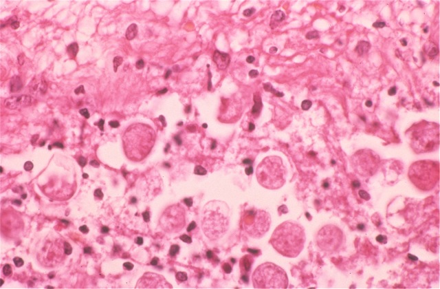
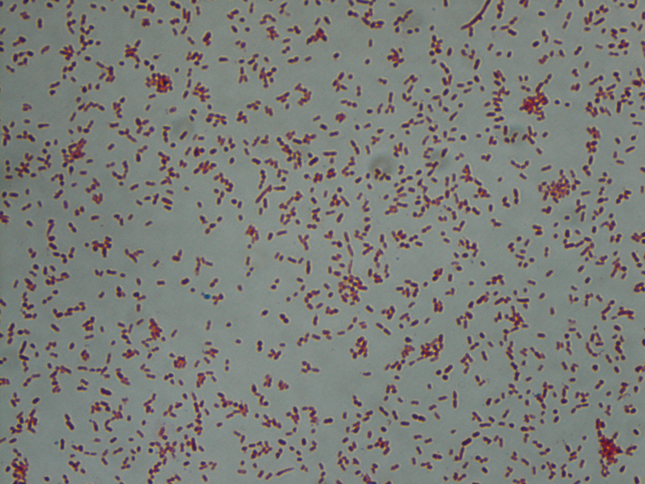
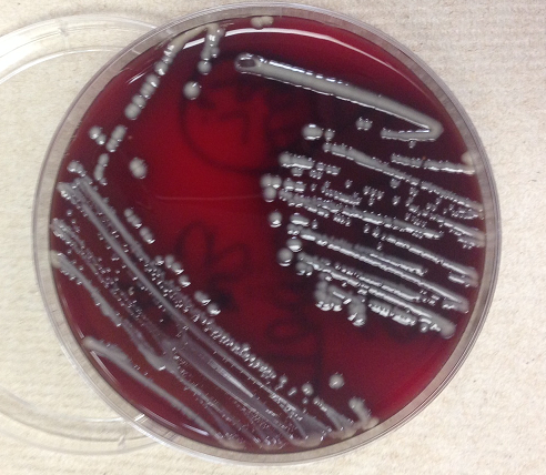
A 38 year old man with history of secondary progressive multiple sclerosis complicated by neurogenic bladder requiring intermittent straight catheterization presented with fevers, chills, and weakness. Clinical evaluation revealed hypotension and leukocytosis concerning for sepsis. His urinalysis was positive for 3+ blood, 1+ leukocyte esterase and nitrites. Urine was sent to our laboratory for culture and grew bacteria with the below gram stain and colony morphology.

Gram stain showing gram negative bacilli.
Sheep blood agar growing nonhemolytic grey-yellow mucoid bacterial colonies.
MacConkey agar growing mucoid lactose fermenting (pink) bacterial colonies.


Laboratory identification:

Klebsiella and Raoultella both are gram negative bacilli that form lactose fermenting, yellow, mucoid bacterial colonies. The bacterial colonies appear mucoid because of the bacteria’s polysaccharide capsule. Distinguishing these two types of bacteria requires molecular analysis as their morphology can be identical. In our initial identification process, only one type of bacteria was detected on the urine culture. Given the gram stain and colony morphology, our differential included Klebsiella, Enterobacter, and Roaultella. The bacteria was verified as Roaultella species using mass spectrometry. However, our laboratory also received two sets of blood cultures which showed the same morphology as above but the bacteria was identified as K. oxytoca. This prompted a review of the urine culture in which a subtle second bacterial colony morphology was seen and confirmed as K. oxytoca.

Discussion:

In summary, our patient had a urinary tract infection caused by Roaultella and Klebsiella which was the source of the patient’s sepsis. Only the Klebsiella was detected in his blood stream. Raoultella and Klebsiella are both gram negative, oxidase negative, non-motile, capsulated, facultative anaerobic bacilli within the Enterobacteriaceae family. Raoultella was initially classified within the Klebsiella genus, but reclassified based on comparative analysis of the 16S rRNA gene and rpoB gene which encodes the β subunit of bacterial RNA polymerase. Members of the Raoultella genus include R. electrica, R. terrigena, R. planticola, and R. ornithinolytica and are found in the environment, specifically in plants, soil, and water. Raoultella species are rare in human disease but have been documented to cause bacteremia, urinary tract infection, conjunctivitis and cholecystitis.

Klebsiella causes human disease much more frequently than Roaultella. Klebsiella are part of human oropharynx and gastrointestinal flora that act as opportunistic pathogens. Mode of transmission may occur endogenously or through person to person spread, hence nosocomial infections are common. K. oxytoca and K. pneumoniae are the most frequently implicated species in human disease and may cause a wide spectrum of infections such as urinary tract infections, respiratory infections, enteritis, meningitis and bacteremia.

For our patient, we reported antibiotic sensitivities that were susceptible to both types of bacteria. The patient was treated with ceftriaxone and then transitioned to cefpodoxime with resolution of his urosepsis. In general, Raoultella’s susceptibility to antimicrobial agents are not well studied and should be based on the antibiotic sensitivities of each strain. In contrast, Klebsiella is well documented to have increasing antimicrobial resistance. Most clinical strains are resistant to ampicillin, carbenicillin and ticarcillin as well as extended spectrum beta-lactam drugs. Resistance to beta-lactam drugs is the result of Klebsiella bacteria that harbor plasmids which produce beta-lactamase enzymes.


-Jill Miller, MD is a 2nd year anatomic and clinical pathology resident at the University of Vermont Medical Center.

Wojewoda-small

-Christi Wojewoda, MD, is the Director of Clinical Microbiology at the University of Vermont Medical Center and an Assistant Professor at the University of Vermont.


Using Evolution to Thwart Resistance

The very act of using antibiotics contributes to antibiotic resistance. Bacteria are exposed to an antimicrobial agent and develop genetic strategies to survive repeated exposures. But what if using antibiotics in a certain sequence could revert resistant strains to the wild type? Researchers from California and Washington DC tested that theory and discovered some promising results.

You can read the PLOS ONE study and the Scientific American article to learn more.

Biomarkers of Renal Disease

When most laboratory professionals think of tests for renal disease, we think of creatinine and blood urea nitrogen (BUN). These two tests have been considered renal function tests for many years (creatinine for over 100 years), and yet neither is a very good marker of early damage to the kidneys.

Creatinine is a biomarker that really needs individual rather than population-based reference intervals. Each person has a range of creatinine values that are “normal” for them, and that individual range is generally much narrower than the population reference interval. Because the reference interval for creatinine is fairly broad, a person can lose 25 – 50% of their renal function before their creatinine rises out of the reference interval for “normal.” Thus creatinine does not detect early renal damage. BUN is also not great for detecting early acute renal damage. BUN concentrations rise when the kidneys sustain damage, but a rise in BUN is not specific to kidney damage. Other causes can elevate BUN as well, such as starvation and increased protein breakdown or intake. In general, BUN and creatinine provide the most useful information in conjunction with each other, and for trending when significant damage to the kidneys has already occurred.

For these reasons, there is a continuous search for better markers of renal damage, especially markers that indicate early renal damage, when perhaps something can be done to reverse it. Cystatin C is another biomarker that is being increasingly used to assess renal damage, and originally was hoped might outperform creatinine in detecting early renal damage. Cystatin C is a small protein which is freely filtered by the glomerulus and doesn’t have many of the drawbacks of creatinine, such as creatinine’s relationship to muscle mass. Unfortunately, although many studies have been done, cystatin C has not been shown to be better than creatinine at indicating early renal damage, and is considered a renal biomarker with uses similar to creatinine and BUN.

Currently, protein or albumin in the urine, and especially very small amounts of protein/albumin in the urine, is probably the earliest indicator of renal damage that is available in the US. Very small amounts of albumin in the urine, what has been called microalbuminuria, is one of the earliest indicators of renal disease.

A new biomarker for early acute renal damage that is gaining the most widespread acceptance is NGAL. Neutrophil gelatinase-associated lipocalin (NGAL) is being extensively studied and has been shown to detect early, acute kidney injury (AKI). In addition, NGAL levels have been reported to be associated with the amount and severity of renal damage. Already in use clinically in Europe, tests for this biomarker are currently working their way through the FDA in the US.

Some other new biomarkers for AKI that are being studied, but are not progressing toward general usage as quickly as NGAL include kidney injury molecule 1 (KIM-1), β-trace protein, liver-type fatty acid-binding protein (L-FABP) and interleukin-18. These are all proteins that appear to be up-regulated in response to AKI. Studies are on-going to see which of these biomarkers may be useful for detecting early AKI and for differentiating between types and causes of AKI. In addition, for all these new kidney biomarkers, studies are needed on the biomarkers’ efficacy in helping with clinical decision-making regarding treatment options and outcomes. It will be interesting to see if any of them become clinically useful tests for the detection of early acute renal damage.

-Patti Jones PhD, DABCC, FACB, is the Clinical Director of the Chemistry and Metabolic Disease Laboratories at Children’s Medical Center in Dallas, TX and a Professor of Pathology at University of Texas Southwestern Medical Center in Dallas.

Ebola and Stress on Health Care Professionals

A recent letter to the editor of CDC’s Emerging Infectious Diseases discusses the psychological stress of caring for an Ebola patient. The authors wondered if the caregivers of patients with Ebola experienced more stress than other providers. You can read the small study–and the surprising results–here.

Histoplasmosis in Montana

NPR did a piece on the recent cases of Histoplasmosis in Montana. Histoplasma is endemic in the Midwest–specifically, the Mississippi and Ohio river valleys–and so this is a bit unusual. If you’re a laboratory professional working in a microbiology department in the western states, brush up on the particulars of this fungus. You can start by reading the NPR article.

Microbiology Case Study–35-Year-Old with a Puncture Wound

Case History:

A 35 year old healthy man presented with a puncture wound of the left hand. He was bit by his neighbor’s dog. Fluid expressed from the wound was sent to the microbiology laboratory with the following gram stain and colony morphology.

past1
Gram stain showing small gram negative coccobacilli.
Sheep blood agar with smooth, gray, non-hemolytic bacterial colonies.


Laboratory Identification:

Bacterial colonies, as shown above, grew on sheep blood and chocolate agars. No growth was present on CNA or MacConkey agars which in combination with the gram stain indicated a type of fastidious gram negative bacteria. Additionally, the bacterial colonies were oxidase positive, catalase positive and indole positive. These findings were consistent with Pasteurella species and confirmed to be P. multocida by mass spectrometry.

Discussion:

Pasteurella species are nonmotile, gram-negative, facultative anaerobic coccobacilli or rods. P. multocida is the most common species involved in human disease. P. multocida is part of the normal oropharyngeal flora in multiple animals including cats, dogs, cattle, horses, rodents and other animals. This bacteria has been reported in up to 70-90% cats and 40-60% of dogs. Humans who have frequent exposure to animals may also harbor P. multocida as part of their normal flora.

Pasteurella are opportunistic pathogens that require mechanical disruption of host barriers. The vast majority of P. multicida related diseases are wound infections and/or cellulitis as a result of a cat bite or scratch. Transmission of Pasteurella also occurs in partially healing wound or regions of poor skin integrity that are licked by a cat or dog. Pasteurella infections may be complicated by septic arthritis and osteomyelitis if wounds are deep and inoculate the underlying soft tissue. Patients who are immunocompromised may develop bacteremia with more widespread infections. The majority of Pasteurella species are susceptible to penicillin, cephalosporins and tetracycline. Our laboratory does not report antibiotic sensitivities for P. multocida for a few reasons: Pasteurella is rarely resistant to penicillin, and bite wounds are generally mixed infections.


-Jill Miller, MD is a 2nd year anatomic and clinical pathology resident at the University of Vermont Medical Center.

Wojewoda-small

-Christi Wojewoda, MD, is the Director of Clinical Microbiology at the University of Vermont Medical Center and an Assistant Professor at the University of Vermont.

Smartphone Diagnostics

Smartphone diagnostics–laboratory tests performed by smartphones–have been popping up lately as a way to bring laboratories to low resource settings or make diagnostics quicker and easier everywhere. These include diagnosing eye disease, diabetes, or kidney damage, ELIZA tests for HIV and syphilis, and bloodborne parasitic infections.

The latest entry in the handheld-laboratory game is a test for the Loa loa parasite, also known as the African eye worm. Traditionally, this parasite can diagnosed with examining a stained blood smear for microfilariae. This, of course, requires reagents, equipment, and time. However, in order to treat patients with onchocerciasis or lymphatic filariasis, it’s imperative to know if they have a concomitant infection with Loa loa. A co-infection can mean serious side effects for the patient once drugs are administered. Using a smartphone attachment and the phone’s camera, a sample can be examined in less than three minutes.

You can read more about this test in this Washington Post article and this abstract.

Swails

Kelly Swails, MT(ASCP), is a laboratory professional, recovering microbiologist, and web editor for Lab Medicine.

Should We Do It Here?

How do you decide whether to bring a test in-house or to send it out (or continue sending it out) to a reference lab? It’s not an easy decision, and it should be one based on as many facts as you can gather. The bullet points below are the considerations I employ for making this decision.

  • Cost: This is almost always the first consideration. Compare what the test currently costs to send out versus what it will cost to run in-house. If it costs more to run in-house than to send out, then there had better be some pretty over-riding reasons for doing it on-site. There are many items that contribute to the cost to run a test including:
    • Basic bottom line cost of the test – i.e. reagents, kit, etc
    • Technologist time – actual time to run the test, and to report the results if it’s not auto-verified
    • Supplies – test tubes, pipets, controls, calibrators, proficiency testing samples, etc.
    • Cost of instrumentation – determine whether a new instrument is needed or whether the test can go on an existing instrument. Either way, depreciation and service contract costs must be considered.
    • Development – for bringing in a newly released, FDA-approved assay on a current platform available in-house, this cost may be minimal, especially if the company brings it in and verifies its performance. For a laboratory developed test, this aspect of cost may be extensive and needs to be considered.
  • Test volume: The number of these tests that are performed will figure into all the rest of the parameters, including the cost of running it and the tech time involved. In addition, the test volume should be high enough to be able to maintain competency in the techs performing it, as well as to be able to keep in-date reagents on hand and instrument maintenance up to date.
  • Turnaround time (TAT): This is often a big factor in the decision to bring a test in-house. Doctors may request that tests be performed in-house to allow for better patient care. For example drug dosing decisions are not optimal if a drug result takes four days to return.
  • Workflow/tech time: How the new test will fit into the current workflow needs to be considered. Will it require additional technologist time or re-shuffling of coverage of other tests? Will it be a random access test or run in batches a few times a week and how will it impact staffing?
  • Patient care: Occasionally the reason for bringing a test in-house may be related to providing the best patient care, despite costs analysis and impact on the lab workflow and staff. An example is pentobarbital. We brought this test in-house even though we perform less than 50 of these a year because results were needed to make decisions related to continuing life support of not. Under these conditions, a 3 to 4 day TAT is not acceptable.

When all these costs are considered, think about your return on investment (ROI). Determine if you will save your institution money, and if so, over what time period. Sometimes it’s not an immediately obvious ROI, but over time you will save money. Sometimes the ROI is actually improved patient care rather than monetary savings. And sometimes the ROI is simply improved relations with the doctors and clinical staff, and that’s not a trivial ROI.

-Patti Jones PhD, DABCC, FACB, is the Clinical Director of the Chemistry and Metabolic Disease Laboratories at Children’s Medical Center in Dallas, TX and a Professor of Pathology at University of Texas Southwestern Medical Center in Dallas.

Make the Diagnosis–A 58 Year Old Male with Abdominal Pain

A 58-year-old male presents with abdominal pain and bloody diarrhea which had a gradual onset over the last 3 weeks. He has lost 8 pounds during this period of time. A cecal biopsy is performed, and a representative section is shown here. What is the most likely diagnosis?

amoebiasis1

  1. Amoebiasis
  2. Ascariasis
  3. Candidiasis
  4. Cryptosporidiosis
  5. Giardiasis

The diagnosis in this case is amoebiasis. The most common cause of amebic enterocolitis is Entamoeba histolytica, an organism spread through ingestion of cysts in contaminated food or water. The trophozoite forms are round, and frequently show intracytoplasmic red cells.

Amoeba with intracytoplasmic red cells

Occasionally, the organisms invade other organs, such as the liver, lung, and heart. Most patients may be treated on an outpatient basis, unless there is severe colitis or extraintestinal disease.

The organisms frequently invade through the mucosa and into the submucosa, often with lateral extension.

"Flask-shaped" ulcer in amoebiasis
“Flask-shaped” ulcer in amoebiasis

Occasionally, the organisms invade other organs, such as the liver, lung, and heart. Most patients may be treated on an outpatient basis, unless there is severe colitis or extraintestinal disease.

Krafts

-Kristine Krafts, MD, is an Assistant Professor of Pathology at the University of Minnesota School of Medicine and School of Dentistry and the founder of the educational website Pathology Student.

World TB Day

Did you know? March 24th was World TB Day. The date commemorates the day in 1882 when Dr. Robert Koch announced he’d discovered the cause of Tuberculosis.

Did you know? Every day there are 24,000 new cases of TB diagnosed. This means more than 2 billion people are infected with TB and 1.5 million people die each year (according to the Stop TB Partnership).

Did you know? The number of people diagnosed with multi-drug resistant TB (MDR-TB) has tripled since 2009 (according to the WHO TB fact sheet).

The burden of TB is highest in resource limited countries and in places where the incidence of HIV is highest. TB is the most common opportunistic infection for individuals with HIV/AIDS and is the most common cause of death in HIV/AIDS patients. In resource limited countries diagnosis and consistent treatment is a challenge. Lack of laboratory resources to use for diagnosis means TB diagnosis are often made based on empirical signs and symptoms.

Once a patient is diagnosed (either clinically or based upon a confirmed laboratory test) given the strenuous treatment, it can be difficult to keep people on a consistent treatment plan. Most treatment lasts six to nine months. In rural settings where access to drugs is limited this can be a prohibitive amount of time. Not only does the cost of the drug add up but the opportunity cost involved in missing work to travel to get the drug, the cost of the travel, and the physical strain of the travel all hamper a patient’s ability and commitment to sticking with the full course.   This can and has led to increased prevalence of MDR-TB.

Unfortunately, the ways to prevent the spread of MDR-TB (quick, accurate diagnosis; taking medication exactly as prescribed; avoiding exposure) aren’t easy in resource limited areas. Basic cell phone technology may be one tool helping to make a difference, however. Some researchers and health care providers are experimenting with sending text message reminders to other health care providers and/or patients to take their medication. Multiple studies are currently underway to determine whether this method of communication has been useful in increasing drug regimen adherence and improving the TB cure rate. If this can solve one of the challenges related to TB, it will be a big step in the right direction.


Levy

-Marie Levy spent over five years working at American Society for Clinical Pathology in the Global Outreach department.