Microbiology Case Study: A 2 Week Old Female with Eye Discharge

Case History

A 2 week old African American female presented to the pediatric emergency department (ED) with erythema, swelling and copious mucopurulent discharge from the right eye. One week earlier, her Mom noted similar symptoms in the left eye which spontaneously resolved. Mom denied fever, irritability, lethargy, rash, and respiratory or urinary symptoms. The baby was born at term through a spontaneous vaginal delivery with no complications. Mom received regular prenatal care and all screening tests were negative. The baby received erythromycin eye ointment at birth prior to initial discharge. Complete blood count showed a slight leukocytosis (WBC 15.7 TH/cm2) and cerebral spinal fluid (CSF) values were unremarkable. A complete sepsis work up was performed with blood, CSF, eye swabs and urine sent for bacterial cultures. Given the high suspicion for a sexually transmitted infection, an eye swab was also collected for Neisseria gonorrhoeae and Chlamydia trachomatis polymerase chain reaction (PCR). Herpes simplex virus PCR from the CSF was also performed. The patient was started on IV ampicillin, cefotaxime and oral erythromycin in the ED.

Lab results

neiss1
Image 1. The eye swab showed growth of glistening, grey bacterial colonies on sheep blood and chocolate agars after 48 hours incubation at 35°C in 5% CO2.
neiss2
Image 2. Gram stain of the bacterial colonies showing uniform Gram negative diplococci.

The organism was positive for both catalase and oxidase and identified by matrix-assisted light desorption ionization- time of flight (MALDI-TOF) as Neisseria meningitidis. The health department also confirmed the identification. PCR of the eye swab was negative for Neisseria gonorrhoeae and Chlamydia trachomatis. Bacterial cultures from the blood, CSF and urine were all negative.

Discussion

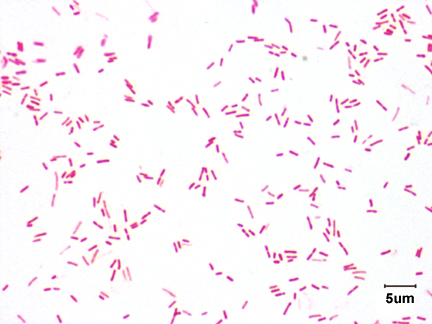
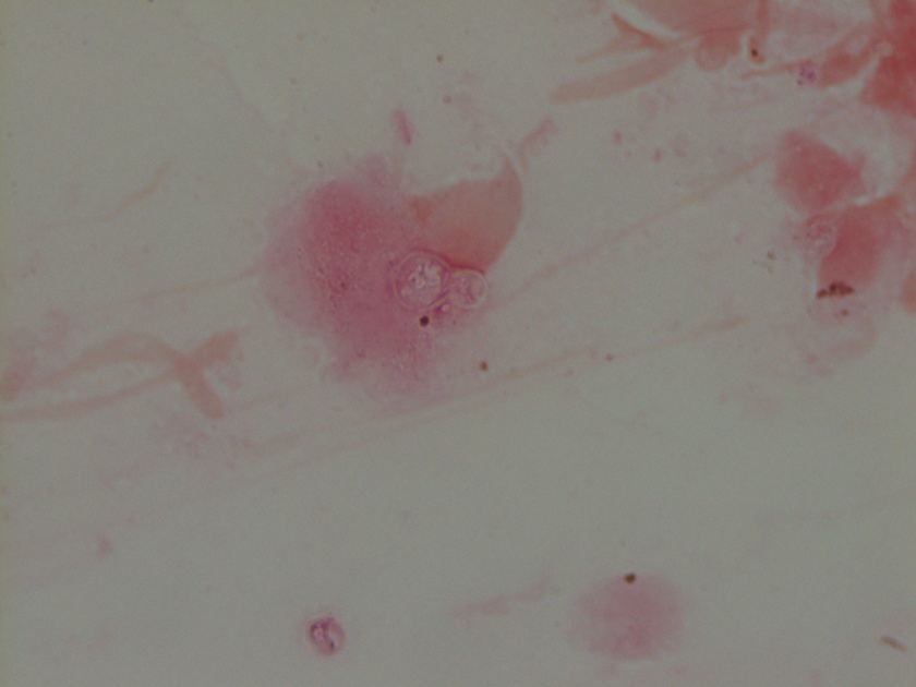
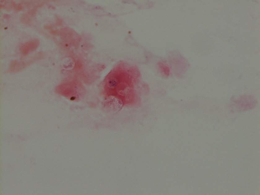
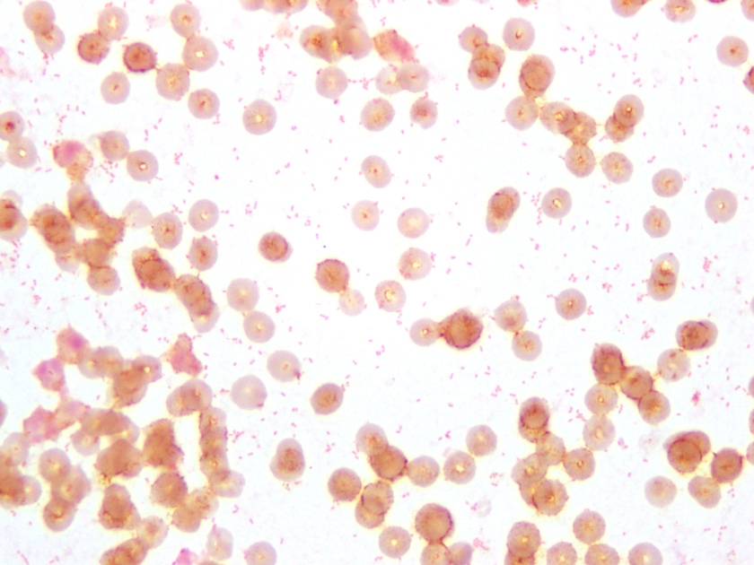
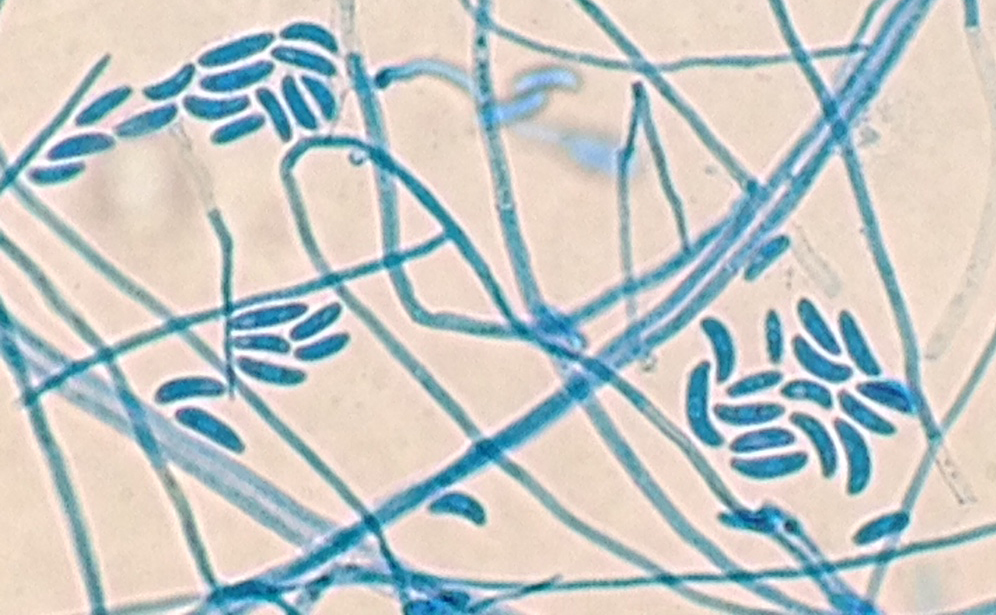
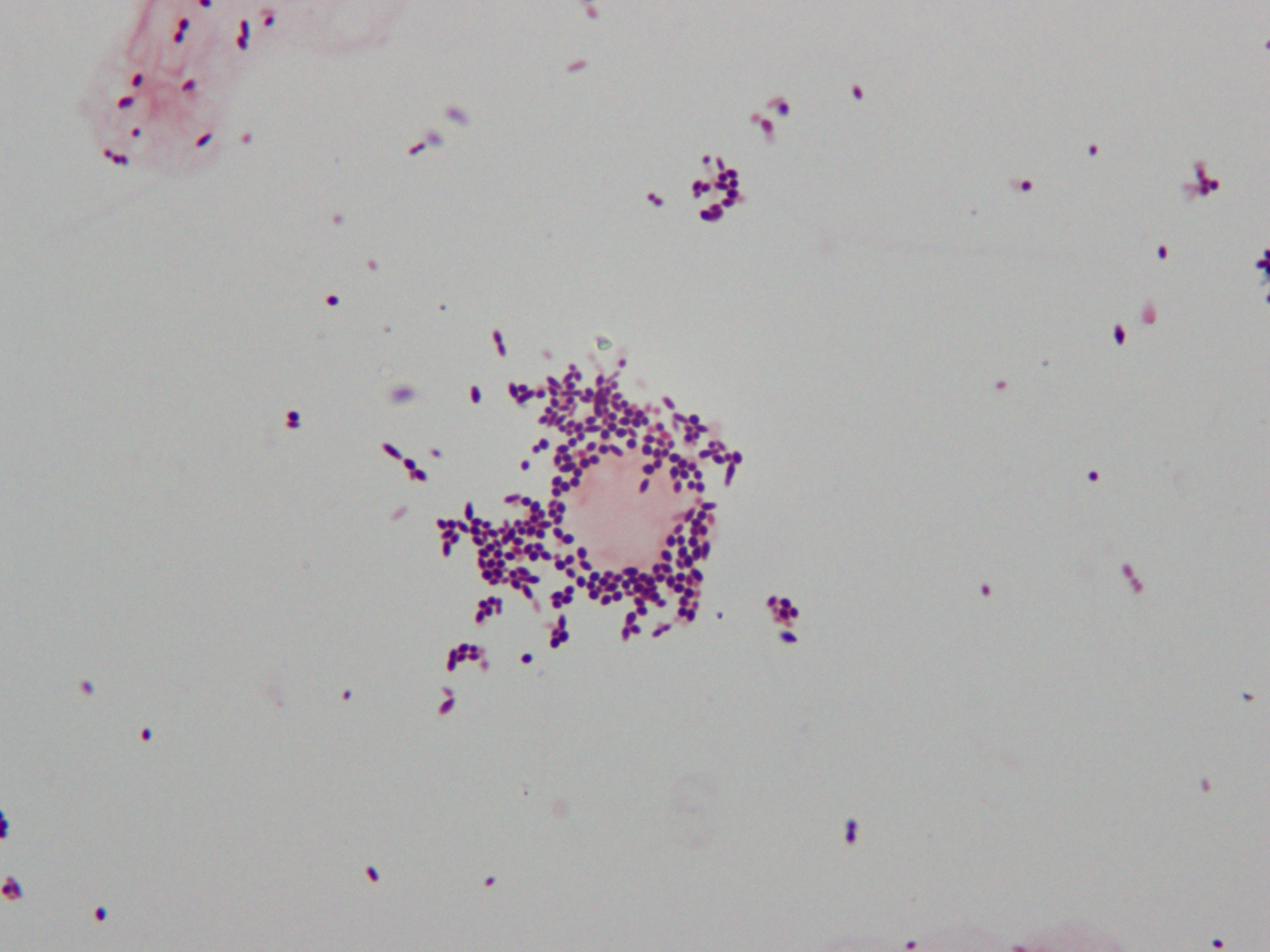
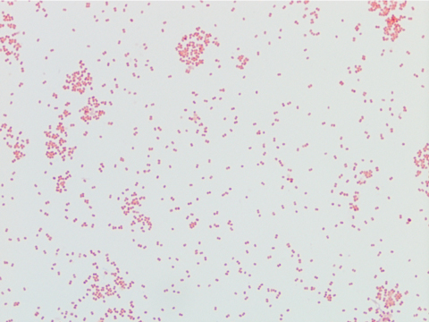
Neisseria meningitidis is an encapsulated Gram negative diplococcus (Image 2) that is usually transmitted through large droplet secretions from the oropharynx from colonized individuals. It can cause invasive meningococcal disease, which can present as meningitis (high fever, stiff neck, and headache), acute sepsis or a combination of both. Waterhouse Friderichsen-syndrome can result in severe dissemination forms of the disease and is characterized by petechial hemorrhages, involvement of the adrenal glands, and disseminated intravascular coagulopathy (DIC). Rarely, N. meningitidis can cause acute bacterial conjunctivitis (1.5 % – 2.5% of cases). Local complications, including corneal ulcers or a more systemic disease, may occur as well.

N. meningitidis produces multiple virulence factors that help cause disease and evade human immune defense mechanisms. The polysaccharide capsule represents the major virulence factor and is also the basis of meningococcal serotyping. Twelve different capsular serotypes can be distinguished, with serotypes A, B, C, W, X, and Y accounting for most invasive disease worldwide. Other virulence factors include pili, which helps the bacteria attach to host surfaces, and IgA protease, an enzyme that cleaves IgA and allows the bacteria to escape the humoral portion of the immune system.

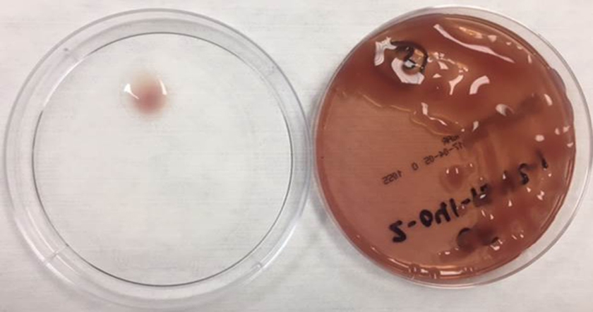
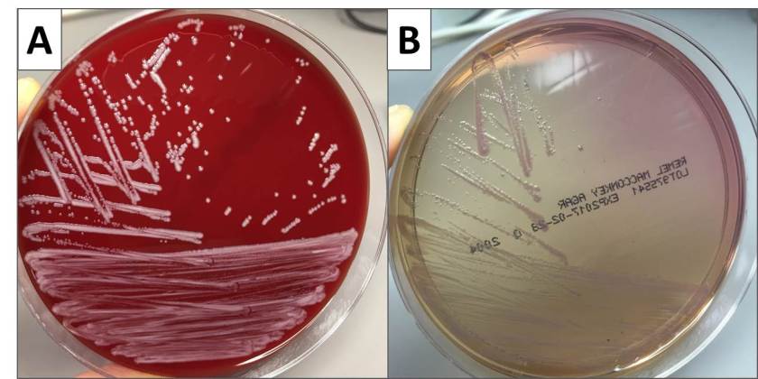
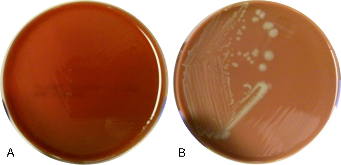
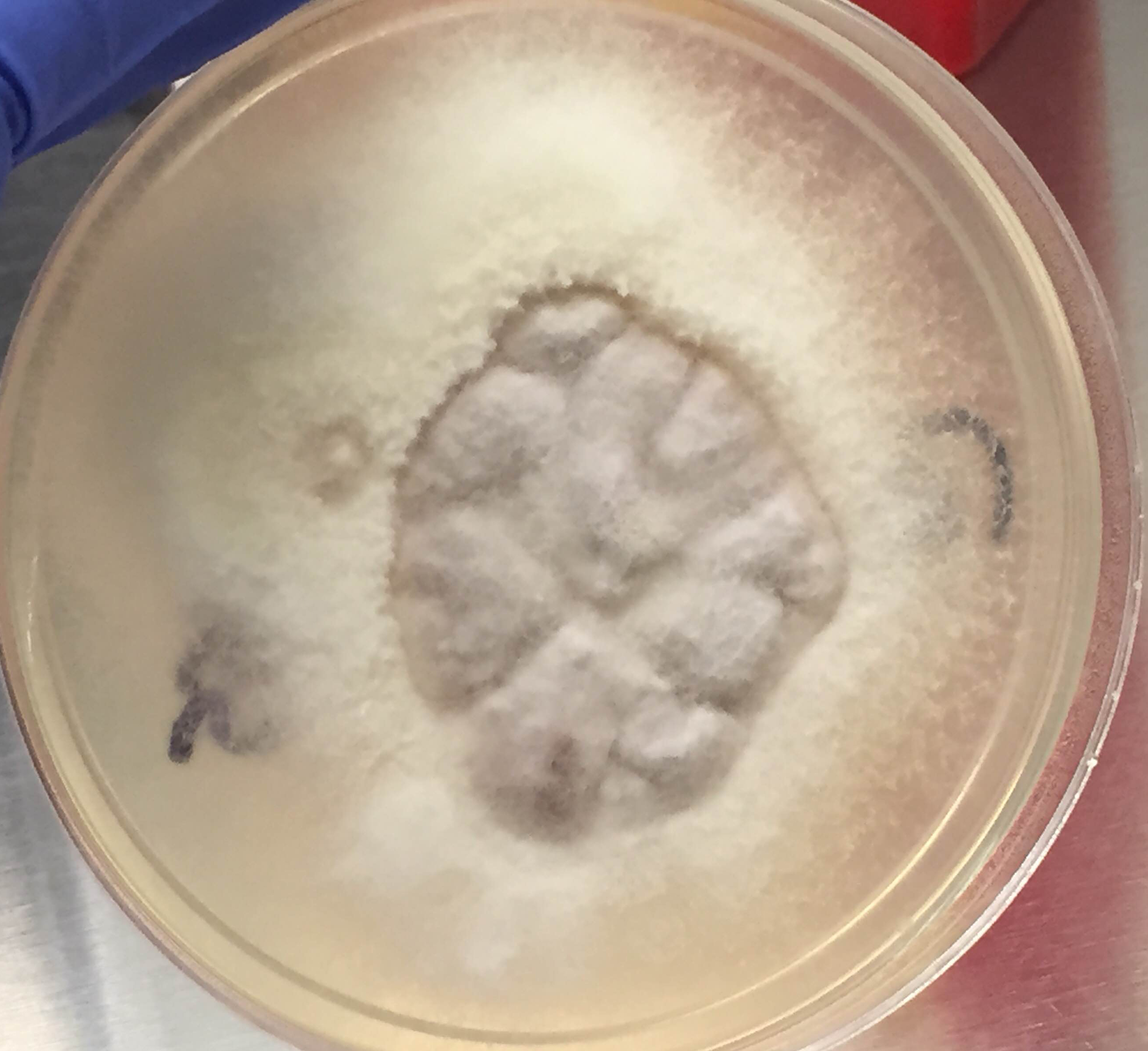
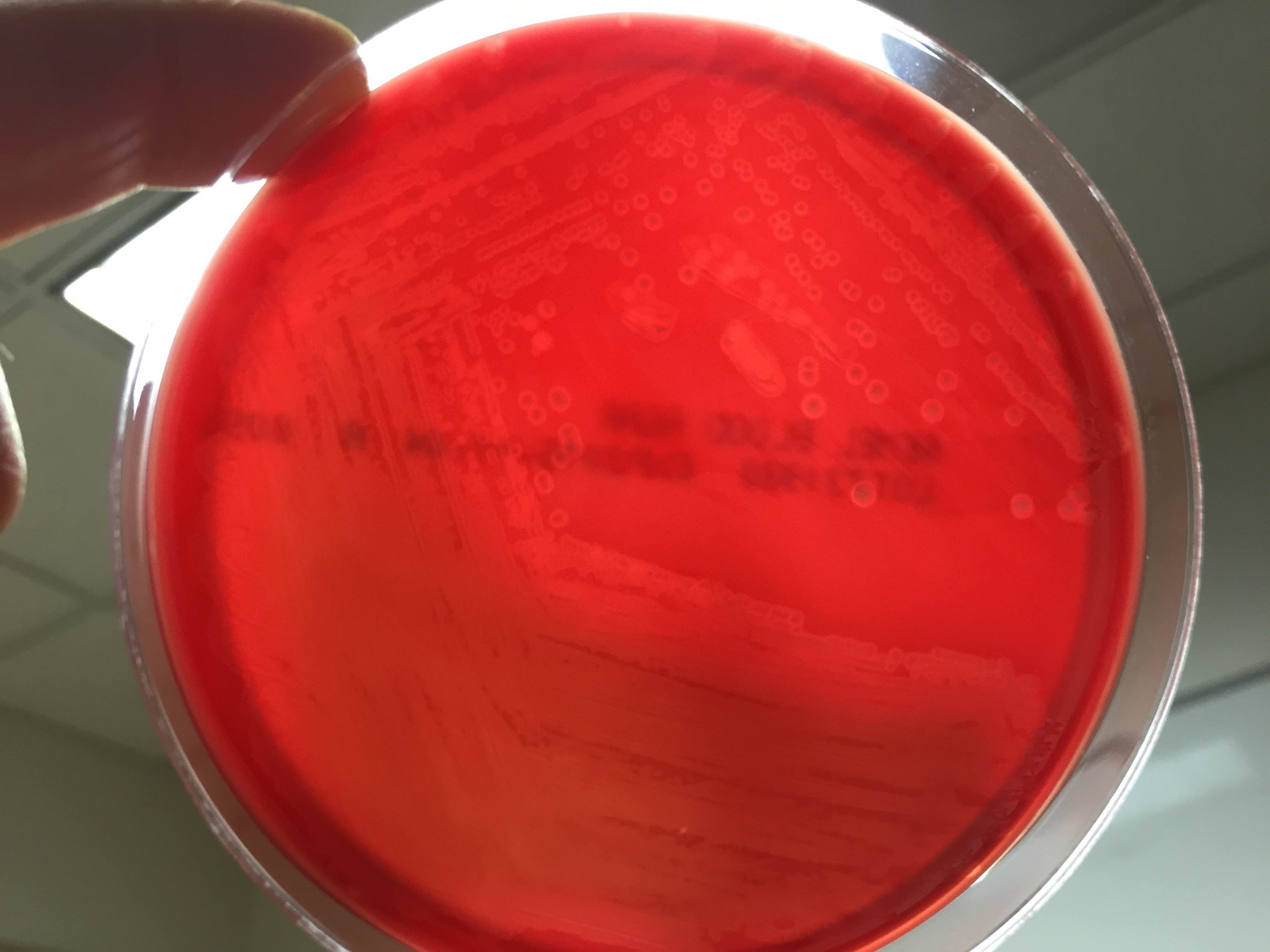
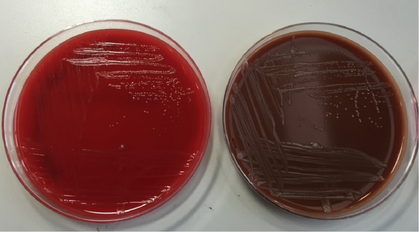
In the laboratory, N. meningitidis grows well on both blood and chocolate agars after 24 hours of incubation (Image 1) and it is positive for both catalase and oxidase. Traditionally, sugar fermentation was used to differentiate Neisseria species from one another. N. meningitidis ferments both glucose and maltose whereas N. gonorrhoeae is only capable of fermenting glucose. Currently, more rapid identification methods (MALDI-TOF, PCR and sequencing) are being increasingly used in most laboratories for a faster and more accurate identification of Neisseria species. The work up of suspected N. meningitidis isolates must be performed using BSL 2 standards, as aerosols created during mobilization from culture plates or performance of biochemical testing has been known to cause invasive disease in laboratory workers.

In general, N. meningitidis is susceptible to penicillin and cefotaxime, but susceptibility testing by disk or gradient diffusion is recommended. Both rifampin and ciprofloxacin can be used for chemoprophylaxis in close contacts of the patient and healthcare & laboratory workers. In addition, a number of meningococcal vaccines are available in the United States (US) and the Centers for Disease Control & Prevention (CDC) recommends vaccinating all adolescents and people at high risk for infection (college students, military recruits, those who had a splenectomy and patients with complement deficiencies). The most common vaccine is a quadrivalent polysaccharide-protein conjugate vaccine which covers serotypes A, C, W and Y. Recently in 2014, the Food and Drug Administration (FDA) approved Trumenba, a vaccine effective against serotype B, which a common serotype causing invasive disease in the US.


AS-small

-Akram Shalaby, MD, is a first year anatomical and clinical pathology resident at the University of Mississippi Medical Center.

Stempak

-Lisa Stempak, MD, is an Assistant Professor of Pathology at the University of Mississippi Medical Center in Jackson, MS. She is certified by the American Board of Pathology in Anatomic and Clinical Pathology as well as Medical Microbiology. She is the director of the Microbiology and Serology Laboratories. Her interests include infectious disease histology, process and quality improvement and resident education.

Microbiology Case Study: A 14 Year Old Cystic Fibrosis Patient

Case History

14 year old cystic fibrosis (CF) patient was admitted to the hospital for a CF exacerbation with a known rhinovirus infection. The patient reported congestion and cough with production of greenish sputum. The following was isolated from a sputum culture.

Laboratory Culture

pseaer1


Image A. MacConkey Agar plate with mucoid Pseudomonas aeruginosa colonies.

pseaer2

Image B. Kirby-Bauer Method of antimicrobial susceptibility.

Discussion

Cystic fibrosis patients have complex polymicrobial respiratory flora. Routine cultures may reveal different bacterial species that can contribute to the difficulty in treating and preventing infections in these patients. Staphylococcus aureus, Haemophilus influenza and Pseudomonas aeruginosa are main contributors to the infections found in CF patients.

In this case, we isolated a mucoid Pseudomonas aeruginosa (Image A). Pseudomonas aeruginosa is an oxidase-positive, gram negative rod-shaped bacterium that is commonly found in the environment. It is considered a severe and frequent pathogen in patients with cystic fibrosis. In chronic infections, it is thought that P. aeruginosa can undergo a “mucoid switch” where the bacterium can acquire mutations that lead to the mucoid phenotype. The phenotype is so impressive that excess polysaccharide will often drip onto the lid of the plate when stored upside down during incubation.

One of the key features of mucoid strains of P. aeruginosa is their ability to form biofilms. Biofilms consist of a matrix of polysaccharide, protein and DNA. This provides not only a protective barrier from antibiotics and the immune system, but also may contribute to the growth of other bacteria within the microenvironment.

With all the excess polysaccharide, it can be difficult to standardize the inoculum of mucoid isolates of P. aeruginosa which is an essential starting point for the microbroth dilution method of antimicrobial susceptibility testing. For this reason, susceptibility testing of mucoid isolates is often performed by Kirby-Bauer (KB) disk diffusion method. The KB method is a test of antimicrobial susceptibility that is based on the zone of inhibition surrounding disks that contain antimicrobial drugs (Image B). The strain of mucoid P. aeruginosa isolated in this case was found to be susceptible to aztreonam, ceftazidime, piperacillin/tazobactam and resistant to amikacin, cefepime, ciprofloxacin, gentamicin, levofloxacin, meropenem and tobramycin, by the Kirby Bauer method.

Case Follow Up

The patient was ultimately treated with IV piperacillin/tazobactam, as well as with chest physical therapy and hypertonic saline inhalation. They clinically improved and were ultimately discharged home after a 2 week hospital stay.

References

The mucoid switch in Pseudomonas aeruginosa represses quorum sensing systems and leads to complex changes to stationary phase virulence factor regulation. Ben Ryall, Marta Carrara, James EA Zlosnik, Volker Behrends, Xiaoyun Lee, Zhen Wong, Kathryn E. Lougheed, Huw D. Williams. PLOS ONE, May 2014, Vol. 9, Iss. 5, Pages 1-11.

Pseudomonas aeruginosa biofilms in cystic fibrosis. Niels Høiby, Oana Ciofu, and Thomas Bjarnsholt. Future Microbiology, November 2010, Vol. 5, No. 11, Pages 1663-1674.

Insights into Cystic Fibrosis Polymicrobial Consortia: The Role of Species Interactions in Biofilm Development, Phenotype, and Response to In-Use Antibiotics. Magalhaes AP, Lopes SP, Pereira MO. Frontiers in Microbiology, January 13, 2017, Vol. 7, Article 2146.

Koneman’s Color Atlas and Textbook of Diagnostic Microbiology. Gary W. Procop, Deirdre L. Church, Geraldine S. Hall, William M. Janda, Elmer W. Koneman, Paul C. Schreckenberger; Gail L. Woods. Seventh Edition. 2017. Pages 343, 1110.


mw

-Megan B. Wachsmann, MD, MSCS, is a 4th year Anatomic and Clinical Pathology Resident and Chief Resident at UT Southwestern Medical Center.

Erin McElvania TeKippe, PhD, D(ABMM), is the Director of Clinical Microbiology at Children’s Medical Center in Dallas Texas and an Assistant Professor of Pathology and Pediatrics at University of Texas Southwestern Medical Center.

Microbiology Case Study: A 10 Year Old Girl with New Onset Fever

A 10 year old girl presented to the ED with several month history of worsening CNS symptoms, including gait instability, left-sided weakness, nausea, vomiting, several witnessed seizure events and loss of consciousness. MRI revealed right frontal mass measuring 7.0 cm in greatest dimension with midline shift. The patient underwent a right-sided craniotomy with tumor resection and was closely followed in the PICU. On post-op day 3, the patient appeared pale and lethargic with a temperature of 39.9 C. Blood cultures were collected and the patient empirically started on ceftriaxone and vancomycin for meningitis coverage. Due to worsening fever and altered mental status, a lumbar puncture was performed with the following results:

  • Opening pressure: 10.5 cm
  • Appearance: slightly cloudy
  • Cells: 91% PMN
  • Gram negative rods were identified on gram smear


OLYMPUS DIGITAL CAMERA
Gram stain shows gram negative rods. Photo credit: Dr. Gary Kaiser.

pseudostut2

Photo credit: University Medical Center of Rotterdam, Dept. of Microbiology and Infectious Disease

Discussion

Pseudomonas stutzeri is an aerobic gram negative bacilli (GNB) with a single polar flagellum that is both catalase and oxidase positive. Several defining features help distinguish P. stutzeri from other Pseudomonas species, including its nutritional versatility (utilizes starch, maltose, and ethylene glycol), its inability to produce fluorescent pigment and its “coral-like” appearance on growth medium.  There are no known virulence factors for P. stutzeri. Hospital distribution rates of P. stutzeri are approximately 1-2% of Pseudomonas spp. Nosocomial infection via medical devices or solutions is the most common route of infection. P. stutzeri isolates are sensitive to beta lactam antibiotics—meropenem is commonly used.


-Christina Litsakos is a Pathology Student Fellow at University of Vermont Medical Center.

Wojewoda-small

-Christi Wojewoda, MD, is the Director of Clinical Microbiology at the University of Vermont Medical Center and an Assistant Professor at the University of Vermont.

Microbiology Case Study: A 14 Year Old Boy with Cystic Fibrosis

Case History

A 14 year-old-boy with Cystic Fibrosis had a respiratory culture collected at his routine clinic visit. It grew abundant mixed respiratory flora, and rare Gram-negative coccobacilli. This organism grew as non-lactose fermenting colonies on MacConkey agar (Figure 1) in approximately 36 hours and was oxidase and catalase positive. The isolate was identified by MALDI-TOF MS with a score of 2.39, which is acceptable for species-level identification.

bord1

Discussion

Our isolate was identified as Bordetella bronchiseptica. Bordetella spp. are small Gram-negative rods that often appear as coccobacilli. Like other Bordetella spp., our isolate was catalase positive. Oxidase results vary across the genus, but B. bronchiseptica is oxidase positive. Some Bordetella spp. including B. pertussis and B. parapertussis, are very sensitive to metabolites and toxic substances found in many types of microbiological media. For the best chance of recovering these fastidious Bordetella spp. in culture, specialized agar such as Regan-Lowe, Bordet-Gengou, or Stainer-Scholte medium along with extended incubation periods are used. Due to the difficulty of culturing B. pertussis and B. parapertussis, these days culture is only performed at large reference laboratories or public health facilities. Testing by PCR is the current clinical practice and has greatly improved the sensitivity of detection for these fastidious organisms. In contrast, other Bordetella spp. including B. bronchiseptica are routinely recovered in culture using standard laboratory methods.

The most clinically relevant Bordetella spp. in humans are B. pertussis, the infectious agent of whooping cough, and B. parapertussis, which causes a similar illness to B. pertussis but generally symptoms are less severe. B. bronchiseptica causes respiratory infections in many animals including cats and dogs, and is the infectious agent of kennel cough.  B. bronchiseptica can cause respiratory infection in humans who acquire the bacterium primarily from their infected pets. Human infection is rare, and most likely to occur in immunocompromised patients such as those with poorly controlled HIV or Cystic Fibrosis.

B. bronchiseptica produces a β-lactamase making the organism resistant to penicillin and many cephalosporins. Most strains are resistant to trimethoprim-sulfamethoxazole as well. In contrast, strains of B. bronchiseptica are generally susceptible to β-lactam/ β-lactamase inhibitor combinations, quinolones, aminoglycosides, and tetracycline.

Our patient was not having an exacerbation at the time of specimen collection, so he continues to do well. We expect to find B. bronchiseptica in his future sputum specimens, but the pathogenicity of B. bronchiseptica in such a low amount compared to respiratory flora is unclear.

Erin McElvania TeKippe, PhD, D(ABMM), is the Director of Clinical Microbiology at Children’s Medical Center in Dallas Texas and an Assistant Professor of Pathology and Pediatrics at University of Texas Southwestern Medical Center.

Microbiology Case Study: A 3 Year Old Girl with Abdominal Pain and Fever

Case History

A 3 year old girl initially presented with abdominal pain and fevers. Ultrasound identified a left kidney mass, leading to a left radical nephrectomy and excision of retroperitoneal mass. Pathology showed a Wilms’ tumor, diffuse anaplasia type. Staging uncovered multiple pulmonary metastases and involvement of a supraclavicular lymph node. She received chemotherapy and radiation without regression of disease.  She then presented to the ED and was admitted for neutropenic fever. She was started on broad-spectrum antibiotics. She continued to spike fevers so an antifungal, micafungin, was added.  While admitted, she developed scattered erythematous papules. Infectious disease was consulted and a skin biopsy from the left forearm was obtained.

Laboratory Results

  • Bacterial cultures, blood: negative
  • Fungal cultures, blood: negative
  • Blastomyces urine antigen: negative
  • Skin biopsy: slight epidermal hyperplasia with follicular dilatation, mild vascular ectasia, and focal erythrocyte extravasation. Negative for organisms.
  • Bacterial culture, tissue: no growth.
  • Gram stain: rare budding yeast forms seen
  • Fungal culture, tissue: no growth to date
blasto-1
The gram smear made from skin biopsy tissue for bacterial culture displayed rare broad-based budding yeast forms, consistent with Blastomyces dermatiditis.

blasto-2

The gram smear made from skin biopsy tissue for bacterial culture displayed rare broad-based budding yeast forms, consistent with Blastomyces dermatiditis.


Discussion

Blastomyces dermatitidis is a dimorphic fungus found in moist soil and decomposing matter. It is endemic within the Mississippi and Ohio River valleys as well as the Great Lakes region and Southern United States.  There are reports of infection in Africa and India. The fungus is transmitted by inhalation of as few as 10-100 conidia. Once in the lungs, the spores convert to yeast and multiply. Infection usually results in a flu-like illness with pulmonary involvement 3-15 weeks post-exposure. Hematogenous spread can further result in involvement of the skin, bone, genitourinary tract, and central nervous system.

The gold standard for diagnosis is culture or cytopathology/histology. However, the organism is a slow grower, which can take 2-4 weeks, and may fail to grow in one-third of cases. On culture at room temperatures (25-30°C), the mold form appears wrinkled and waxy and is cream to tan in color. Microscopically, they form septate hyphae with short or long conidiophores bearing small round to pear-shaped conidia (2-10 microns). This arrangement of the mold is described as a “lollipop” appearance. At 35-37°C, the fungus is a yeast (8-10 microns) with classic broad-based budding and double contoured walls.

Antigen testing is available on urine, serum, bronchoalveolar lavage fluid, and CSF. Antigen testing is more rapid, utilizing enzyme immunoassay, but has a lower sensitivity. Antigen testing is most sensitive in patients with isolated pulmonary disease. Serial urine antigen testing can be used to indicate disease regression or relapse.

A real-time PCR assay is available for confirmation of B. dermatitidis. The probe targets the promoter region of the BAD1 gene, which encodes an adhesin molecule and virulence factor. This method can be performed in five hours, but is only available at reference laboratories.

Mild to moderate pulmonary and extrapulmonary blastomycosis can be treated with oral itraconazole. Severe cases, CNS involvement, or infection of immunosuppressed patients, pregnant women, or children require amphotericin B followed by step-down therapy with itraconazole for 6-12 months.

Upon report of the mold on gram smear, micafungin was discontinued and amphotericin B treatment initiated. Her fever and rash resolved. The patient was transitioned to oral itraconazole prior to discharge and will remain on therapy for 12 months.

REFERENCES

  1. https://www.cdc.gov/fungal/diseases/blastomycosis/index.html
  2. Frost HM, Novicki TJ. Blastomyces Antigen Detection for Diagnosis and Management of Blastomycosis. Journal of Clinical Microbiology. 2015;53(11):3660-3662. doi:10.1128/jcm.02352-15.
  3. Sidamonidze K, Peck MK, Perez M, et al. Real-Time PCR Assay for Identification of Blastomyces dermatitidis in Culture and in Tissue. Journal of Clinical Microbiology. 2012;50(5):1783-1786. doi:10.1128/jcm.00310-12.
  4. Chapman SCAW, Dismukes WE, Proia LA, et al. Clinical Practice Guidelines for the Management of Blastomycosis: 2008 Update by the Infectious Diseases Society of America. Clinical Infectious Diseases. 2008;46(12):1801-1812. doi:10.1086/588300.


-Prajesh Adhikari, MD is a 2nd year anatomic and clinical pathology resident at the University of Vermont Medical Center.

Wojewoda-small

-Christi Wojewoda, MD, is the Director of Clinical Microbiology at the University of Vermont Medical Center and an Assistant Professor at the University of Vermont.

Microbiology Case Study: A 4 Day Old with Eye Drainage

A 4 day old infant presented to the ED with copious amounts of left eye drainage. He was born at 38 weeks via vaginal delivery and was discharged home on day 2 of life. On day 3 of life the baby had acute onset of left eye drainage. On day 4 of life the discharge had become so profuse that the mother was cleaning the baby’s eye several times per hour and his eye was red. In the ED, the discharge was collected and sent to the lab for culture. The specimen Gram stain was reported as many polymorphonuclear leukocytes (pmns) and rare gram negative diplococci. Two days later the following growth was noted on chocolate, 5% sheep blood, and Thayer Martin agars (Figure 1).

ngono1

Figure 1. Growth of organism on (A) chocolate, (B) 5% sheep blood, and (C) Thayer Martin agars

Discussion

The organism was identified as Neisseria gonorrhoeae. N. gonorrhoeae are gram negative diplococcic that grow on solid media in 24-48 hours when incubated under aerobic conditions at 35-37°C with 5% CO2. They exhibit more robust growth on chocolate agar, but can grow on 5% sheep blood agar as well (Figure 1A and B). Like most other Neisseria spp., N. gonorrhoeae are oxidase and catalase positive. Species-level identification can be made by testing carbohydrate utilization; N. gonorrhoeae can only utilize glucose while N. meningitidis can utilize glucose and maltose.

To increase the sensitivity of N. gonorrhoeae detection from mucosal (non-sterile) sites, selective media such as Thayer Martin agar is commonly used (Figure 1C). Selective media for N. gonorrhoeae contains colistin, vancomycin, and antifungal agents to suppress gram negative bacteria, gram positive bacteria, and yeast, respectively, along with other inhibitory agents. N. gonorrhoeae are fastidious bacteria that are at great risk for dying in transport. For maximal recovery of N. gonorrhoeae, special transport packs have been developed. Figure 2 is an image of a Thayer Martin transport pack including agar plate, CO2 generating tablet, and plastic bag for transport back to the lab.

ngono2

Figure 2. Components of the Thayer Martin transport system for bedside plating of specimen.

Neisseria spp. reside in the mucosal membranes of animals including humans and most species are considered normal flora of the upper respiratory tract. N. gonorrhoeae is an exception and is always considered a pathogen no matter what amount or from what location it is isolated. N. gonorrhoeae is spread by sexual transmission where it infections the mucosal surfaces of the urethra, cervix, rectum, and pharynx. Infection presents as acute urethritis in men with symptoms of urethral discharge and sometimes dysuria, but infection is asymptomatic in up to 10% of cases. In women, N. gonorrhoeae infection is most often asymptomic. If symptoms are present, they are generally mild and non-specific including increased vaginal discharge, dysuria, and intermenstrual bleeding. Delay in treatment due to lack of recognition of infection can lead to assentation of the bacteria resulting in pelvic inflammatory disease. Rectal and pharyngeal infections occur in both men and women and are generally asymptomatic. N. gonorrhoeae can cause conjunctivitis which leads to corneal ulceration. In adults conjunctivitis is caused by autoinoculation of the eye. In newborns, such as our case patient, it is transmitted from infected mother to child as the baby moves through the birth canal. In the olden days N. gonorrhoeae infection of newborns was a cause of blindness, but now it is standard to administer 1% aqueous solution of silver nitrate or antibiotic ointment containing erythromycin to newborns just after birth to prevent infection.

These days, molecular detection of N. gonorrhoeae from urine is the most common method used to identify N. gonorrhoeae infection in adult males and females. One of the benefits of molecular detection is that it does not require live bacteria for detection. Many reference laboratories and public health clinics have validated their molecular assays for detection of N. gonorrhoeae from rectal and pharyngeal swabs. Traditionally for sites such as eyes, molecular detection was not available and culture was the only means of N. gonorrhoeae detection. This is a rapidly changing area of microbiology as we were able to get our patient’s eye drainage tested by transcription-mediated amplification (APTIMA) at our reference lab and the specimen was positive.

Our patient was treated with cefotaxime and is being followed outpatient by his pediatrician and ophthalmology clinic.

References

  1. MCM, 10th edition
  2. CDC (https://www.cdc.gov/std)


-Erin McElvania TeKippe, PhD, D(ABMM), is the Director of Clinical Microbiology at Children’s Medical Center in Dallas Texas and an Assistant Professor of Pathology and Pediatrics at University of Texas Southwestern Medical Center.

Microbiology Case Study: A 21 Month Old Boy with Diaper Rash and Diarrhea

Case history

The patient was in his otherwise healthy state of being until 3 days prior when he developed non-bloody diarrhea. On the morning of presentation the stool had become bloody. The patient was afebrile, had some reduced intake of food, but drinking fine. Most notably, he periodically stops walking and bends over as if he is in pain. This happened 3-4 times the previous day and these episodes tended to last for about one minute, after which the boy would continue to play. He does not attend daycare and his immunizations are up to date.

Stool culture was sent and a predominant organism was an oxidase negative, lactose-fermenting, Gram-negative rod (Figure 1). The organism was non-sorbitol fermenting based on growth on Sorbitol-MacConkey agar, and grew as mauve colonies on E. coli 0157 screening agar. MUG testing was negative.

O157.png
Figure 1: Subculture of the disease causing organism on (A) MacConkey, (B) Sorbiol-MacConkey, (C) and E. coli O157 screening agars.

Discussion

The isolate was E. coli O157. Isolates of E. coli O157 commonly produce shiga toxins (sxt1 and sxt2) which are responsible for diarrhea, hemorrhagic colitis, and most famously hemolytic-uremic syndrome (HUS). Typical illness starts with non-bloody diarrhea which becomes bloody after 2-3 days due to onset of hemorrhagic colitis. Often severe abdominal pain and low grade fever are present as well. HUS is a serious complication of E. coli O157 infection which results in acute renal dysfunction. HUS most often occurs in children < 5 years of age, of which 15% of those with laboratory confirmation of E. coli O157 developing this complication, compared to 6% in the general population. It is possible for other E. coli to produce shiga toxins, with 1% of HUS is caused by non-E. coli O157 infection.

It is recommended that all patients with suspected HUS should have stool cultured on selective and differential media for detection of E. coli O157 and direct shiga toxin detection should be performed to identify non-E. coli O157 isolates that are producing toxin. E. coli 0157 isolates look exactly the same as non-E. coli O157 normal fecal flora on 5% sheep blood, chocolate, and MacConkey agars. All E. coli ferment lactose on MacConkey agar (Figure 1A). E. coli O157 can be differentiated from other E. coli strains by growth on Sorbitol-MacConkey (SMAC) agar; E. coli O157 is a non-sorbitol fermenter while most other E. coli will ferment sorbitol (Figure 1B). Chromagenic agar for E. coli O157 is another option to screen stool specimens for E. coli O157. E. coli O157 grow mauve colored colonies on this particular agar (BBL CHROMagar O157 , Becton Dickinson) (Figure 1C). A summary of this data can be found in Table 1.

Growth of organisms suspicious for E. coli O157 on any media requires confirmation prior to reporting. Biochemical confirmation tests include E. coli O157 antiserum or latex agglutination and 4-methylumbelliferyl-beta-D-glucuronide (MUG) testing. For latex agglutination or antisera testing, it is essential to test the isolate of interest with the E. coli O157-specific reagent as well as a non-specific control to exclude non-specific binding. Unlike most E. coli strains, E. coli-O157 does not express beta-glucuronidase and is therefore MUG test negative (Table 1).

Table 1. Characteristics of E. coli O157 in comparison to other E. coli strains

Test Non-E. coli O157 E. coli O157
Appearance on MacConkey agar Lactose fermenter Lactose fermenter
Appearance on Sorbitol-MacConky agar Sorbitol fermenter Non-sorbitol fermenter
MUG testing Positive Negative

For direct detection of shiga toxin, there are several commercially available immunoassays available for detection of shiga toxin protein. New on the market are multiplex gastrointestinal panels that can be used for molecular based detection of shiga toxin genes sxt-1 and sxt-2 among a host of other agents of gastrointestional disease.

E. coli O157 is spread via fecal oral route. It can be acquired directly from person to person or indirectly through food and water sources contaminated with fecal matter from infected humans and animals. Classic scenarios are undercooked ground beef, leafy greens, unpasteurized milk and juice, petting zoos, and contaminated drinking water. The incubation period prior to symptoms is 3-4 days (range 1-8 days).

Treatment for E. coli O157 is largely supportive consisting of fluids to prevent dehydration. The role of antibiotics is controversial with some studies suggesting antibiotics increase the risk of developing HUS while others found no association between the their use and increased HUS.

Following our patient’s stool culture result for E. coli O157, he was recalled to the Emergency Department for evaluation. He was still having diarrhea and vomiting, but it was reduced compared to the previous day. The patient was given fluids and sent home without antibiotic treatment and via phone conversation with his mother, his symptoms resolved a few days later.

-Erin McElvania TeKippe, PhD, D(ABMM), is the Director of Clinical Microbiology at Children’s Medical Center in Dallas Texas and an Assistant Professor of Pathology and Pediatrics at University of Texas Southwestern Medical Center.

Microbiology Case Study: 6 Year Old Girl with Headache

A 6 year-old girl with a history of posterior fossa ependymoma presented with a one month history of fever, headaches, vomiting and more recently, neck stiffness. Additional history includes remote tumor resection followed by radiation and chemotherapy resulting in remission, with a residual ventriculoperitoneal shunt (VPS). Her parents reported she was in good health until approximately 1 month prior to presentation and is up to date on her immunizations. She was previously seen by her primary care physician for her symptoms and treated her with amoxicillin for suspected strep throat. Upon admission, she received supportive therapy for her symptoms after she was found to have tumor recurrence on imaging. The patient was scheduled for resection approximately two weeks after discharge and on post-operative day two she developed fever, vomiting and neck stiffness again. At this time, blood cultures were drawn and a lumbar puncture (LP) was performed. Cerebrospinal fluid (CSF) from both the LP and VPS submitted for fluid analysis (Table 1) and culture.

Table 1: Cerebrospinal Fluid Analysis

Spinal Fluid LP VPS
Appearance Clear Clear
Nucleated cells 1075 cells/μL 628 cells/μL
RBC 150 cells/μL 35 cells/μL
Polys 94% 87%
Lymphs 2% 6%
Mono/Macrophage 4% 7%
Glucose 68 mg/dL 13 mg/dL
Protein 69 mg/dL 164 mg/dL
haem1
Figure 1. Gram stain of the pathogen isolated from aerobic blood culture, showing gram-negative coccobacilli, sometimes in pairs. The same organism was seen on the patient’s CSF Gram stain.
haem2
Figure 2. Aerobic blood culture on (A) 5% sheep blood agar plate (BAP), showing no growth and on (B) chocolate agar plate (CAP), showing round, smooth, opaque grey-yellow colonies.


Culture results:

The CSF Gram stain showed rare, paired, Gram-negative diplococci, which could raise suspicion for Neisseria meningitidis, however the typical flattened sides of adjacent bacteria were not observed. Rather, the morphology was more consistent with Gram-negative coccobaccilli, which is better demonstrated on Gram stain of the blood culture (Figure 1). Culture of both the CSF and blood specimens grew fairly large, smooth, round, opaque grey-yellow colonies on CAP, however showed no growth on BAP (Figure 2), suggesting a fastidious organism requiring growth factors. The colonies were both catalase and oxidase positive. The organism was identified as Haemophilus influenzae by MALDI-TOF MS (matrix-assisted laser desorption/ionizations time-of-flight mass spectrometry). This H. influenzae isolate was non-typeable by slide agglutination serotyping performed at the state public health laboratory.

Discussion:

H. influenzae are small, pleomorphic, gram-negative rods or coccobacilli that are non-motile. They are facultative anaerobes that grow best between 35-37°C with 5% CO2. H. influenzae is a fastidious species, requiring hemin (X factor) and nicotinamide-adenine-dinucleotide (NAD/V factor) for growth, which are both available in chocolate agar, but not blood agar. On chocolate agar, the colonies are non-hemolytic, typically large, smooth, round and convex with an opaque, colorless or grey hue. Encapsulated strains, including H. influenzae serotype b (Hib), appear mucoid and are typically small, grey colonies on CAP. Isolates are catalase and oxidase positive. H. influenzae displays the “satellite phenomenon” when grown near Staphylococcus aureus. This occurs when colonies of S. aureus lyse nearby red blood cells releasing hemin and NAD in the media. The presence of extracellular hemin and NAD allow colonies of H. influenzae to grow in the immediate vicinity of S. aureus.

H. influenzae is widely distributed in humans, colonizing the nose and throat and is spread from person-to-person via direct contact or respiratory droplets. Severe infections, including pneumonia, bacteremia and meningitis, affect predominantly infants and children. The American Academy of Pediatrics recommends routine vaccination with the Hib conjugate vaccine for infants aged 2 through 6 months (2 or 3 doses, depending on vaccine product) followed by a booster dose at age 12 through 15 months. Hib is the only serotype preventable by vaccine. Prior to routine vaccination in the US, approximately 20,000 children under the age of 5 were infected with H. influenzae and 3-6% died each year.

References:

1. Ledeboer N, Doern G. 2015. Haemophilus, p 667-684. In Jorgensen J, Pfaller M, Carroll K, Funke G, Landry M, Richter S, Warnock D (ed), Manual of Clinical Microbiology, 11th Edition. ASM Press, Washington, DC.
2. http://www.cdc.gov/meningitis/lab-manual/chpt09-id-characterization-hi.html
3. http://www.cdc.gov/hi-disease/index.html


-Petra Rahaman, MD is a 4th year Anatomic and Clinical Pathology resident at UT Southwestern Medical Center.

-Erin McElvania TeKippe, Ph.D., D(ABMM), is the Director of Clinical Microbiology at Children’s Medical Center in Dallas Texas and an Assistant Professor of Pathology and Pediatrics at University of Texas Southwestern Medical Center.

Microbiology Case Study: Immunocompromised Boy with Skin Nodules

An elementary school aged boy with a history of pre-B cell acute lymphocytic leukemia with a failed bone marrow transplant was transferred to a regional children’s hospital for leukodepletion and participation in an experimental clinical trial. At that time, his CBC was significant for 10% polymorphonuclear cells and 50% blasts. He was subsequently transferred to the ICU in respiratory failure and developed papulonecrotic lesions on his face, trunk, and bilateral legs. Prior to this, he was pancytopenic with no blasts present with cell counts of 100 WBC, hemoglobin 8.3 and 37,000 platelets. His Fungitell assay, which detects (1-3)-β-d-glucan, was positive.

Routine blood culture, fungal culture from the endotracheal tube, and fungal culture from the skin lesion biopsy specimens all had fungal elements on KOH stain. Young growth of a whitish, fluffy mold was present on all cultures within two days. Histopathology on the punch biopsy of a skin lesion on the thigh showed septate hyphae within the dermis, epidermis, and invading the vasculature that was particularly apparent with GMS stain (Figure 1a and 1b). Within a few days, the fungal cultures showed septate hyphae with microconidia using lactophenol cotton blue tape preparation, and shortly thereafter the mold developed into macroconidia with multiple septations taking on canoe-like forms (Figure 2). The white, cotton-like colonies developed a pink tinge (Figure 3). These characteristics allowed for the identification of the growth as Fusarium sp.

Septate hyphae on GMS stained section of the skin punch biopsy.
Microscopic identification of Fusarium by lactophenol cotton blue stain.
Colony of Fusarium growing on inhibitory mold agar (IMA).

Fusarium is an opportunistic hyaline mold with infection most commonly seen in immunocompromised hosts. It can cause keratitis through contamination of contact lenses, penetration due to trauma, or use of immunosuppressive steroid ophthalmic solution. It is increasingly becoming the cause of disseminated infection in neutropenic hosts with a broader spectrum of disease, which includes: skin lesions, fungemia, rhinocerebral involvement and pneumonia. In these cases, without an immune system to fight the infection, mortality is high. Inhalation of airborne conidia, ingestion from water sources or access through mucosal membranes are all potential points of entry.

The colony growth on plated fungal media is rapid, usually maturing within four days. On microscopic examination, Fusarium hyphae are septate, approximately 3-6 microns wide with acute angle branching. Microconidia are small, oval-shaped, and no larger than 4 x 8 microns in size. These can look like Acremonium sp. Macroconidia are canoe- or sickle-shaped with the largest dimension being about 80 microns in length, exhibiting 3-5 septatations.


Jodi Music, MD, is an AP/CP resident at UT Southwestern Medical Center.

-Erin McElvania TeKippe, Ph.D., D(ABMM), is the Director of Clinical Microbiology at Children’s Medical Center in Dallas Texas and an Assistant Professor of Pathology and Pediatrics at University of Texas Southwestern Medical Center.

Microbiology Case Study: An 8-Week-Old Female with Pallor, Vomiting, Fever, and Blue Feet

Clinical History

An 8 week old female was brought to an outside hospital due to pallor, decreased eating over several days, vomiting, fever, and blue color in the feet. The patient had received her two month vaccinations the day prior to presentation. Her past medical history was significant for being born at 32 weeks gestation, followed by an uneventful 4 week NICU stay. At the outside hospital the patient was in respiratory distress, tachycardic, with pallor. She was intubated and transferred to our institution due to concern for an ALTE (apparent life-threatening event). Blood and CSF specimens were drawn. Upon presentation, the patient had a white blood cell count of 19,600/mm3. Her CSF had 63 nucleated cells/mm3 (30% neutrophils, 49% lymphocytes, 11% monocytes), glucose of 23 mg/dL and protein of 212 mg/dL. Blood and CSF cultures were performed with the following results:

Gram stain of cerebral spinal fluid (CSF) specimen showing Gram postive cocci in singles and pairs
Gram stain of cerebral spinal fluid (CSF) specimen showing Gram positive cocci in singles and pairs
Gram stain of positive blood culture broth showing Gram positive cocci in chains
Small zones of beta hemolysis around colonies growing on 5% sheep blood agar

Laboratory Identification

CSF Gram stain was read as many Gram-variable coccobacilli (Image 1) and blood culture Gram stain was resulted as Gram-positive cocci in pairs and chains (Image 2). The Nanosphere Verigene Gram-positive blood culture molecular assay was performed on the positive blood culture broth immediately following Gram staining. CSF and positive blood culture specimens were plated to 5% sheep blood, chocolate, MacConkey, and CNA agars. In >24 hours colonies from both specimens grew a single organism on sheep blood, chocolate, and CNA agars. On blood agar, colonies exhibited a soft zone of b-heomlysis (Image 3). Colony Gram stains showed Gram-positive cocci in chains, catalase testing was negative, and the organism typed in Lancefield antigen group B. Verigene identified the organism directly from the positive blood culture broth as Streptococcus agalactiae (aka Group B Streptococcus or GBS) and MALDI-TOF mass spectrometry confirmed the identification of S. agalactiae.

Discussion

Laboratory Considerations

As our CSF specimen demonstrated, streptococci can be difficult to interpret from specimen Gram stains. Organisms are often are pleomorphic in size and shape and they have a tendency to stain Gram-variable. This lead to the report of Gram-variable coccobacilli on our patient’s CSF culture.

S. agalactiae produces a soft zone of b-heomlysis on sheep blood agar. Unlike Streptococcus pyogenes (aka Group A Streptococcus) which produces a wide zone of b-hemolysis, soft b-hemolysis can often be very subtle, especially with young growth. Soft zones b-hemolysis can best be seen by holding plates up to a light source or my moving a colony out of the way to observe if hemolysis is present underneath. The pattern of S. agalactiae b-hemolysis is very similar to that produced by Listeria monocytogenes. Streptococcal isolates that type as Lancefield Group B, but produce large zones of b-hemolysis can create confusion and are most likely not S. agalactiae, but S. porcinus or S. pseudoporcinus.

Clinical Significance

S. agalactiae is the cause of significant neonatal disease. Early-onset infection presents as systemic infection, respiratory distress, apnea, shock, and pneumonia within the first 24 hours of life (range, 0–6 days). Meningitis is less common in early-onsetS. agalactiae infections, found in just 5-10% of cases. Late-onset disease presents at 3 to 4 weeks of age (range, 7–89 days) as meningitis and/or sepsis with other focal infection. Approximately 50% of survivors of early- or late-onset meningitis have long-term neurologic sequelae.

S. agalactiae colonizes the urogenital or gastrointestinal track of 10-30% of pregnant women. Being born to a S. agalactiae colonized mother is the most significant risk factor for development of disease in neonates. For this reason, women are screened for “Group B Strep” colonization between 35 and 37 weeks of pregnancy. Women found to be colonized receive prophylactic antibiotics immediately prior to delivery to prevent transmission to the child. Since implementation of these practices in 1996, there has been a substantial decline in early-onset S. agalactiae infections. Interestingly, these measures have not affected the incidence of late-onset disease.

Treatment

Until recently S. agalactiae was considered universally susceptible to penicillin. There have now been a few reports of S. agalactiae isolates with increased penicillin MICs due to mutations in the penicillin binding protein Pbp2x. The detection of these isolates is still extremely rare, so much so that S. agalactiae susceptibility testing for penicillin and other b-lactams is not considered necessary at this time. Penicillin and its derivatives are the preferred treatment option.

Patient follow-up

Our patient had a complicated course of late-onset Group B Streptococcal meningitis including multifocal cerebral infarctions and seizures. She was treated with a 28 day course of ampicillin. Blood cultures taken 24 hours after the start of antibiotics were negative and her CSF culture was negative when rechecked 5 days after her presentation. Due to her complicated course, the patient was hospitalized for 4 weeks. Follow-up appointments have shown the patient’s MRI is nearly normal 8 weeks post infection and the patient is doing well, although she is still followed by neurology to assess for long term sequelae.

Our patient’s mother had an unknown Group B Strep carrier status at the time of her birth, as she was born at 32 weeks, which is before routine screening occurs for pregnant women. Either way, the mother’s status would not have affected the patient’s risk for late-onset Group B Streptococcal infection.

References:

  1. Red Book 2015
  2. Journal of Clinical Microbiology, 10th edition

-Erin McElvania TeKippe, Ph.D., D(ABMM), is the Director of Clinical Microbiology at Children’s Medical Center in Dallas Texas and an Assistant Professor of Pathology and Pediatrics at University of Texas Southwestern Medical Center.