Microbiology Case Study: A 21 Year Old With Chronic Kidney Disease

Case History

The patient is a 21 year old male with a history of developmental delay and chronic kidney disease secondary to posterior urethral valves, status post kidney transplant at age 14, who presents for a routine office visit with his pediatric nephrologist. In the past year, he has had chronic antibody-mediated transplant rejection despite immunosuppression. In addition, he drinks 1-1.5 gallons of water daily, self-catheterizes every three hours, and has an indwelling Foley at night. During the office visit, he denies any urinary symptoms, including dysuria, hematuria, cloudy urine, reduced output, or fever. However, given his significant risk factors for urinary tract infection, his provider orders a urinalysis and urine culture.

Laboratory Identification

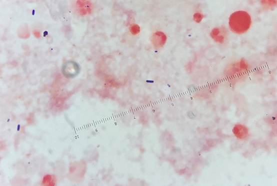
The urine was noted to be cloudy, was positive for nitrites and leukocyte esterase, and had 11-50 white blood cells per high-powered field. 

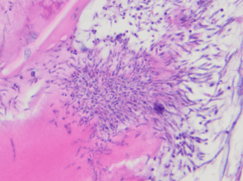
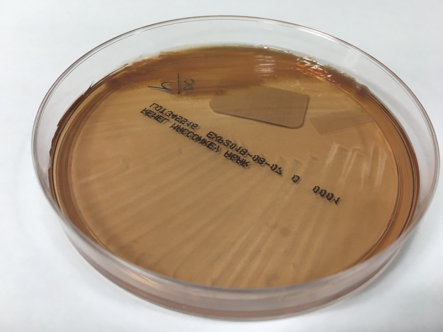
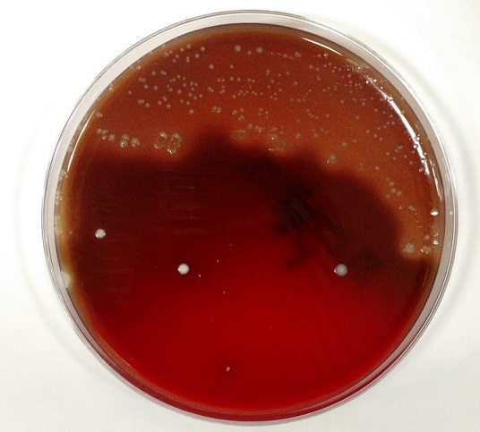
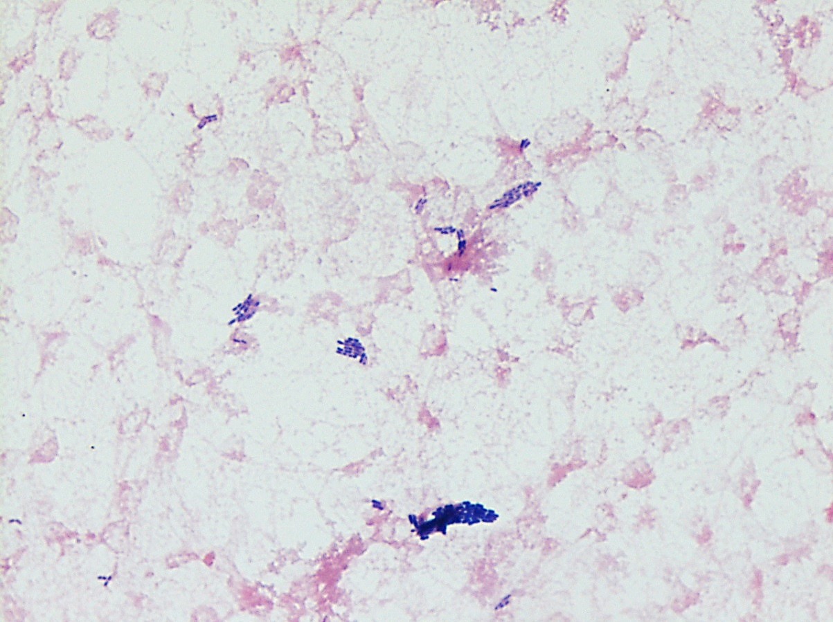
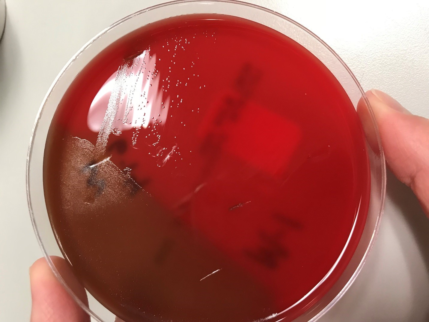
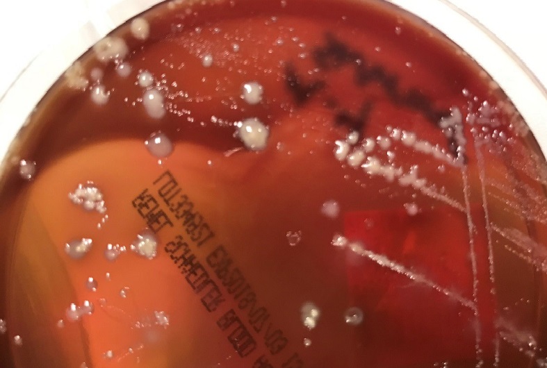
Urine culture demonstrated the growth of two organisms, one of which was identified to be greater than 100,000 CFU of Proteus miribalis, and the second of which grew 10,000-100,000 CFU, was isolated, and is shown below:

Image 1. Appearance of the second organism’s growth on blood agar after the bile solubility test.
Image 2. Gram stain showing gram positive diplococci.

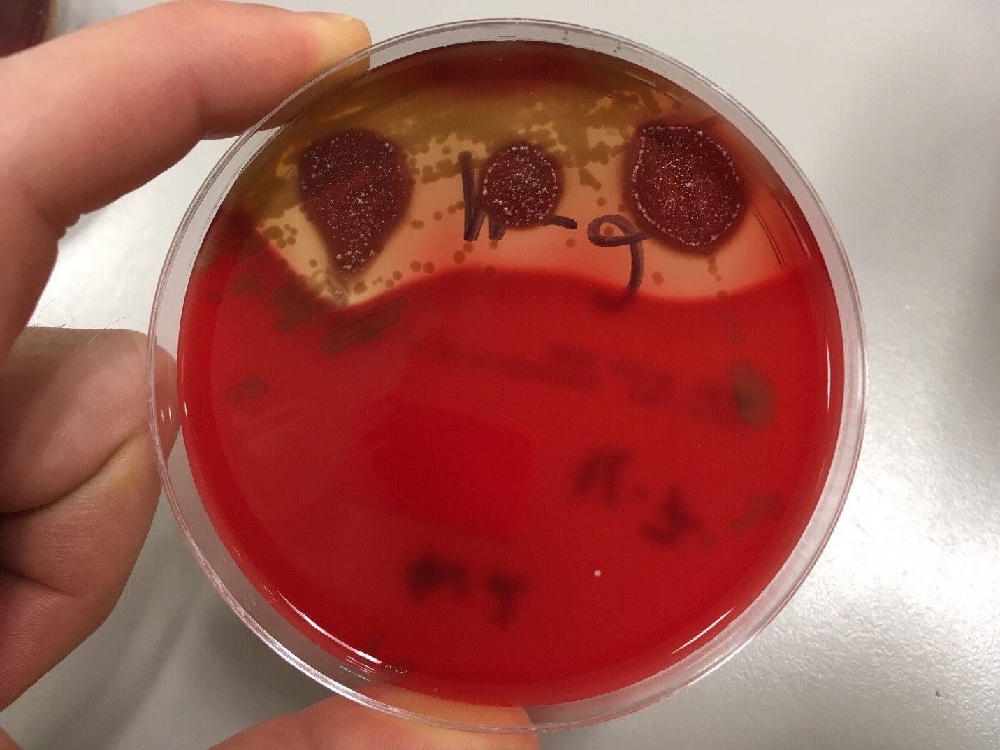
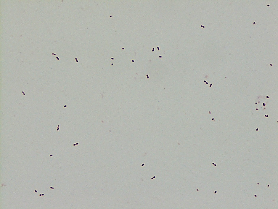
Mass spectrometry by MALDI-TOF confirmed that this second organism is Streptococcus pneumoniae, a bile-soluble gram positive diplococci. 

Discussion

S pneumoniae is implicated in a number of diseases, but it is an uncommon pathogen in the urine. Several case-series and case reports have been published demonstrating a predilection of pathogenic urinary S pneumoniae for pediatric patients with urinary tract abnormalities. In one series, 26 urine cultures from 18 patients were identified as growing S pneumoniae, with CFU counts ranging from 100 to 100,000. Sixteen of the 26 cultures grew only S pneumoniae. Of the 18 patients, only six were adults, eight had had a kidney transplant, and four others had chronic problems with their kidneys (1). In another series of three pediatric cases, one patient had congenital bilateral duplication of the renal collecting system, one had a “congenital imperforate anus (high type 1A) with a rectovesical fistula and grade 4 bilateral vesicoureteral reflux,” and the third had bilateral renal dysplasia (2). Neither case series was able to identify a specific serotype of S pneumoniae responsible for these infections.

As discussed by Choi et al, the altered flow dynamics of the abnormal urinary systems in these patients may be compromising normal host immune clearance mechanisms, thereby increasing the susceptibility to infection (2, 3). However, it is unclear why S pneumoniae infections have a predilection for congenital urinary tract abnormalities, as opposed to all urinary tract abnormalities. Choi et al postulate that some of the gene polymorphisms known to predispose individuals to UTI or pneumococcal infections could be genetically linked to genes responsible for urinary tract abnormalities, thus increasing the probability that an individual with a congenital urinary tract abnormality would have an S pneumoniae urinary tract infection (2,4).

Given the patient’s history and risk factors, the presence of S pneumoniae in his urine was found to be significant. Treatment of both organisms and appropriate follow-up was recommended.

References

  1. Burckhardt, Irene, Jessica Panitz, Mark van der Linden, and Stefan Zimmermann.  “Streptococcus pneumoniae as an agent of urinary tract infections – a laboratory experience from 2010 to 2014 and further characterization of strains.”  Diagnostic Microbiology and Infectious Disease.  2016; 86: 97-101.
  2. Choi, Rihwa, Youngeun Ma, Kyung Sun Park,  Nam Yong Lee, Hee Yeon Cho, and Yae-Jean Kim.  “Streptococcus Pneumoniae as a uropathogen in children with urinary tract abnormalities.”  The Pediatric Infectious Disease Journal.  2013; 32(12): 1386-1388.
  3. Bogaert, D, R de Groot, PWM Hermans.  “Streptococcus pneumoniae colonization: the key to pneumococcal disease.”  The Lancet Infectious Diseases.  2004; 4(3): 144-154.
  4. Yuan, Fang Fang, Katherine Marks, Melanie Wong, Sarah Watson, Ellen de Leon, Peter Bruce McIntyre, John Stephen Sullivan.  “Clinical relevance of TLR2, TLR4, CD14, and Fc gamma RIIA gene polymorphisms in Streptococcus pneumoniae infection.”  Immunology and Cell Biology.  2008; 86(3): 268-270.

-Fritz Eyerer, MD is a first year Anatomic and Clinical Pathology Resident at the University of Vermont Medical Center.

-Christi Wojewoda, MD, is the Director of Clinical Microbiology at the University of Vermont Medical Center and an Associate Professor at the University of Vermont.

Microbiology Case Study: A 44 Year Old Man with Severe Back Pain

Case History

A 44 year old male presented to the emergency department with severe, throbbing back pain in his mid-thoracic spine. He states the pain began a couple weeks ago and noted no recent fevers or night sweats, but does admit to chills. His past medical history is significant for end stage renal disease requiring dialysis, insulin dependent diabetes mellitus, and multiple amputations. On physical examination, there was tenderness to palpation along spine in mid-thoracic region. Lab work showed a normal white blood cell count, C reactive protein of 0.90 mg/dL (0.00 – 0.50 mg/dL), and an erythrocyte sedimentation rate of 60.0 mm/hr  (0.0 -10.0 mm/hr). MRI of the spine was consistent with discitis and osteomyelitis at T7-8 with compression fractures causing spinal stenosis and cord compression. Given the concern for an infection process, blood cultures were collected and interventional radiology performed a bone biopsy. The specimen was sent for bacterial, fungal, and AFB cultures as well as for histology.

Laboratory Identification

Image 1. Discrete, creamy colonies growing on blood agar (left) and Sabouraud dextrose agar (right) after 48 hours of incubation at 35°C.
Image 2. Fragments of bone with focal necrosis, foci of acute and chronic inflammation with clusters of yeast forms and pseudohyphae consistent with a Candida spp. infection.

The organism grew as discrete, creamy colonies growing on blood agar and Sabouraud dextrose agar after 48 hours of incubation at 35°C and resembled a yeast. MALDI-TOF mass spectrometry identified the isolate as Candida parapsilosis.  Similarly, the surgical pathology specimen showed necrotic bone with inflammation and yeast forms and pseudohyphae consistent with a Candida spp. infection. Blood cultures were negative. On chart review from an outside hospital, it was discovered the patient had an episode of candidemia ten months ago which was thought to be related to his dialysis port.

Discussion

Yeasts are ubiquitous in the environment and make up the normal microbiota of human skin as well as the oral cavity, gastrointestinal tract and genitourinary tract. In general, when Candida spp. cause infections it is thought to an opportunistic infection acquired endogenously and due to exposure to prolonged antibiotics, suppressed immune system, or as a result of intravascular catheters. Those with diabetes mellitus, mucositis, bowel perforations, and intravenous drug users are most susceptible. Infections with Candida parapsilosis are becoming more common, and have the potential to cause invasive disease, such as fungal endocarditis and severe infections in the neonatal population. 

In the microbiology laboratory, C. parapsilosis grows rapidly as discrete, creamy colonies on a variety of agars. On cornmeal-Tween 80 agar, C. parapsilosis grows as short, curved pseudohyphae with blastoconidia arranged singly or in small clusters at points of constriction. The arrangement is sometimes described as resembling a sage bush. C. parapsilosis is germ tube negative and is negative for urease. In many laboratories currently, identification is achieved by automated methods, such as Vitek 2, or mass spectrometry, allowing for more rapid and accurate identification.  

Anti-fungals, such as echinocandins, azoles, and amphotericin B, are all potential therapeutic options to treat C. parapsilosis infections. CLSI C.parapsilosis specific breakpoints exist for fluconazole, voriconazole,micafungin, caspofungin, and anidulafungin in the M27-S4. Susceptibility testing should be performed on significant isolates from normally sterile sites.

In the case of our patient, infectious disease was consulted and he was started on IV micafungin and then transitioned to oral fluconazole. He had a transesophgeal echo and eye exam performed to ensure he didn’t have endocarditis or an invasive eye infection due to hematogenous spread of the yeast. He was discharged home on long term oral fluconazole.  

-Rim Alkawas, MD, is a second year Anatomic and Clinical Pathology resident at the University of Mississippi Medical Center. 

-Lisa Stempak, MD, is an Assistant Professor of Pathology at the University of Mississippi Medical Center in Jackson, MS. She is certified by the American Board of Pathology in Anatomic and Clinical Pathology as well as Medical Microbiology. She is the Director of Clinical Pathology as well as the Microbiology and Serology Laboratories.  Her interests include infectious disease histology, process and quality improvement, and resident education.

Microbiology Case Study: A Young Adult with A Skin Lesion

Case History

A young adult female presents to an urgent care clinic with the chief complaint of a “bump and surrounding redness” on her right medial thigh. The patient reports the bump had been present without change for 1 year. Approximately 2 days prior to presenting at the urgent care clinic the patient states she nicked the bump while shaving, and subsequently the bump became tender with surrounding erythema and produced purulent drainage. The patient denies any similar prior lesions and denies any significant past medical history. On physical exam, the lesion measured 1 cm with the surrounding erythema measuring 5cm. The urgent care physician performed an incision and drainage and noted a scant amount of white purulent material within the lesion. A cyst wall was identified and was removed as much as possible. A swab of the purulent material was collected and submitted to microbiology for culture.

Laboratory Identification

The primary gram smear of the swab specimen was interpreted as no bacteria or polys seen. Routine culture media including blood, chocolate, MacConkey, and CNA agar were inoculated and incubated aerobically. Following incubation, the blood agar showed few gram positive cocci consistent with usual skin flora and few single morphology of medium to large sized gray colonies without hemolysis. On the MacConkey agar, few single morphology non-lactose fermenting colonies were identified. The gray colonies identified on the blood agar gram stained as gram negative bacilli with unremarkable morphology. An oxidase test was performed and the bacteria was found to be oxidase positive. The key biochemical and physiologic characteristics of the isolate included: good growth on thiosulfate citrate bile salts and sucrose (TCBS) agar with yellow colonies, good growth in 6% NaCl nutrient broth, and no growth in 0% NaCl nutrient broth. The organism was identified by matrix-assisted laser desorption/ionization time of flight (MALDI-TOF) as Vibrio alginolyticus.

vibalg
Image 1. Blood agar isolate of medium sized gray colonies.

vibalg2
Image 2. MacConkey agar with non-lactose fermenting colonies.

Discussion

Vibrio spp. are water organisms commonly found in marine or brackish water environments. These organisms are gram negative bacilli which classically have “comma” shaped morphology on gram smear, though this is not an absolute. On sheep blood agar, these organisms are variably beta hemolytic medium to large gray colonies and on MacConkey agar are non-lactose fermenting (with the exception of Vibrio vulnificus)Vibrio spp. are oxidase positive, ferment glucose, and readily grow on most isolation media with growth being enhanced with the addition of 1% NaCl to the media. The growth characteristics on media containing different concentrations of NaCl can be used in differentiating the different Vibrio spp. Thiosulfate Citrate Bile Salts and Sucrose (TCBS) agar is both selective and differential for Vibrio spp. with sucrose fermentation being detected as yellow colony formation.

Vibrio angiolyticus typically causes extraintestinal infections, with wound infections and otitis externa being the most frequent. Transmission frequently occurs via traumatic aquatic injuries in contaminated water. Vibrio angiolyticus rarely causes intestinal disease and is isolated in less than 5% of stool cultures in patients with Vibrio associated diarrhea. Growth characteristics of Vibrio alginolyticus include yellow colonies on TCBS due to its ability to ferment sucrose and good growth on 6% NaCl and no growth on 0% NaCl. Additional key biochemical characteristics of Vibrio alginolyticus include oxidase positivity, nitrite positivity, negative for myo-Inositol fermentation, negative for arginine dihydrolase, positive for lysine decarboxylase, and variable positivity for ornithine decarboxylase. Most wound infections due to Vibrio alginolyticus are non-severe, and most mild infections will clear without antibiotic therapy.

References

  1. Procop GW, Koneman EW. Koneman’s Color Atlas and Textbook of Diagnostic Microbiology, North American Edition. LWW; 2016.
  2. Morris, J., Calderwodd, S., and Bloom, A. Minor Vibrio and Vibrio-like species associated with human disease. In: UpToDate, Post, TW (Ed), UpToDate, Waltham, MA, 2017.


-Justin Rueckert, DO is a 3nd year anatomic and clinical pathology resident at the University of Vermont Medical Center.

Wojewoda-small

-Christi Wojewoda, MD, is the Director of Clinical Microbiology at the University of Vermont Medical Center and an Associate Professor at the University of Vermont.

Gram Stain Examination – Beyond Infectious Organisms

Case History

A 72 year old female with past medical history of stage IV ovarian adenocarcinoma treated with chemotherapy and interval debulking surgery, presented to emergency room with a one week history of confusion and worsening balance.

CT scan of the head showed new communicating hydrocephalus.  A magnetic resonance imaging couldn’t be performed initially because of patient’s uncontrolled agitation.  Lumbar puncture (LP) was performed.  Following this procedure the patient’s mental status showed some improvement and therefore neurosurgery team decided to insert ventriculoperitoneal (VP) shunt to treat her hydrocephalus and prevent recurrence of seizures.

It was Friday afternoon when a microbiology technologist brought the patient’s cerebrospinal fluid (CSF) gram stain to be reviewed.  It was confirmed that no inflammatory cells and organisms were present.  However, cells in the background looked very atypical (Image 1a, b).

CSF1
Image 1:  Gram stain of CSF showing atypical epithelial cells at (a) 40x and (b) 100x with oil.

CSF2
Image 1b.

Discussion

The gram stain is used to provide preliminary information about the microorganism present in the specimen.  Gram stain differentiates bacteria into two fundamental varieties of cells.  Bacteria that retain the initial crystal violet stain (purple) are said to be “Gram-positive,” whereas those that are decolorized and stain red with carbol fuchsin (or safranin) are said to be “Gram-negative” (1).  An adequate examination of a gram-stained smear includes observing numerous representative fields and the fields containing neutrophils yield the most information (2).  Gram stain provides information about number of bacteria present, gram reaction and shape of the bacteria.  In background we can also see epithelial cells and inflammatory cells.  However, it’s a good practice to also appreciate and investigate any odd looking findings.

To investigate further, we visited the hematology laboratory to view their CSF slide to determine if these cells were a processing artifact.  After it was confirmed that hematopathology saw the same atypical cells, a cytopathologist was requested to review the gram stain since patient’s CSF cytology specimen was to be processed after the weekend.  Cytopathologist favored our suspicion and decided to process the cytology specimen late in the day on Friday and it was confirmed that those atypical cells were consistent with the metastatic adenocarcinoma.

Neurosurgery team was immediately contacted to reconsider insertion of the VP shunt as the shunt would drain fluid from the CSF into the peritoneal cavity and thus there was concern for transferring of malignant cells from central nervous system into abdomen/pelvis. However, after consulting oncology team it was later decided to proceed with the procedure since patient’s primary tumor originated in abdomen/pelvis and current intraabdominal tumor burden was not significant as compared to the symptoms driven by CNS involvement. Proceeding with this procedure was considered to be palliative and the best course of action to improve the patient’s quality of life.

References

  1. Beveridge TJ. Use of the gram stain in microbiology. Biotech Histochem.2001 May;76(3):111-8.
  2. Barenfanger J, Drake C. Interpretation of gram stains for the nonmicrobiologist. 2001 July;32(7):368–375.

KM

-Kiran Manjee, MD, is a 1st year anatomic and clinical pathology resident at University of Chicago (NorthShore).

-Erin McElvania, PhD, D(ABMM), is the Director of Clinical Microbiology NorthShore University Health System in Evanston, Illinois. Follow Dr. McElvania on twitter @E-McElvania. 


Microbiology Case Study: A 64 Year Old Man with Metastatic Colon Cancer

Case History

A 64 year old man with metastatic colon cancer and a history of recent motor vehicle collision with polytrauma presented from a rehabilitation facility with fever up to 105 degrees Fahrenheit. Two months prior to admission he was hospitalized for the motor vehicle collision in which he sustained orthopedic injuries to multiple extremities. In addition to external fixation of several injuries, he returned to the operating room on multiple occasions for additional incision and drainage of a wrist wound which demonstrated gross purulence with cultures growing Enterococcus, Prevotella, and an extended spectrum beta-lactamase-producing Morganella morganii. Antimicrobial regimen initially consisted of surgically placed antibiotic beads and broad-spectrum therapy with vancomycin, piperacillin-tazobactam, and then meropenem. The patient was eventually transitioned to an oral antibiotic regimen consisting of linezolid and ciprofloxacin for an anticipated course of six weeks and he was discharged to a rehabilitation facility.

After several weeks at the rehabilitation facility, the patient became febrile and was admitted for workup of his fever. Initially the fever was of uncertain origin with malignancy (rectal cancer with metastasis to the liver), drug fever (linezolid), and wound site infection on the differential. Linezolid was discontinued, daptomycin initiated, and ciprofloxacin maintained. Fever persisted and ciprofloxacin was discontinued as another possible source of drug fever. Ertapenem was initiated. Initially, prior wounds and surgical sites appeared well-healing. Blood cultures all yielded no growth. However, on day five of this hospitalization, purulent drainage was noted from the site of a left leg surgical wound. Arthrocentesis yielded 0.5 mL of bloody fluid which was sent for cell count, differential, and culture.

Laboratory Findings

Initial Gram stain showed few polymorphonuclear leukocytes with no bacteria seen. Cell count was unable to be performed due to viscosity of the specimen but differential showed 80% neutrophils. There was no growth on aerobic blood, chocolate, or MacConkey agars. Anaerobic Schaedler (non-selective) agar grew 1-2 mm brownish colonies (Image 1). Gram stain of this isolate revealed gram variable bacilli forming long filaments (Image 2). The isolate was identified using MALDI-TOF MS (Vitek) as Clostridium ramosum.

cloran1
Image 1. 1-2 mm brownish colonies on anaerobic Schaedler agar

cloran2
Image 2. Gram stain showing gram variable bacilli in filamentous chains

The patient was taken to the operating room for incision and drainage of the left knee with two additional samples sent for culture which grew Clostridium ramosum.

Discussion

Clostridium species are anaerobic, spore-forming, gram positive bacilli. C. ramosum is non-motile and is normally found in the human colon and the environment. One study identified C. ramosum in the feces of 83% of sampled adults. Former names include Eubacterium filamentosum, Ramibacterium ramosum, Actinomyces ramosus, and Eubacterium ramosum. Figure 2 demonstrates a notable characteristic of C. ramosum, i.e. its variable appearance on Gram stain. The morphology here may be described as gram negative or “over-decolorized”, though gram positive bacilli are clearly seen forming many of the filaments. Its terminal endospores are often difficult to identify on Gram stain and this is true of Figure 2. These characteristics on Gram stain have historically made identification difficult, though use of MALDI-TOF MS facilitated identification in our case. Biochemically, C. ramosum ferments glucose and hydrolyzes esculin; it is negative for lecithinase and lipase.

C. ramosum possesses an IgA protease though it is not commonly pathogenic. When it is pathogenic, the spectrum of disease overlaps with that of other anaerobes and includes deep-seated abscesses, e.g. intra-abdominal abscess secondary to trauma. Osteomyelitis and primary bacteremia are also possible, particularly in immunocompromised patients. Otitis media in children is another possible clinical scenario.

Septic arthritis due to Clostridium ramosum

A 2016 case report described two cases of septic arthritis due to C. ramosum. In one case, a patient with rheumatoid arthritis on methotrexate and prednisone and history of revision knee arthroplasty eight years prior presented with knee swelling. Synovial fluid aspirate was consistent with an infectious process; the prosthesis was removed but synovial and intraoperative cultures were negative. Antimicrobial therapy with linezolid and ciprofloxacin was administered for six weeks with clinical improvement. Two weeks after discontinuation of antibiotics the patient became febrile. Blood cultures were negative but culture of synovial fluid grew C. ramosum. The patient required multiple operations due to joint destruction and was ultimately managed with intravenous penicillin and clindamycin with transition to oral metronidazole for three months of therapy. The second case of C. ramosum septic arthritis presented in this report was ultimately managed with surgical debridement and amoxicillin-clavulanate. Both cases presented in patients with immunocompromising comorbidities and the course of their septic arthritis was chronic, recurring, and destructive but non-fatal with both patients dying from other causes.

These clinical and laboratory characteristics are consistent with the case of C. ramosum septic arthritis identified at our institution. The case of septic arthritis presented here involved an immunocompromised host (malignancy) with history of trauma, foreign body placement (external fixator), and long-term antibiotic therapy. This patient’s wound required debridement in the operating room on three occasions. Once clinically stable, the patient was discharged to a subacute rehabilitation facility and continued on ertapenem with amoxicillin for an expected duration of six weeks with the plan to switch to amoxicillin-clavulanate and ciprofloxacin for suppressive therapy.

References

  1. Forrester JD, Spain DA. Clostridium ramosum bacteremia: case report and literature review. Surg Infect (Larchmt). 2014 Jun;15(3):343-6. doi: 10.1089/sur.2012.240. Epub 2013 Nov 27. Review. PubMed PMID: 24283763.
  2. García-Jiménez A, Prim N, Crusi X, Benito N. Septic arthritis due to Clostridium ramosum. Semin Arthritis Rheum. 2016 Apr;45(5):617-20. doi: 10.1016/j.semarthrit.2015.09.009. Epub 2015 Oct 1. PubMed PMID: 26546506.
  3. Procop, Gary W et al. Koneman’s Color Atlas and Textbook of Diagnostic Microbiology. Seventh ed., 2017.


-Benjamin F. Smith is a Pathology Student Fellow at University of Vermont Medical Center.

Wojewoda-small

-Christi Wojewoda, MD, is the Director of Clinical Microbiology at the University of Vermont Medical Center and an Associate Professor at the University of Vermont.

Microbiology Case Study: A 37 Year Old Man with Endocarditis

Case History 

A 37 year old African American male was transferred from an outside hospital due to mitral valve endocarditis. His past medical history was significant for diabetes mellitus type II and end stage renal disease requiring long-term dialysis. Four months prior, he was bacteremic with methicillin resistant Staphylococcus aureus and received IV vancomycin therapy. At admission, his temperature was 102.1°F and labs revealed a white blood cell count of 25.3 TH/cm2 with 95% neutrophils and a left shift, a normocytic anemia, and a creatinine of 5.39 mg/dL. Physical exam revealed a severe mitral valve regurgitation. Various imaging modalities showed vegetations on the mitral valve, complete occlusion of the distal infrarenal abdominal aorta, several subacute infarcts in the brain, multiple sites of osteomyelitis of the spine, and a pelvic bone abscess. After collecting initial blood cultures, vancomycin and cefepime were started.

Laboratory Identification

visa1.jpg
Image 1. Large, whitish-yellow colonies grew from a positive blood culture bottle that showed gram positive cocci in clusters. These features are consistent with Staphylococcus aureus

visa2.jpg
Image 2. Susceptibility testing of the isolate revealed a vancomycin Etest minimum inhibitory concentration (MIC) of 3 µg/ml.

Multiple blood cultures were positive and Staphylococcus aureus was identified by MALDI-TOF mass spectrometry (Image 1). Susceptibility testing of the isolate showed resistance to cefoxitin and oxacillin, consistent with methicillin resistant S. aureus (MRSA). Due to a vancomycin MIC of 2 ug/ml by broth microdilution, an Etest was set up and results showed an MIC of 3 ug/ml (Image 2). Rounded up to next doubling dilution, this resulted in a MIC of 4 ug/dl, which was concerning for a vancomycin intermediate S. aureus (VISA). Repeat identification and susceptibility testing confirmed these findings. For this reason, the isolate was sent to the department of health for confirmatory testing.

Discussion

 Vancomycin is the first line agent for treating infections caused by methicillin resistant S. aureus (MRSA). When MRSA isolates show reduced susceptibility results to vancomycin, they are classified as vancomycin intermediate S. aureus (VISA) or vancomycin resistant S. aureus (VRSA). This phenomenon is concerning, as it leaves clinicians with relatively few therapeutic options. Broad spectrum antibiotics such as daptomycin, linezolid, or 5th generation cephalosporins (ceftaroline & ceftobiprobe) are potential treatment selections in these cases.

The Clinical and Laboratory Standards Institute (CLSI) has set the following MIC breakpoints for vancomycin in relation to S. aureus: ≤2 ug/ml susceptible, 4-8 ug/ml intermediate, and ≥16 resistant. If elevated vancomycin MICs are encountered in the laboratory, the isolate should be checked for purity, the organism identification should be confirmed, and susceptibility testing repeated. The laboratory should notify the state health department and hospital infection prevention team if a VISA or VRSA is suspected. Further testing of the isolate by the health department and/or CDC is required. Appropriate infection control measures, such as wearing gowns & gloves and adherence to hand hygiene, should be taken to decrease the spread of VISA/VRSA, as treatment options are limited.

While not fully understood, the reduced vancomycin susceptibility in VISA isolates is thought to be due to an abnormally thickened peptidoglycan cell wall that makes it more difficult for vancomycin to reach the cell membrane and inhibit cell growth. On the other hand, VRSA isolates most commonly acquire the vanA vancomycin resistance gene from Enterococcus faecium, which confers high-level vancomycin resistance. Usually those with VRSA infections previously have been infected with both VRE and MRSA. This co-infection allows for the vanA gene to be transferred by a plasmid or transposon from the VRE to the MRSA isolate, resulting in a S. aureus isolate that is now resistant to vancomycin.

In the case of our patient, further testing by the health department showed his MRSA isolate had a vancomycin MIC of 2 ug/ml by broth microdilution (susceptible) and an MIC 3 ug/ml by Etest. It was noted these results were within a single doubling dilution. As broth micro dilution is the reference method, his isolate was considered susceptible to vancomycin. However, the elevated MICs by both methods suggest the isolate is developing reduced susceptibility to vancomycin. Due to septic emboli in various organs, he was not a surgical candidate and was managed medically with ceftaroline and linezolid.

JK.jpg

-Jaswinder Kaur, MD, is a fourth year Anatomic and Clinical Pathology chief resident at the University of Mississippi Medical Center. 

Stempak

-Lisa Stempak, MD, is an Assistant Professor of Pathology at the University of Mississippi Medical Center in Jackson, MS. She is certified by the American Board of Pathology in Anatomic and Clinical Pathology as well as Medical Microbiology. She is the Director of Clinical Pathology as well as the Microbiology and Serology Laboratories.  Her interests include infectious disease histology, process and quality improvement, and resident education.

Microbiology Case Study: 64 Year Old Male with Pleuritic Chest Pain and Fevers

Case History

The patient is a 64 year old male with a history of diabetes mellitus and hypertension who presented as a transfer from an outside hospital with a 2 week history of chest pain and pressure, as well as recurrent fevers, rigors, and soaking sweats, and an echocardiogram concerning for a pericardial effusion. He was also found to have markedly elevated CRP, and mildly elevated troponins, was diagnosed with pericarditis, and was started on colchicine. He continued to have fevers, and developed diarrhea and was transferred for elevation of care. C. difficile PCR was negative, and since the onset of diarrhea coincided with the initiation of colchicine, that was determined to be the cause. Blood cultures on arrival grew a Gram positive rod and a transesophageal echocardiogram was done which again showed pericardial thickening with small effusion, and fluid with fibrinous appearance. There was no evidence of valvular vegetation. At this point the patient was started on IV meropenam as he is allergic to penicillin’s and sulfa drugs. The pericarditis seemed to improve with colchicine so a non-infectious process was favored and a repeat ANA was recommended when he has recovered from his current infection.

Laboratory Identification

listermono1
Image 1. Gram Stain of blood culture showing gram positive palisading rods.

listermono2
Image 2. Gray-white colonies with soft β-hemolysis on blood agar.

Blood cultures grew grey-white colonies that are gram positive, catalase positive rods with soft beta-hemolysis on the blood agar plate, and tumbling motility under light microscopy. CAMP testing would be positive with Staphylococcus aureus. This was identified as Listeria monocytogenes by the MALDI. 

Discussion

Listeria monocytogenes is a gram positive rod that can be found in the soil, water, sewage, vegetation, and as part of the fecal flora of animals. It is facultative intracellular pathogen that is able to invade and survive in human cells including macrophages (1). They possess a surface protein called internalin that interacts with E-cadherin on human cells resulting in endocytosis (1). Once within the cell the bacteria can produce listeriolysin O and other phospholipases which allow it to escape from the phagosome before it fuses with the lysosome, which prevents intracellular killing of the bacteria (2). L. monocytogenes is a common contaminant of food products as it can form biofilms on the food surfaces. Listeria also has the ability to grow a 4°C so it can continue to grow on refrigerated foods (1). Foods such as raw milk, raw vegetables, fish, poultry, and fresh and processed meats are the highest risk for contamination.

Ingestion during pregnancy can result in a flu like illness, occasionally with vaginal discharge, diarrhea, and urinary tract symptoms (1). Infection during pregnancy is particularly dangerous as occult bacteremia with transplacental transmission may occur (2). Infection in utero may result in premature labor and birth of an infected or stillborn fetus. Prognosis is highly dependent on the gestational age at infection.

Non- pregnant adults can also become infected by Listeria. The most common results of ingestion of contaminated food in immunocompetent patients is a transient asymptomatic carrier state, and can be excreted in the feces. Less commonly, febrile gastroenteritis can occur. Immunocompromised patients or those with underlying malignancy tend to present with acute sepsis, meningitis, or meningoencephalitis.  Focal infections such as cutaneous infection, abscesses, arthritis, peritonitis, liver/splenic abscess, cholecystitis, artificial joint/graft infections, osteomyelitis, and myo- and endocarditis can be seen and typically occur in immunocompromised patients as a results of hematogenous spread.1 Treatments includes ampicillin with or without an aminoglycoside. Occasional resistance to tetracyclines has been reported.2

Regarding the patient’s Listeria bacteremia, the patient reported no exposures to the common carriers of Listeria. It is possible that is was translocated from his gut during his diarrhea illness or could have been the cause of his diarrhea, although blood cultures at the outside hospital were negative.

References

  1. Winn, Washington C., et al. Color Atlas and Textbook of Diagnostic Microbiology. Lippincott Williams & Wilkins, 2006.
  2. Tille, Patricia M. Bailey & Scotts Diagnostic Microbiology. 13th ed., Elsevier, 2014.


-Casey Rankins, DO, is a 1st year Anatomic and Clinical Pathology resident at the University of Vermont Medical Center.

Wojewoda-small

-Christi Wojewoda, MD, is the Director of Clinical Microbiology at the University of Vermont Medical Center and an Associate Professor at the University of Vermont.

Microbiology Case study: 48 Year Old Male with Multiple Injuries Following a Motorcycle Accident

Case History

The patient is a 48 year old male who presented after a motorcycle crash where he slide sideways into a ditch. He did not lose consciousness. At presentation he complained of pain across the abdomen, right wrist and left shoulder. He also had shortness of breath and chest wall pain. He denied hitting his head and was helmeted. He underwent exploratory laparotomy with repair of traumatic diaphragmatic hernia and left chest tube placement with post-operative course complicated by significant leukocytosis and bandemia on post-operative day 5 which triggered CT re-imaging of the chest/abdomen/pelvis. This study demonstrated intraperitoneal free air that prompted repeat exploratory laparotomy with subtotal colectomy with the abdomen open and wound vac in place. On post-operative day 9 the patient had an increasing white count that prompted return to OR with bowel staple line demonstrating leak, which prompted a small bowel resection, and ileostomy formation. After leaving the OR, the patient experienced worsening septic shock. Aggressive antibiotic therapy and IV fluid resuscitation was continued. Blood cultures taken on post-operative day 9 grew a large Gram positive rod. Growth of this organism was seen both aerobic and anaerobically.

Laboratory Identification

closter1
Image 1. Gram stain of the anaerobic blood culture bottle showing gram positive/variable rods.


closter2.jpg
Image 2. Aerobic blood agar plate showing small clear colonies.

Blood cultures showed box shaped gram positive/ variable rods that were growing on blood agar plates both aerobe and anaerobically. It was motile, indole and catalase negative, and esculin positive, and was identified by the MALDI to be Clostridium tertium.

Discussion

Clostridium tertium is an aerotolerant gram positive rod that is widely distributed in the soil and can also be found the GI tract of animals and humans (1). C. tertium is non-toxin producing and produces terminal spores in anaerobic conditions (2). Infection with C. tertium is uncommon though it has been increasing in frequency as a cause of bacteremia, especially in patients with neutropenia, the immunocompromised, those with hematologic malignancy, those with inflammatory bowel disease, and in people with abdominal trauma or who have undergone abdominal surgery. Less commonly, C. tertium can be isolated in spontaneous bacterial peritonitis, enterocolitis, meningitis, septic arthritis, necrotizing fasciitis, post-traumatic brain abscess, and complicated pneumonia in mono- or polymicrobial infections (1). Additionally there has been a link between C. tertium infection and attempted suicide with the herbicides containing Glyphosate, as it causes GI toxicity which alters the gut environment (2). As with Clostridium difficile, the use of broad-spectrum antibiotics such as third-generation cephalosporins might predispose to intestinal colonization with C. tertium (1).

Increases in the diagnosis of C. tertium may be related to better diagnostic equipment such as the MALDI as it can easily be confused with a facultative anaerobic Bacillus or Lactobacillus species due to its gram variable appearance and ability to grow in aerobic conditions. A distinguishing feature between Bacillus species and C. tertium is negatively of the catalase reaction as Bacillus should be positive. Lactobacillus can still be a challenge as they are also catalase negative.

The treatment of C. tertium infection is complicated due to resistance to various antibiotics, including various beta-lactam antibiotics (such as third- and fourth-generation cephalosporins), clindamycin, daptomycin, and cotrimoxazole. Older reports state resistance to metronidazole, but this has not been confirmed in more recent publications. Available data indicate sensitivity to vancomycin, carbapenems, and quinolones (1)(3).

References

  1. Salvador F, Porte L, Durán, L, Marcotti A, Pérez J, Thompson L, Noriega LM, Lois V, Weitzel T. Breakthrough bacteremia due to Clostridium tertium in a patient with neutropenic fever, and identification by MALDI-TOF mass spectrometry. International Journal of Infection Disease. 2013;17:11 (1062-1063). https://doi.org/10.1016/j.ijid.2013.03.005
  2. You M-J, Shin G-W, Lee C-S. Clostridium tertium Bacteremia in a Patient with Glyphosate Ingestion. The American Journal of Case Reports. 2015;16:4-7. doi:10.12659/AJCR.891287.
  3. Miller D, Brazer S, Murdoch D, Reller LB, Corey, GR. Significance of Clostridium tertium Bacteremia in Neutropenic and Nonneutropenic Patients: Review of 32 Cases. Clinical Infectious Diseases. 2001; 32:975–8


-Casey Rankins, DO, is a 1st year Anatomic and Clinical Pathology resident at the University of Vermont Medical Center.

Wojewoda-small

-Christi Wojewoda, MD, is the Director of Clinical Microbiology at the University of Vermont Medical Center and an Associate Professor at the University of Vermont.

Microbiology Case Study: A 3 Year Old Boy with Gastrointestinal Illness and Severe Dehydration

Case History

A 3 year old Caucasian male presented to the hospital due to a gastrointestinal illness and severe dehydration. Per parents, the patient started having diarrhea 4 days prior to admission. Initially, the diarrhea was watery, yellow, and foul-smelling but then became bloody and intermixed with clots. Diarrhea episodes were occurring every 20 minutes and associated with severe abdominal cramping. Additional symptoms included vomiting and malaise, but no fever was noted. There was no history of travel and no one else in the family or at daycare had similar symptoms. On physical examination, there was diffuse abdominal tenderness, signs of dehydration and altered mental status. Vitals showed an elevated blood pressure (144/97) and tachycardia (140 beats/minute). Initial lab work revealed a marked leukocytosis with left shift (78.9 TH/cm2, 62% neutrophils, 11% bands), normal hemoglobin & hematocrit, and a slight thrombocytopenia (156,000 TH/cm2). Notably, the renal panel showed kidney injury (BUN 79 mg/dL and creatinine 3.29 mg/dL) and a significantly elevated LDH (9,408 U/L).  Blood and stool specimens were collected for culture and the patient was started on fluids for rehydration therapy.

Laboratory Identification

O157-1.png
Image 1.The stool culture identified a lactose positive (pink colonies on MacConkey agar, left), sorbitol negative (clear colonies on MacConkey-Sorbitol, right) gram negative rod.

O157-2
Image 2. The Shiga toxin lateral flow assay was positive for Shiga toxin 2.

The isolate was identified as Escherichia coli O157 by Vitek and latex agglutination was positive. Additionally, the patient had a GI PCR panel performed which was also positive for E. coli O157 and Shiga-like toxin-producing E. coli stx1/stx2.  The specimen was sent to the department of health which also confirmed the identification of E. coli O157:H7 by molecular methods. All blood cultures remained negative. Based on the clinical presentation and culture results, the child was diagnosed with hemolytic uremic syndrome (HUS).

Discussion

Escherichia coli is a gram negative rod that a member of the Enterobacteriaceae family. General features of the genus include fermentation of glucose, a negative oxidase reaction, and most strains having the ability to ferment lactose. E. coli is a normal component of gastrointestinal flora, but pathogenic strains can cause illness due to ingestion of contaminated food or water, contact with animals, or person to person spread. In the case of E. coli O157:H7, the infectious dose can be as small as 10-100 CFU.

The clinical syndromes caused by E. coli O157:H7 are commonly referred to as Shiga toxin-producing E. coli (STEC) or enterohemorrhagic E. coli (EHEC).  Most commonly, E. coli O157:H7 is due to ingestion of undercooked ground beef. Presentations can range from mild non-bloody diarrhea to severe hemorrhagic colitis with hemolytic uremic syndrome (HUS). HUS is characterized by a hemolytic anemia, decreased platelets and organ damage, including renal failure and altered mental status. While only 4% of those infected with E. coli O157:H7 develop HUS, at least 80% of HUS cases in North America are due to E. coli O157:H7.

When a stool culture in received in the laboratory, a selective and differential agar to identify E. coli O157:H7 is always used. This is due to the fact that the majority E. coli O157:H7 do not ferment sorbitol. If a sorbitol negative E. coli is identified, latex agglutination is used to confirm the isolate is O157. In addition, lateral flow assays for the detection of the Shiga toxin 1 & 2 proteins are also performed as part of the routine stool culture to detect E. coli isolates that ferment sorbitol or are serotypes other than O157. Molecular testing to detect Shiga toxins provides a sensitive method of detection with an improved turnaround time. Presumptive isolates should be sent to the state department of health for confirmation and monitoring of potential outbreak situations.

Treatment for E. coli O157:H7 is generally supportive as it has been reported that certain antibiotics may stimulate further toxin production, thereby increasing the risk of HUS and as such, no routine susceptibility testing is recommended for STEC strains. Patients may need dialysis in the event of renal failure.

In the case of our patient, he had a prolonged disease course that necessitated dialysis and was complicated by a bowel perforation and pancreatitis. While his condition improved, he still requires dialysis three times a week.


AlAl

-Aljunaid Alhussain, MD, is a first year Anatomic and Clinical Pathology resident at the University of Mississippi Medical Center. 

 Stempak

-Lisa Stempak, MD, is an Assistant Professor of Pathology at the University of Mississippi Medical Center in Jackson, MS. She is certified by the American Board of Pathology in Anatomic and Clinical Pathology as well as Medical Microbiology. She is the Director of Clinical Pathology as well as the Microbiology and Serology Laboratories.  Her interests include infectious disease histology, process and quality improvement, and resident education.

Microbiology Case Study: a 77 Year Old Woman with a Splenic Abscess

Case History

The patient is a 77 year old woman with a past medical history significant for hypertension, hyperlipidemia, diverticulitis, and advanced vascular disease with mesenteric ischemia and post-prandial pain who presented to an outside hospital with severe diffuse abdominal pain. Of note she was scheduled to undergo endovascular repair of a known occluded celiac artery. Imaging at the outside hospital showed intraperitoneal free air and a fluid collection by the tail of the pancreas. The inferior mesenteric artery was patent but the celiac and superior mesenteric artery were occluded. The lesion at the tail of the pancreas was previously known to the patient, work up showed it was benign. The patient was transferred to our facility and was taken urgently to the OR for exploratory laparotomy for diffuse peritonitis. During surgery the patient was found to have an infarcted spleen with a splenic abscess; no ischemic bowel was seen. A surgical mesh was in place from a previous hernia surgery and was removed. A drain was placed in the abscess and the abdomen was closed. The patient was placed on Zosyn intraoperatively and remained on Zosyn following transfer to the ICU. On post-operative day 6, Zosyn was switched to Ertepenam due to IV and dosing problems.

Laboratory Identification

closperf1
Image 1. Box shaped gram positive rod identified on Gram stain of splenic abscess culture (100x oil immersion).

closperf2
Image 2. Mixed culture anaerobic growth. Pathogen of interest is clear and spreading causing a double zone of beta-hemolysis (Schaedler blood agar plate).

An aspirate from the abscess was sent to the lab for aerobic and anaerobic culture. Cultures from the abscess showed mixed gram positive and gram negative aerobic and anaerobic organisms. Of note, there was a gram positive rod which showed a double zone of beta hemolysis on the Schaedler blood plate, which grew only in anaerobic conditions. The colonies are clear/gray. The organism is catalase negative and indole positive, and was identified by the MALDI as Clostridium perfringens.

Discussion

C. perfringens is an anaerobic gram positive spore forming rod. This organism is known to cause myonecrosis, gas gangrene, gangrenous cholecystitis, bacteremia, food poisoning, and is a worldwide cause of necrotizing enterocolitis (1). The main virulence factor is the alpha toxin, which is a hemolytic toxin with both phospholipase C and sphingomyelinase activities and is essential in disease. The toxin can rapidly breakdown phospholipid membranes, which is particularly dangerous because it can cause massive intravascular hemolysis in bacteremic patients (1, 2). It is important to recognize the signs of infection early as bacteremia can have a mortality rate of up to 75% (1). Splenic abscess with C. perfringens is rare with only a few case reports in the literature. Typically, splenic abscess are caused by aerobes such as Escherichia coli, Staphylococcus ssp., Streptococcus ssp., and Salmonella ssp. Infection with anaerobes account for only 10% of splenic infections (3). Those with clostridial infections typically had a predisposing condition such as colitis, diabetes, trauma, or malignancy (3).


References

  1. Hashiba M, Tomino A, Takenaka N, et al. Clostridium Perfringens Infection in a Febrile Patient with Severe Hemolytic Anemia. The American Journal of Case Reports. 2016;17:219-223. doi:10.12659/AJCR.895721.
  2. Awad MM, Bryant AE, Stevens DL, Rood JI.  Virulence studies on chromosomal alpha-toxin and theta-toxin mutants constructed by allelic exchange provide genetic evidence for the essential role of alpha-toxin in Clostridium perfringens-mediated gas gangrene. Molecular Microbiology. 1995;15(2):191.
  3. Chalasani, Rajendra MD; Siripurapu, Shantipriya MD; Hasan, Saqib MD. Splenic Abscess due to Clostridium perfringens: A Rare Entity. Infectious Diseases in Clinical Practice. 2007;15(2);137-138.


-Casey Rankins, DO, is a 1st year Anatomic and Clinical Pathology resident at the University of Vermont Medical Center.

Wojewoda-small

-Christi Wojewoda, MD, is the Director of Clinical Microbiology at the University of Vermont Medical Center and an Associate Professor at the University of Vermont.