Chemistry Case Study: Hypercalcemia in Sarcoidosis

Our patient is a 47-year-old female with a history of type II diabetes mellitus, hypertension, pancreatic insufficiency, systemic sarcoidosis with lung and liver involvement. She was admitted into the ED for severe hypercalcemia, hypokalemia and hypomagnesemia. Her total calcium concentration was at 15.1 mg/dL (ref range: 8.3-10.2 mg/dL, critical: >13.0) at admission and albumin was low. Further testing revealed 25-hydroxy vitamin D (25(OH)D) of 23.2 ng/mL, which is considered insufficient, and decreased PTH of < 15 pg/mL (ref range: 15 – 65). From these results, primary hyperparathyroidism was ruled out. PTH related peptide (PTHrp) was tested given her sarcoidosis history.

PTHrp is produced by some cancers, especially kidney, breast and lung cancers, and as well as lymphoma and leukemia. It has the same N-terminal and binds to the same receptor as PTH, therefore sharing some functions of PTH. In patients with hypercalcemia associated with malignancy, PTHrP may be evaluated. There are also case reported sarcoidosis-related hypercalcemia due to production of PTHrp. In the case, PTHrp was normal at 0.4 pmol/L (ref range: < 2.0).

Further tests showed that 1, 25-dihydroxyvitamin D (DHVD) was elevated at a concentration of 93.3 pg/mL (ref: 18.0 – 78.0). In the presence of decreased 25(OH)D, this result suggested that the 1-alpha-hydroxylase could be the cause of hypercalcemia. DHVD is the active form of vitamin D. It promotes intestinal calcium absorption and, in concert with PTH, skeletal calcium deposition. 25(OH)D converts to DHVD via 1-alpha-hydroxylase, which is almost exclusively expressed in the kidney, but can also be found in some extrarenal tissues, including inflammatory cells of the monocyte/macrophage lineage commonly seen in sarcoidosis and other granulomatous diseases. DHVD produced in extrarenal tissues is PTH-independent, and moreover, elevated calcium induced by extrarenal DHVD can inhibit PTH production via calcium-sensing receptor (CaSR) on parathyroid cells.

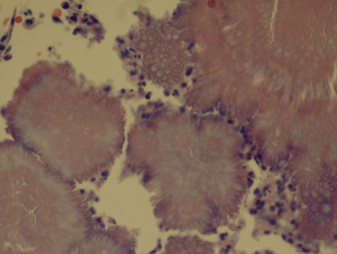
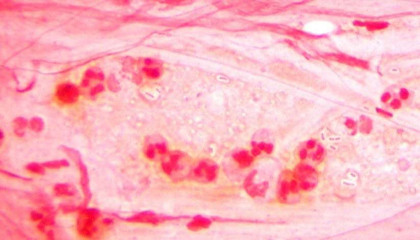
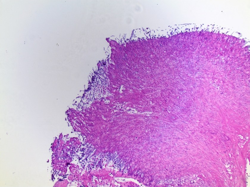
Sarcoidosis is a multisystem inflammatory disease of unknown etiology manifests as granulomas found predominantly in the lungs and lymph nodes. Hypercalcemia is seen in about 10-13% of patients. Overproduction of 1-alpha-hydroxylase and production of PTHrp can both contribute to the hypercalcemia in some patients with sarcoidosis. In this case, PTHrp was normal and elevated 1-alpha-hydroxylase was found to be the cause of hypercalcemia.

In addition to treatment of the underlying disorder, treatment of hypercalcemia in sarcoidosis is aimed at reducing intestinal calcium absorption and DHVD synthesis. Besides dietary interventions, glucocorticoids and bisphosphonates have also been used successfully to treat hypercalcemia in sarcoidosis:

  • Glucocorticoids: inhibit DHVD synthesis by the activated mononuclear cells (major contribution), inhibit intestinal calcium absorption and osteoclast activity
  • Bisphosphonates: inhibit the resorption of bone by osteoclasts


References

  1. https://emedicine.medscape.com/article/301914-overview
  2. https://www.uptodate.com/contents/hypercalcemia-in-granulomatous-diseases
  3. https://www.uptodate.com/contents/diagnostic-approach-to-hypercalcemia
  4. https://www.ncbi.nlm.nih.gov/books/NBK109831/


Huang

-Rongrong Huang, PhD is a first year clinical chemistry fellow at Houston Methodist Hospital. Her interests include general clinical chemistry, genetic biochemistry and applications of mass spectrometry in clinical laboratories.

Xin-small

-Xin Yi, PhD, DABCC, FACB, is a board-certified clinical chemist, currently serving as the Co-director of Clinical Chemistry at Houston Methodist Hospital in Houston, TX and an Assistant Professor of Clinical Pathology and Laboratory Medicine at Weill Cornell Medical College.

Microbiology Case Study: An Elderly Woman with a Cough Develops Skin Nodules

Case History

Our patient is an 83 year old female with previous history of arterial hypertension, atrial flutter and chronic obstructive pulmonary disease who presented with dry cough (~2 weeks), fever (102ºF), and cutaneous ulcerated plaques with elevated borders on forearm, foot, leg, and neck. Chest radiographs and chest CT scan showed numerous bilateral nodular consolidations compatible with pneumonia. Additionally, mild leukocytosis (14,200 cells /mm3) and hypohemoglobinemia (10.9 mg/dl) were documented. A skin biopsy was taken from the forearm lesion. Periodic acid–Schiff (PAS) and Grocott-Gomori’s (or Gömöri) methenamine silver (GMS) stains identified rare budding yeast (PAS, Image 1). Acid-fast bacilli (AFB) and Gram stains were negative for mycobacteria and bacteria, respectively.

blasto
Image 1. Budding yeast with a thick double cell wall were identified microscopically surrounded by prominent supurative granulomatous reaction (PAS, 100x).

Discussion

A diagnosis of disseminated blastomycosis was made based broad based budding yeast seen on PAS stain (Image 1).

Blastomycosis infection most commonly affects persons living in the Mississippi and Ohio River valleys, Great Lakes Region of the United States, and southern Canadian provinces. It is a fungal infection that can cause asymptomatic infection, isolated pulmonary disease, or serious and potentially fatal disseminated disease. B. dermatitidis can infect every organ of the body giving great variety of clinical manifestations, which is the reason why it is known as “the great pretender.” More than half of infected patients are asymptomatic. Symptomatic patients generally present with pulmonary symptoms, and the development of disseminated disease after hematogenous spread is common (~25 to 40% of symptomatic cases). The most common extra-pulmonary locations are: skin, bone, genitourinary tract, and central nervous system (CNS). Unlike histoplasmosis, most cases of blastomycosis are seen in immunocompetent patients, although immunocompromised patients may be at higher risk to develop severe forms of the disease.

Blastomyces is a thermally dimorphic fungus that grows as a yeast in the body and as filamentous fungi with septate hyphae in the environment. Recent phylogenic analysis has divided the Blastomyces genus into 2 species, B dermatitidis and B gilchristi . Culture of B. dermatitidis from the environment is extremely difficult, and much of what we know is conjecture from a few documented outbreaks, of which several occurred in wooded areas near waterways. These investigations found that exposure to dust clouds associated with construction or crop harvesting were the only identified risk factors for infection. Blastomycosis infection occurs through aerosolization of conidia from the environment causing respiratory infection or less commonly through direct inoculation into cutaneous abrasions. Once in the host, the conidia transform into yeast. The specific proteins expressed during the yeast phase allow the evasion of phagocytic and CD4+ cells.

Laboratory diagnosis

The most expedient method to diagnose blastomycosis remains examination of stained fluid or tissue specimens. Yeast are 8-15 µm in size with broad based buds of 4-5 µm and have a characteristic refractory double cell wall. Fluid can be stained with 10% potassium hydroxide plus calcofluor white, whereas formalin fixed paraffin embedded tissue samples can be stained with GMS or PAS. B. dermatitidis yeast can be difficult to visualize with Gram or hematoxylin and eosin (H&E) stains, but if found, the characteristic broad-based budding pattern of yeast can lead to presumptive diagnosis before culture and non-culture based diagnostic test results are available.

Culture of B. dermatitidis provides a definitive diagnosis of pulmonary and extra-pulmonary disease. B. dermatitidis grows well on routine fungal media such as Sabouraud dextrose agar, potato dextrose agar, and brain–heart infusion media. The yeast phase is inhibited by media containing cyclohexamide. Culture typically demonstrates growth in as little as 4-7 days. Colonies will initially appear yeast-like, but then develop white cottony aerial mycelium and turn tan with age. Mature growth is achieved around day 14 and the reverse of the colony is a tan color. At 25-30°C, B. dermatitidis forms septate hyphae with round or pear-shaped conidia attached to the hyphae by short or long conidiophores. This gives the characteristic appearance of “lollipops.” Scedosporium spp. and Chrysosporium spp. are common confounders because they make similar structures. Definitive identification of Blastomyces sp. can be made by conversion of the mold phase to the yeast phase by incubation at 37°C. An alternative to conversion is using a DNA probe assay.


References

  1. Medically Important Fungi, 5th edition
  2. Principles and Practices of Infectious Disease, 7th edition

Julio picture

-Julio Diaz-Perez, MD is a 1st year anatomic and clinical pathology resident at University of Chicago (NorthShore).

-Erin McElvania, PhD, D(ABMM), is the Director of Clinical Microbiology NorthShore University Health System in Evanston, Illinois.

Microbiology Case Study: 42 Year Old Female with HPV

Case History

A 42 y/o female G2P2002 patient presented to her Ob/Gyn for Colposcopy for monitoring of persistent High-Risk HPV. She was originally found positive for HPV in 2015, but has had never had a Pap with a squamous intraepithelial lesion, abnormalities on colposcopy, or dysplasia seen on endocervical curettage. Additionally, she endorsed a complaint of vague diffuse pelvic/lower abdominal pain for approximately the last 2 months. She states that the pain is mild and comes and goes and is not associated with anything in particular. She has noticed some clear to gray-white discharge now and then since she first noticed the pain, but nothing that really worried her. Pt denies changes in bowel or bladder habits, denies nausea, fever, or chills. Pt has been in a monogamous relationship with her partner for the last 12 years. She had a Mirana IUD placed 4 years prior, without complication, and has not had menses since placement. Prior to that, the patient had normal, regular cycles. She has 2 children with the same father, both were delivered by spontaneous vaginal delivery without complications. She has mild anxiety and depression for which she is treated, but no other medical problems. There is no surgical history. She has 1-2 glasses of red wine every week, denies tobacco use, and denies illicit drug use.

Pelvic exam revealed a benign appearing cervix that was not painful to touch or motion. There was a clear to white mild discharge that was suspected to be normal vaginal secretions. IUD strings were noted. Colposcopy revealed an easily appreciated transformational zone without any obvious lesions. A routine endocervical curettage (ECC) was performed followed by observed increased clear discharge from the cervical os. ECC was sent for routine pathology:

actinomyces1
Actinomyces, H&E, 20x
actinomyces2
Actinomyces, H&E, 40x

Discussion

Actinomycosis is an infection by a species within the Actinomyces genus, generally seen in dental and other oropharyngeal abscess formations. However, rare occurrences of pelvic Actinomycosis can be seen in women with intrauterine devices in place. Pelvic infections can result in cervicitis and endometritis and progress into abscess formation within the fallopian tubes and the ovaries along with salphigitis. The more profound disease consisting of abscess formation generally presents with fever, specific lower abdominal tenderness, and elevated WBCs, thus can mimic acute appendicitis, ovarian torsion, or ectopic pregnancy (1). The first case reported in the literature was in 1967 (2).

Three main species of Actinomyces have been found to be associated with IUD-associated pelvic infection: A. naeslundii, A. odontolyticus (3), and A. hongkongensis (4). All of these species are obligate to facultative anaerobes, catalase negative, and nitrate reducing. A sub-species group of A. naeslundii, however, can be catalase positive and is CAMP test-positive. All members of A. naeslundii are urease positive while A. odontolyticus and A. hongkongensis are urease negative.

References

  1. Joshi et al. Pelvic Actinomycosis: a Rare Entity Presenting as Tubo-ovarian Abscess. Arch Gynecol Obstet. 2010, 281:305-306.
  2. Brenner et al. Pelvic Actinomycosis in the Presence of an Endocervical Contraceptive Device. Obstet Gynecol. 1967, 29: 71-73.
  3. Woo et al. Diagnosis of Pelvic Actinomycosis by 16S ribosomal RNA Gene Sequencing and its Clinical Significance. Diagnostic Microbiology and Infectious Disease. 2002; 43: 113-118.
  4. Flynn et al. Identification by 16S rRNA Gene Sequencing of an Actinomyces hogkongensis Isolate Recovered from a Patient with Pelvic Actinomycosis. J. Clin. Microbiol. 2013, 51(8):2721. DOI: 10.1128/JCM.00509-13.


-Jeff Covington, MD, PhD, is a 1st year anatomic and clinical pathology resident at the University of Vermont Medical Center.

Wojewoda-small

-Christi Wojewoda, MD, is the Director of Clinical Microbiology at the University of Vermont Medical Center and an Associate Professor at the University of Vermont.


Microbiology Case Study: A 36 Year Old Man with Symptoms of Bowel Obstruction

Case History  

A 36 year old Caucasian male presented to the hospital with symptoms of a bowel obstruction.  His past medical history was significant for a gunshot wound to the abdomen followed by the development of colon cancer with metastasis to the liver. Recently, he had an intestinal stent placed in order to relieve an obstruction from tumoral growth. During the current admission, he was taken to the operating room for a diverting colostomy and two days later developed a fever of 101.1°F and increasing abdominal pain. Due to a concern for sepsis, blood cultures were collected and piperacillin-tazobactam was added to his antibiotic therapy regimen which already included vancomycin and ciprofloxacin.


Laboratory Identification

aero1
Image 1. Gram stain from the blood bottle showing many gram negative rods (100x oil immersion).
aero2
Image 2. The organism grew as mucoid colonies on blood, chocolate and MacConkey agars after 24 hours of incubation at 35°C in 5% CO2.


aero3
Image 3. The organism exhibited beta hemolysis on blood agar when held up to a light source.

The blood culture signaled positive after less than 24 hours on the automated instrument and Gram stain revealed gram negative rods (Image 1). The organism grew as mucoid colonies on blood, chocolate and MacConkey agars after 24 hours of incubation at 35°C in 5% CO2 (Image 2). When the blood agar plate was examined with a light source, the organism clearly illustrated beta hemolysis (Image 3). Rapid biochemical testing was positive for catalase, oxidase and indole. The Vitek II identified the isolate as Aeromonas veronii bv. sobria.

Discussion 

Aeromonas spp. are facultative anaerobic gram negative rods. They are inhabitants of aquatic ecosystems and as such can cause wound infections in people exposed to polluted lakes or brackish water with fresh breaks in their skin. Additionally, gastroenteritis is common with Aeromonas spp. and are often acquired through ingestion of unpurified water or to a lesser extent by consumption of contaminated meats, fresh produce or dairy products. Extraintestinal infections, including sepsis and meningitis, can result by spread from GI sources or wound infections. Interesting, medicinal leeches, used in the post-operative setting to increase blood flow to the surgical site, are colonized with Aeromonas spp. (most commonly Aeromonas veronii bv. sobria) and can result in systemic infections in the patient.

In the laboratory, Aeromonas spp. grow readily from stool, wound and blood sources on commonly used media and exhibits beta hemolysis on blood agar. In addition, Aeromonas spp. will grow on CIN agar (at room temperature as well as incubator temperature) as colonies with a pink center surrounded by a white apron and are indistinguishable from Yersinia spp. Aeromonas spp. is positive for catalase, oxidase and indole by rapid testing. In most cases, identification of Aeromonas spp. to the complex level can be accomplished by biochemical testing (esculin, VP), automated instrumentation or MALDI-TOF mass spectrometry. The three clinically relevant complexes include: A. hydrophila complex, A. caviae complex and A. veronii complex.

With regards to susceptibility testing for Aeromonas spp., the CLSI M45, 3rd edition provides guidelines for the three complexes discussed above. Third or fourth generation cephalosporins, fluoroquinolones and trimethoprim-sulfamethoxazole are recommended as antibiotics for primary testing for isolates from extraintestinal sites. Aeromonas spp. are uniformly resistant to ampicillin, amoxicillin-clavulanate and cefazolin and many strains may possess various inducible beta lactamases.

In the case of our patient, with the laboratory identification of Aeromonas veronii bv. sobria, his gram negative coverage was switched to ciprofloxacin for a 10 day course. His PICC was removed and on further imaging studies he was found to have a large abdominal abscess which required surgical drainage. Subsequent cultures from the abdominal abscess were negative.


Stempak

-Lisa Stempak, MD, is an Assistant Professor of Pathology at the University of Mississippi Medical Center in Jackson, MS. She is certified by the American Board of Pathology in Anatomic and Clinical Pathology as well as Medical Microbiology. She is the director of the Microbiology and Serology Laboratories. Her interests include infectious disease histology, process and quality improvement, and resident education.

Chemistry Case Study: Conjugated Bilirubin in Neonatal Jaundice

Case History

Patient was a 1-week-old infant in the level 2 NICU born at 37 weeks. This infant was initially born with indirect hyperbilirubinemia but now also has increasingly elevated level of direct bilirubin (see measurements in table below). Neonatologist requested conjugated and unconjugated bilirubin test due to increasing elevated level of direct bilirubin. Conjugated bilirubin test is not routinely performed in our hospital laboratory and needs to be send out.

Question: What’s the difference between conjugated bilirubin and direct bilirubin? When does conjugated bilirubin need to be assessed?

Ref Range 3/6/18 3/7/18 3/9/18 3/10/18 3/12/18
Bilirubin total, neonatal 1.0-10.5 mg/dL 9.2 8.7 10.8 10.2 8.6
Bilirubin direct, neonatal 0.0 – 0.6 mg/dL 0.5 0.7 1.8 1.8 2.1

Discussion

Neonatal jaundice is commonly seen in newborns in the first few days of life, mainly due to increased bilirubin formation from break down of red blood cells and limited conjugation of bilirubin. Total bilirubin normally peaks at day 2-3 and should decline by day 4-5. Sample is collected via heelstick in green top tube and protected from light. Measurement of total bilirubin is interpreted based on the Bhutani Nomogram to assess risk of hyperbilirubinemia. Most often, unconjugated bilirubin is elevated in neonatal jaundice owing to hemolytic causes. In cases with prolonged jaundice, conjugated bilirubin needs to be determined to rule out cholestasis.

Conjugated bilirubin refers to bilirubin conjugated with one or two glucuronic acid, and this term “conjugated bilirubin” is often used interchangeably with direct bilirubin. Direct bilirubin refers to bilirubin fractions that can directly react with diazo reagent without the addition of accelerator, such as methanol or ethanol. This fraction usually includes conjugated bilirubin and delta bilirubin. Delta bilirubin is formed by covalent bonding between conjugated bilirubin and albumin, and has a similar half-life as albumin, 21 days. Therefore, direct bilirubin measurement overestimate conjugated bilirubin and in cases with persist or atypical jaundice, clear differentiation between conjugated and direct bilirubin is important. Clinician should know what the laboratory is measuring when interpreting the bilirubin fraction results.

In laboratories, conjugated bilirubin can be assessed by the VITROS BuBc dry slide, which simultaneously measures unconjugated (Bu) and conjugated (Bc) bilirubin by use of a mordant. In the presence of the mordant, the visible spectra of conjugated and unconjugated bilirubin are different, allowing measurement of both species from a single slide. Fractions of bilirubin can also be separated by HPLC, but this is not practical to use in a routine clinical laboratory. In this case, conjugated bilirubin was measured by VITROS BuBc slide test, and result came back elevated at 1.0 mg/dL (ref range: < 0.3 mg/dL).


Ketcham

-Megan Ketcham, MD is a 4th year anatomic and clinical pathology resident at Houston Methodist Hospital. She will be completing both hematopathology and dermatopathology fellowships. Her interests include pathology resident and medical student education and skin lymphomas.

Xin-small

-Xin Yi, PhD, DABCC, FACB, is a board-certified clinical chemist, currently serving as the Co-director of Clinical Chemistry at Houston Methodist Hospital in Houston, TX and an Assistant Professor of Clinical Pathology and Laboratory Medicine at Weill Cornell Medical College.

Microbiology Case Study: A 79 Year Old Male with Rheumatic Heart Disease

Case History

The patient is a 79 y/o male with past medical history of rheumatic heart disease, permanent atrial fibrillation, mechanical aortic and mitral valves (2004), status post single chamber pace maker for bradycardia (2010), and prostate adenocarcinoma treated in 2000. He had new MRI compatible pace maker placed on Oct 19, 2017. During follow-up he was noted to have a hematoma over the incision site. He had a revision done on Nov 3, 2017. At that time, the blood from the incision site was sent for culture. 

Laboratory Identification

Gram stain showed moderate amount of polys with no bacteria seen. The isolate was a gram-negative rod that was identified on the MALDI-ToF as Burkholderia multivorans.


burkmult1
Image 1: Semi-mucoid, yellow-grey colonies on Chocolate agar and on Blood agar plates.

Discussion

The Burkholderia genus appears as gram-negative medium-sized straight rods, with the exception being B. mallei which is a coccobacillus. The will grow on blood, chocolate, and MacConkey agar. Oxidative-fermentative-base-polymyxin B-bacitracin-lactose (OFPBL) agar can be used to isolate B. cepacia and Ashdown medium can be used to isolate B. pseudomallei. They are non-lactose fermenters on MacConkey, but B. cepacia can turn into a dark pink to red due to oxidation of lactose after 4-7 days.

B. multivorans is a species within the Burkholderia genus, which are normal to plant, soil, and water, but not normally considered common human flora. Formerly of the Pseudomonas genus, B. cepacia, B. mallei, and B. pseudomallei are the most commonly seen as infections in humans. Further, B. cepacia and B. mallei are not typically human pathogens in a healthy human host. Because of the rarity of this genus to infect humans, their pathogenicity is not well known; but, importantly, they are intrinsically resistant to many antibiotics and can thus be associated with hospital acquired infections.

Of this genus, very little literature is present on B. multivorans specifically, and of the literature that does exist, most of it is in relation to cystic fibrosis patients. Taxonomic advances has shown that B. cepacia complex is a cluster of nine closesly related genomic species or genomorvars (1).  B. multivorans represents genomorvar II. Hospital acquired clinical infections from this complex (but perhaps not specifically from this particular genomorvar) has been seen following catheterization, cystoscopy, heart surgery, and with contaminated ventriculoatrial shunt (2). B. multivorans biochemically is oxidase positive, catalase positive, lipase positive, nitrate-reducing, urease positive, resistant to colistin, and can grow at 42C (3, 4).

A recent comparative genomic study showed that B. multivorans is a highly evolutionarily preserved genome with genomic characteristics from the environment and isolated from cystic fibrosis patients to be similar, and that isolates from different continents are also similar (5). Further, a murine model for pulmonary infections showed that B. multivorans could persist in the host by establishing an intracellular presence within macrophages, which could explain the persistence of this pathogen in cystic fibrosis patients (6). Importantly though, due to the conserved and common genomic structure, there rests a possibility for potential vaccination for cystic fibrosis patients against B. multivorans.

The patient was prescribed a single dose of oral Bactrim and then advised to come into the hospital for admission for IV antibiotics. IV ceftazidime was started with pending blood cultures, which are negative at the time of this documentation.

References:

  1. Coenye T. et al. Taxonomy and identification of the Burkholderia cepacia complex. J Clin Microbiol 2001;39:3427-3436.
  2. Pallent LJ. et al. Pseudomonas cepacia as contaminant and infective age. J Hosp Infect 1983;4:9-13.
  3. Henry DA. et al. Phenotypic methods for determining genomovar status of Burkholderia cepacia complex. J Clin Microbiol 2001;39:1073-1078.
  4. Vandamme P. et al. Occurrence of multiple genomovars of Burkholderia cepacia in patients with cystic fibrosis and proposal of Burkholderia multivorans sp. nov. Int J Syst Bacteriol 1997;47:1188-1200.
  5. Peeters C. et al. Comparative genomics of Burkholderia multivorans, a ubiquitous pathogen with a highly conserved genomic structure. PLoS One. 2017, 21; 12 (4): e0176191.
  6. Chu KK. et al. Persistence of Burkholderia multivorans with the Pulmonary Macrophage in the Murine Lung. Infect Immun 2004; 72 (10): 6142-6147.


-Jeff Covington, MD, PhD, is a 1st year anatomic and clinical pathology resident at the University of Vermont Medical Center.

Wojewoda-small

-Christi Wojewoda, MD, is the Director of Clinical Microbiology at the University of Vermont Medical Center and an Associate Professor at the University of Vermont.

Microbiology Case Study: A Middle-Aged Man with Malaise, Shaking, and Chills

Case history

A middle-aged male presented to the hospital emergency room with the complaints of malaise, shaking and chills for the last two days. He denied any runny nose, cough, abdominal pain, nausea, vomiting, headache or known sick contacts. His past medical history was significant for alcohol use disorder. Imaging of the abdomen revealed an ill-defined region of decreased attenuation in the right lobe of the liver measuring 4.8 x 4.7 x 2.2 cm. The Gram stain of the abscess showed 4+ WBCs (PMNs) and 4+ gram negative rods with a very large capsule surrounding them (Image 1).  The organisms grew very mucoid colonies on 5% sheep blood, chocolate, and MacConkey agars (Image 2).  A string test performed on the mucoid bacterial colonies was >5 mm (Image 3).

kleb1
Image 1. Gram stain of abscess showing 4+ WBCs and 4+ GNR with large capsule.
kleb2
Image 2. Cultures showed mucoid colonies on the chocolate and MacConkey agars.
kleb3
Image 3. A string test was performed on the mucoid colonies and was positive (mucoid capsule “string” > 5mm).

Discussion

The organism was identified as Klebsiella pneumoniae by MALDI-TOF MS.  Based on the mucoid capsule and positive string test, this organism was further identified as hypermucoviscous K. pneumoniae.

Hypermucoviscous K. pneumoniae is a relatively newly recognized hypervirulent variant of K. pneumoniae. It was first described in the Asian Pacific rim and is now increasingly recognized in Western countries. Defining clinical features include serious, life-threatening community-acquired infection in younger healthy hosts, an unusual feature for enteric gram negative bacilli in the non-immunocompromised population. It can cause a variety of diseases including, but not limited to liver abscess, pneumonia, meningitis, osteomyelitis, necrotizing fasciitis and endophthalmitis.

Intestinal colonization, appears to be a critical step leading to infection. It is seen mostly in Asians, raising the issue of a genetic predisposition vs. geospecific strain acquisition.  The increased virulence might be due to the ability to more efficiently acquire iron and perhaps an increase in capsule production, which confers the hypermucoviscous phenotype to the organism. The vehicles for acquisition and subsequent colonization appear to be food and water, person-to-person transmission (e.g., close contacts such as family members or sexual partners) or animal-to-person transmission (e.g., between pets and their owners).

To date, most strains of hypermucoviscous K. pneumoniae have been very susceptible to antimicrobials except ampicillin.  However, in recent literature, propensity for hypermucoviscous Klebsiella pneumoniae to become multi-, extreme or pandrug-resistant, including the acquisition of extended-spectrum β-lactamases (ESBL) and carbapenemases has been reported. Since hypermucoviscous K. pneumoniae strains often cause abscesses, source control is a major aspect of the overall management plan and a need to drain abscesses and closed space infections is essential for optimal outcome.

References

  1. Alyssa S. Shon, Rajinder P.S. Bajwa and Thomas A. Russo; Hypervirulent (hypermucoviscous) Klebsiella pneumonia: A new and dangerous breed; Virulence 4:2, 107–118; February 15, 2013; 2013 Landes Bioscience
  2. Bonnie C Prokesch, Michael TeKippe, Jiwoong Kim, Prithvi Raj, Erin McElvania TeKippe, David E Greenberg; Primary osteomyelitis caused by hypervirulent Klebsiella pneumonia; The Lancet Infectious Diseases , Volume 16 , Issue 9 , e190 – e195


MA

-Muhammad Ahmad, MD is a 2nd year anatomic and clinical pathology resident at University of Chicago (NorthShore) program based at Evanston Hospital, Evanston, IL. His academic interests include breast pathology and cytopathology.

-Erin McElvania, PhD, D(ABMM), is the Director of Clinical Microbiology NorthShore University Health System in Evanston, Illinois.

Microbiology Case Study: A 28 Year Old Female with Cough.

Case History

A 28 y/o female with a past medical history of chronic eosinophilic pneumonia, chronic persistent asthma, and elevated IgE status post Xolair therapy presented with a cough. She is a former smoker and a former IV drug user. She has been having a productive cough since March and has not improved despite multiple courses of antibiotic therapy. She coughs mostly in the morning and describes her sputum as thick and greenish. She does not have any associated fevers and does not feel that her rescue inhalers help much. She was given a course of doxycycline for 10 days, and sputum was sent for culture.

Laboratory Identification

pasmul1
Image 1: Gram stain showed many polys, moderate mixed gram positive and gram negative organisms. Sputum culture was reported out as mixed gram negatives.
pasmul2
Image 2: Chocolate and blood agar plates of the mixed gram positive and gram negative organisms.

One of the gram negative rods was identified by the MALDI-ToF as Pasteurella multocida.

Discussion

The genus Pasteurella consists of multiple identified species with the one most commonly seen in the clinical setting as Pasteurella multocida. As a genus, they are typically gram-negative straight bacilli that are nonmotile, oxidase-positive, catalase-positive, nitrate reducing, and ferment glucose. They will grow on blood and on chocolate agars, but importantly will not grow on MacConkey. Their colony morphology on blood agar is generally convex, smooth, and nonhemolytic.

Infections with Pasteurella are classically associated with animal bites, such as from a dog or cat. However, prior cases in the literature have shown that pulmonary infection with Pasteurella can be associated with other chronic pulmonary diseases such as COPD (1). The choice for using doxycycline is supported in the literature and was specifically discussed in a prior case with improvement (2).

References:

  1. Klein NC. et al. Pasteurella multocida pneumonia. Semin Respir Infect 1997; 12 (1): 54-56.
  2. Bhat S. et al. A case of lower respiratory tract infection with canine-associated Pasteurella canis in a patient with chronic obstructive pulmonary disease. J Clin Diagn Res 2015; 9 (8): DD03-DD04.


-Jeff Covington, MD, PhD, is a 1st year anatomic and clinical pathology resident at the University of Vermont Medical Center.

Wojewoda-small

-Christi Wojewoda, MD, is the Director of Clinical Microbiology at the University of Vermont Medical Center and an Associate Professor at the University of Vermont.

Microbiology Case Study: A 59 Year Old Female with Fevers, Weakness, and Altered Mental Status

Case History

A 59 year old African American female presented to the emergency department with fevers, weakness, fatigue and altered mental status. Her past medical history was significant for hypertension, diabetes mellitus (type 2) with end stage renal disease and a recent cerebrovascular accident the month prior. Her surgical history included a mitral valve repair surgery three years ago and a renal transplant two years ago. Current medications included prednisone, mycophenolate and tacrolimus immunosuppressive agents. Physical examination was unremarkable except for a temperature of 101°F and she was oriented to person, place and time. Pertinent labs included a WBC count of 13.2 TH/cm2, microcytic anemia, and a creatinine of 1.51 mg/dL. Due to previous cardiac surgery, a transesophageal echocardiograph (TEE) was performed and showed a large vegetation (1.6 x 1.5 cm) on the mitral valve.  A diagnosis of endocarditis was made and the patient was started on broad-spectrum antibiotics & taken to surgery for a mitral valve replacement. Multiple blood cultures were negative to this point. Portions of the mitral valve were submitted to surgical pathology and the microbiology laboratory for bacterial, fungal and AFB cultures.

Laboratory identification

Surgical pathology received an aggregate of tan-yellow, fibrous tissue fragments (3.1 x 1.5 x 1.1 cm). Histologic assessment showed a confluent mass containing abundant narrow, septate hyphae consistent with a fungal infection (Image 1). No definitive pigment was identified. Grocott’s methenamine silver (GMS) stain also highlighted the narrow hyphae with numerous septations (Image 2). In the microbiology laboratory, a darkly pigmented mold grew after 5 days of incubation on Sabouraud dextrose agar (Image 3). Lactophenol cotton blue prep showed pigmented, curved conidia with 2-3 transverse septations consistent with Curvularia spp (Image 4). All blood cultures were finalized as no growth after 5 days. Fungitell was found to be greater than 500 pg/ml and Aspergillus galactomannan was negative (<0.5).

curve1.jpg
Image 1. Sections of mitral valve tissue showed a confluent mass of abundant hyphal elements (H&E, 4x).
curve2.jpg
Image 2. Special stains of the fungal mass highlighted narrow hyphae with numerous septations and acute angle branching (GMS, 4x).
curve3.png
Image 3. A darkly pigmented mold grew of Sabouraud dextrose agar after 5 days of incubation at 25°C.
curve4.jpg
Image 4. Many pigmented, curved conidia with multiple transverse septations were seen (lactophenol cotton blue prep, low power).

Discussion

Curvularia spp. belong to a heterogeneous group of dematiaceous or black molds. The presence of pigment in this category of molds is due to melanin in the hyphae. Dematiaceous molds are ubiquitous in nature and can occasionally cause human infections.  These molds have a characteristic dark appearance on fungal media that is often dark gray, brown or black in color. In addition, when the reverse of the plate or slant is examined, the under surface is also pigmented. Based on their growth rate, the dematiaceous fungi are divided into the fast growers, such as Curvularia, Bipolaris and Alternaria spp., which are mature in 5-7 days. The second group is slow growers that take between 7-25 days to fully mature. Examples of slow growers include Phialophora, Exophila/Wangiella, Cladosporium, Fonsecaea and Rhinocladiella spp.

Most commonly, dematiaceous molds infections usually present as phaeophyphomycosis, chromoblastomycosis or mycetomas. These three entities are cutaneous or subcutaneous mycoses that are obtained by traumatic implantation but vary from one another based on clinical features and histologic features of the mold in tissues. They are most frequently cause infection in male agricultural workers in rural areas of tropical or subtropical climates.  These infections are indolent in nature but can lead to significant morbidity over time, as they are difficult to treat effectively.

In addition to the above superficial infections, Curvularia spp. has also be known to cause keratitis, sinusitis and wound infections. In immunosuppressed individuals, disseminated infections with spread to the lungs and brain have been documented. Endocarditis due to Curvularia spp. is quite rare with very few cases previously reported in the literature. On those documented, Curvularia spp. infections tend to have a predilection for prosthetic heart valves or occur after cardiac surgery. Diagnosis of infective endocarditis is difficult as symptoms are indolent and blood cultures do not have a high yield. Therefore, culture of the vegetation may be the only way to make a diagnosis.

In the microbiology laboratory, Curvularia spp. will grow on routine fungal media as a darkly pigmented mold in a relatively short time. On lactophenol cotton blue prep, Curvularia spp. produce large conidia that usually contain 4 cells that are divided by transverse septations. The conidia take on a curved appearance due to swelling of the subterminal cell, which is often the largest and most deeply pigmented. If identification is necessary beyond the genus level, panfungal PCR assays followed by sequencing of ribosomal genes may be useful in providing a species level diagnosis from fresh or paraffin embedded tissue.

For localized infections, surgical treatment alone may be adequate in some cases.  In infections that are extensive or if there is dissemination, treatment with newer triazoles, such as posaconazole or voriconazole, have shown a broad spectrum of activity against dematiceous molds. Amphotericin B is also another effective option. While susceptibility testing can be performed on clinically significant Curvularia spp. infections, interpretative breakpoints have not been defined and clinical correlation is lacking.

In the case of our patient, she remained on a ventilator following surgery and with the identification of mold on histology, she was started on micafungin. She was switched to amphotericin B after the mold was classified as Curvularia spp. Her condition did not improve despite therapy and she died 3 weeks after surgery.


-Azniv Azar, MD, is a fourth year anatomical and clinical pathology resident at the University of Mississippi Medical Center.

Stempak

-Lisa Stempak, MD, is an Assistant Professor of Pathology at the University of Mississippi Medical Center in Jackson, MS. She is certified by the American Board of Pathology in Anatomic and Clinical Pathology as well as Medical Microbiology. She is the director of the Microbiology and Serology Laboratories. Her interests include infectious disease histology, process and quality improvement, and resident education.

Hematology Case Study: The Race to Save a 48 Year Old Man from a Rare Disease

A 48-year-old Caucasian male presented to a Baltimore Emergency Room complaining of fever, chills, and aches. He stated he had not been feeling well for the past week. His symptoms had progressed rapidly over the last 3 days to include night sweats, nausea and excessive somnolence. History taken in the ER revealed the patient had returned 10 days prior from a Safari in Botswana and Zambia. The patient was admitted to the ICU, in shock, with a BP of 75/50. Even though the patient had taken anti-malarial medication, the doctors suspected malaria. Blood was sent to the lab for a blood parasite exam and treatment for malaria was started while the doctors waited for the confirmation.

In the Hematology laboratory, technologists perform microscopy of thick and thin blood smears to look for malarial parasites. The thin smear is a typical Wright Giemsa stained wedge smear, and the thick smears are prepared and stained so that the red blood cells are lysed, and the sample is concentrated, making examination easier. Thorough, careful examination of the thick smear is aimed to identify whether a particular parasite is present, but they require a long drying period and take several hours to prepare and read. Thin smears can detect the parasites but also permit identification of particular species of malaria. While the thick smears were drying the technologist examined the thin smear.

The technologist who examined this patient’s thin smears saw parasites (image 1) under her microscope. She consulted with a supervisor and pathologist to confirm, and the patient’s doctor was notified that the patient did not have malaria, but instead, had Trypanosoma! This was an exciting find in the laboratory, as there have been only 40 cases seen in the US in the past 50 years.

tryp1
Image 1. This slide shows the parasite, in dark blue. The parasite causes
African trypanosomiasis, also known as sleeping sickness
(Courtesy of Greater Baltimore Medical Center).

The race for diagnosis and treatment did not stop there, as there are 2 types of African trypanosomiasis, or African sleeping sickness, and effective and appropriate treatment must be started in a timely fashion. Both types look identical on a blood smear and both are considered universally fatal, if not treated. West African trypanosomiasis and East African trypanosomiasis are caused by the tsetse fly, which only lives in rural Africa. The patient stated he did remember being bitten by tsetse flies, and because there had been such a short span of time between being bitten and the onset of symptoms, doctors concluded that the patient had the rarer and fast-acting East African trypanosomiasis, which can kill within months.

Epidemiologists at CDC were contacted, who then consulted other infectious disease specialists at CDC. There are 2 treatments depending the stage of the disease. Surinam is the first line of defense, but melarsoprol, which is arsenic-like and very toxic, must be used if the parasites have reached the central nervous system. Because of the urgent need to start treatment, emergency shipments of both drugs were flown to Baltimore. The patient was started on Surinam to reduce the number of parasites in his blood to a level low enough to allow a spinal tap to be performed. After confirming that the CSF showed no signs of the parasite, treatment with surinam was continued and the patient was discharged a week later and has made a full recovery.

Because of the excellent work done by the medical technologists who made the first discovery, the speed with which the critical calls were made, the actions of the doctors involved, and the cooperation of the CDC, this patient received his first dose of Surinam a little over 24 hours after his blood was sent to the lab. This case shows the importance of a thorough medical and travel history in differential diagnosis. It also illustrates the importance of the competency evaluations and surveys in which all laboratory professionals are required to participate. None of the technologists, doctors or scientists involved had ever actually seen a case of African Trypanosomiasis, they had only read about it in books and seen it on competency assessments.

This case is based on an actual case from 2016. My coworker, Gail Wilson, was the technologist who first saw the Trypanosoma on the slides. Many thanks to Gail for her keen eye and attention to detail!

tryp2
Image 2: L&R: Trypanosoma brucei in thin blood smears stained with Giemsa. Center: A close up of a tsetse fly. Credit: DPDx


References 

  1. Jon E. Rosenblatt Barth Reller Melvin P. Weinstein.pages 1103-1108, Laboratory Diagnosis of Infections Due to Blood and Tissue Parasites Clinical Infectious Diseases, Volume 49, Issue 7, 1 October 2009; retrieved March 2018 from https://academic.oup.com/cid/article/49/7/1103/316703
  1. Ivo Elliott, Trupti PatelJagrit Shah, and Pradhib Venkatesan. West-African trypanosomiasis in a returned traveller from Ghana: an unusual cause of progressive neurological decline BMJ Case Rep. 2014; 2014: bcr2014204451. Published online 2014 Aug 14.doi: 1136/bcr-2014-204451; retrieved March 2018 from https://www.ncbi.nlm.nih.gov/pmc/articles/PMC4139564/
  1. Lena H. Sun. Medical Detectives raced to save a man from a rare, ‘universally lethal’ disease; retrieved March 2018 from https://www.washingtonpost.com/news/to-your-health/wp/2016/12/22/medical-detectives-raced-to-save-a-man-from-a-rare-universally-lethal-disease/?utm_term=.16d7b136bc47
  1. Parasites – African Trypanosomiasis (also known as Sleeping Sickness). Retrieved March 2018 from https://www.cdc.gov/parasites/sleepingsickness/
  1. DPDx- Laboratory Identification of parasites of Public Health Concern; retrieved March 2018 from https://www.cdc.gov/dpdx/


Socha-small

-Becky Socha, MS, MLS(ASCP)CM BB CM graduated from Merrimack College in N. Andover, Massachusetts with a BS in Medical Technology and completed her MS in Clinical Laboratory Sciences at the University of Massachusetts, Lowell. She has worked as a Medical Technologist for over 30 years. She’s worked in all areas of the clinical laboratory, but has a special interest in Hematology and Blood Banking. When she’s not busy being a mad scientist, she can be found outside riding her bicycle.