Peripheral Smear Review: Inexpensive Test to Establish Diagnosis of a Rare Disease

29-year-old woman with a history of ITP (immune mediated thrombocytopenia) diagnosed in 2008. She had previously been treated with steroids, IVIG (intravenous immunoglobulin) and splenectomy. She also received Romiplostim for 1 year prior to its discontinuation. She had also been diagnosed with lupus one year previous.

Follow up visit after having her platelets evaluated in the office 2 weeks ago revealed a platelet count of 9 K/uL on CBC which was flagged by the instrument due to platelet clumping.

  • WBC: 7.5 K/ul
  • HGB: 12.2 g/dl
  • HCT: 37.3 %
  • PLT: 9 K/ul *

*On manual count the platelet count was deemed to be 74 K/uL.

Peripheral smear review:

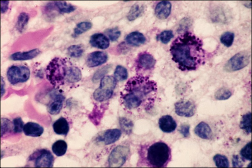
Numerous large and some giant platelets, platelet clumping and basophilic Dohle body like inclusions in nearly all neutrophils.

mayheg1

Characteristic giant platelet with poorly defined granulation. Normal-sized platelet is also present. Neutrophil contains large, well-defined, basophilic, peripherally placed cytoplasmic inclusion body (resembling Döhle body).

mayheg2

The value of peripheral smear review is very well highlighted by this case. As the smear was never reviewed earlier in the disease course, diagnosis of MHA was not made and the patient was diagnosed as ITP entirely based on clinical presentation. Perhaps earlier review of peripheral smear would have significantly altered the clinical management of the case.

After discussion with pathologist who reviewed the peripheral blood smear, diagnosis of May-Hegglin anomaly was confirmed and appropriately documented in the patient’s chart.

May-Hegglin anomaly (MHA):

May-Hegglin anomaly (MHA) is a rare autosomal dominant disorder characterized by various degrees of thrombocytopenia that may be associated with purpura and bleeding; giant platelets containing few granules; and large (2-5 um), well-defined, basophilic, cytoplasmic inclusion bodies in granulocytes that resemble Döhle bodies.

MHA is one of a family of macrothrombocytopenias characterized by mutations in the MYH9 gene present in chromosomal region 22q12-13. The mutation results in disordered production of nonmuscle myosin heavy-chain type IIA, which leads to invariable macrothrombocytopenia secondary to defective megakaryocyte maturation.

Clinical features:

The rarity of MHA has led to conflicting literature regarding the risk for bleeding. Asymptomatic patients have been described however, abnormal bleeding has also been documented. The bleeding risk is increased by taking drugs that decrease platelet function. The risk for excess bleeding with surgical procedures is unclear. Rare reports have described arterial thrombotic events associated with May-Hegglin anomaly, though the risk remains unclear. Patients are often asymptomatic. The bleeding tendency associated with MHA is generally mild and is thought to mainly depend on the degree of thrombocytopenia.

Clinical Features of MYH9 -Related Thrombocytopenias

Condition Macrothrombocytopenia Granulocyte inclusions Nephritis and Deafness Cataracts
MHA Yes Döhlelike No No
Epstein syndrome Yes Absent or faint Yes No
Fechtner syndrome Yes Spherical granules Yes Yes
Sebastian syndrome Yes Spherical granules No No

Differential diagnosis:

In addition to acute immune thrombocytic purpura, the differential diagnosis for thrombocytopenia associated with large platelets (elevated mean platelet volume) includes Bernard-Soulier syndrome, Montreal platelet syndrome, gray-platelet syndrome, and Alport syndrome.

The differential diagnosis for thrombocytopenia due to ineffective thrombopoiesis includes Bernard-Soulier syndrome, Wiskott-Aldrich syndrome, Greaves syndrome, thrombopoietin deficiency, and megaloblastic anemia.

The differential diagnosis for leukocytic inclusions, sometimes called Döhle bodies, includes septicemia, myeloproliferative disorders, and pregnancy.

Laboratory investigation:

  • The complete blood count (CBC) is essential in assessing MHA. The platelet count is decreased (usually in the range of 40-80 ´ 109/L), but the degree of thrombocytopenia varies.
  • The disorder is also characterized by giant platelets. Platelets are enlarged (>15 µm in diameter), and the mean volume of MHA platelets can be as high as 30 fL. Platelet morphology is otherwise normal.
  • On electron microscopy, platelets are seen to contain normal organelles (alpha granules, dense granules, lysosomes, and mitochondria). The most conspicuous ultrastructural feature of the platelets is an increased amount of disorganized microtubules.
  • Cytoplasmic inclusion bodies particularly in the neutrophils but also in monocytes, eosinophils, and basophils. The inclusions are large (>5 µm), spindle-shaped, pale, blue-staining bodies that consist of ribosomes, segments of endoplasmic reticulum, and microfilaments. They are located in the periphery of the cytoplasm and resemble Döhle bodies.
  • Ultrastructural studies reveal that these bodies consist of clusters of ribosomes oriented along parallel myosin heavy-chain filaments 7–10 nm in diameter. Neutrophil function is considered to be normal, and patients have no increased susceptibility to infections.
  • Immunocytochemistry can detect NMMHCIIA complexes within the leukocytes and is a useful confirmatory test.
  • The bleeding time is prolonged in concordance with the degree of thrombocytopenia.
  • Platelets usually aggregate normally in response to various agonists. The glycoprotein composition of the platelet surface is normal.

Management

Most patients with MHA do not appear to have clinically significant bleeding problems, and specific treatment is not required.

  • Corticosteroids and splenectomy are ineffective
  • In rare patients with severe bleeding, platelet transfusion may be required
  • Bleeding risk is not significantly increased by normal vaginal delivery
  • For patients scheduled to undergo surgery, intravenous desmopressin acetate (DDAVP) may be valuable; routine prophylactic platelet transfusions are not usually indicated, but platelets should be kept available
  • Depending on circumstances, refraining from participation in contact or collision sports may be prudent.

Vajpayee,Neerja2014_small

-Neerja Vajpayee, MD, is an Associate Professor of Pathology at the SUNY Upstate Medical University, Syracuse, NY. She enjoys teaching hematology to residents, fellows and laboratory technologists. 

Hematology Case Study: 60-Year-Old Male with Hives

A 60-year-old male presents with hives, skin flushing, and headaches. After an appropriate preliminary work-up, a bone marrow biopsy is performed. A representative section from the bone marrow biopsy is shown here. What are the granulated cells at the center of this image?

mast

A. Megakaryocytes
B. Promyelocytes
C. Mast cells
D.Myeloma cells
E. Adenocarcinoma cells

The granulated cells in this image are mast cells, which are identified by their abundant, metachromatic granules. This patient was diagnosed with systemic mastocytosis, a clonal disorder of mast cells and their precursors.

Mastocytosis is actually a spectrum of rare disorders, all of which are characterized by an increase in mast cells. Most patients have disease that is localized to the skin, but about 10% of patients have systemic involvement, like the patient in this case. There is a localized, cutaneous form of mastocytosis called urticaria pigmentosum that happens mostly in children and accounts for over half of all cases of mastocytosis.

Clinically, the skin lesions of mastocytosis vary in appearance. In urticaria pigmentosum, the lesions are small, round, red-brown plaques and papules. Other cases of mastocytosis show solitary pink-tan nodules that may be itchy or show blister formation. The itchiness is due to the release of mast cell granules (which contain histamine and other vasoactive substances).

In systemic mastocytosis, patients have skin lesions similar to those of urticaria pigmentosum – but there is also mast cell infiltration of the bone marrow, lymph nodes, spleen and liver. Patients often suffer itchiness and flushing triggered by certain foods, temperature changes, alcohol and certain drugs (like aspirin).

Krafts

-Kristine Krafts, MD, is an Assistant Professor of Pathology at the University of Minnesota School of Medicine and School of Dentistry and the founder of the educational website Pathology Student.

Advantages of Resident Engagement: BE PROACTIVE AND GET INVOLVED!

So, I was recently in Boston at the start of the USCAP Annual Meeting in the midst of our American Society for Clinical Pathology (ASCP) Resident Council meeting and we were talking about a topic close to my heart, that of resident engagement. As I’ve mentioned before, one of the greatest benefits of becoming a resident member (often for free as a resident) of organized pathology organizations are the opportunities for engagement. So, what do I mean by engagement?

Pathology is a small world. And in the past, we’ve been stereotyped as likely to be the more introverted out of our physician counterparts. And there may be some truth to that stereotype. But I’ve always been pretty involved and vocal since my high school days when I worked with grassroots organizing groups in the minority and immigrant communities in Chicago. Even now as a resident I’m involved as one of the five elected (and only non-primary care specialty) delegates for my hospital’s resident union, the Committee of Interns and Residents (CIR) which is affiliated with the Service Employees International Union (SEIU).

I had originally contacted our CIR/SEIU contract organizer because I was one of the many residents who paid my parking fee early and before they realized that the parking office was overcharging at the non-resident rate. Within this conversation, I had mentioned that I knew three key members of CIR/SEIU who are still active in the organization now from when I was the American Medical Student Association’s (AMSA) Race, Ethnicity, and Culture in Health (REACH) national chair in charge of AMSA’s immigrant and minority health equity campaigns and education during medical school. We even held one of our Health Equity Leadership Institute where participants came from as far as France to attend, at the CIR/SEIU office in NYC when we organized it during medical school.

Fast forward a few conversations later and I had agreed to have my name on the delegate election ballot since they were a delegate and two alternates short. We didn’t have speeches or bios, only our house staff pictures. I was stuck grossing during elections and didn’t even get to vote nor did I tell my co-residents to go vote. But despite four write-in candidates, I was surprised to learn that I had been elected. And when I asked the contract organizer how this happened she said that residents from other departments said they knew me because I had been helpful to them – either when they came to the pathology department because they needed help with an issue or when I took extra time to answer their questions after a tumor board or presentation. My point is, even when we’re not trying, the impressions we can leave on others can have an unexpected, indelible impact.

What’s even more powerful is the next step – that of joining your voice together with others to make a stronger collective voice for our profession. I cannot quantitate what I’ve received in return when I chose to become engaged, and not with a Machiavellian “ends justify the means” mentality where I do something or manipulate people to receive a gain. I don’t believe in that. But the wonderful surprises of seeing work (especially in health policy) that I’ve either participated in or supported becoming a reality, the totally unexpected relationships I’ve developed where pathology leaders saw something in me that they thought to invest in by thinking of and providing me with opportunities (eg – fellowships, work on publications, etc), and relationships with other residents that I met either through ASCP, CAP, or this very blog. I’ve kept in touch with those of you through email, social media, and texts and I appreciate that you privilege me by asking my opinions on CVs, fellowship applications, etc. Thank you and I hope that I was helpful.

These are the types of non-quantifiable benefits I’ve experienced by being more engaged. Of course, there is the satisfaction of contributing not to just a collective voice but also to a larger work to impact our profession (most of my focus has been in the realm of graduate medical education). Life seems fuller when I am involved. Our ASCP Resident Council, for instance, will have a number of members graduated and spots to fill in the next coming months so make sure that your program’s ASCP Resident Representative keeps you updated as to the application deadline. We also have a resident representative on many of ASCP’s commissions and committee where you can represent the resident voice (and your travel costs are all paid for if you can get the time off to attend meetings). So please feel free to email me at chungbm@rwjms.rutgers.edu if you want to get more involved and I’ll get your info to the correct people.

We’ve already planned the sessions for 2015 ASCP Annual Meeting in Long Beach – don’t forget to register early at http://ascp.org/2015-Annual-Meeting/index.html – and it’ll be around Halloween so I promise it will be fun! And it will be in conjunction with the Society of Hematopathology and European Association for Hematopathology (http://www.sh-eahp.org/meetings/details/19-SH-EAHP%202015%20Workshop) at the Long Beach Convention Center for those hemepath people like me =) …but if you want to help go through abstracts and chose session speakers for the 2016 Annual Meeting in Las Vegas (Sin City baby!), then email me and indicate if you have an area of interest and I’ll pass along your info. If you’d like to blog for the Lablogatory, also let me know. We are working to get more residents engaged in ASCP and I promise that we are in the planning stages now to provide more resident focused time (on top of the subspecialty sessions and mini-boards course sessions) during our Annual Meeting in Long Beach. We want to provide a physical space for those of you who are resident reps to come together to not only be recognized but also to talk together about issues important to us…so stay tuned! And let me know if you have any ideas for a resident rep session at the Annual Meeting or would like to become more involved with ASCP in any way!

Positive change takes time and persistence. Don’t just complain but get involved in a collective to bring about the change you want to see.

Chung

-Betty Chung, DO, MPH, MA is a third year resident physician at Rutgers – Robert Wood Johnson University Hospital in New Brunswick, NJ.

Microbiology Case Study–Pleural Thickening in Lung Transplant Patient

Case history:

A 71-year-old man with a past medical history of idiopathic pulmonary fibrosis and asbestosis status post recent single lung transplant presented with worsening dyspnea. He had a right pleural catheter since the time of his lung transplant surgery five months ago. A chest CT scan was performed and revealed a right pleural effusion with pleural thickening. A sample of the pleural fluid was sent to the microbiology laboratory with the following gram stain and colony morphology:

Gram stain showing gram positive bacilli with subtle palisading arrangements and formations that resemble Chinese letters.
Blood agar plate with gray-white, moist, smooth, non-hemolytic bacterial colonies.


Laboratory Identification:

The pleural fluid grew bacterial colonies on blood agar plates as shown above. No growth was present on MacConkey agar (selective for gram negative bacteria). The colony morphology and gram stain was suggestive of Corynebacterium species. Mass spectrometry confirmed the bacteria as Corynebacterium striatum.

Discussion:

Corynebacterium striatum are gram-positive bacilli that are normal skin and mucosal membrane flora. C. striatum is commonly regarded as a contaminant but may be an opportunistic pathogen in immunocompromised patients such as the patient presented in the above case. Transmission of C. striatum most likely occurs when the patient’s endogenous strain gains access to a normally sterile site of the body. C. striatum has also been documented to spread nosocomially in patients with severe chronic obstructive pulmonary disease. C. striatum is associated with a spectrum of diseases including infectious endocarditis, bacteremia, pneumonia, lung abscess, arthritis, chorioamnionitis and foreign medical device infections. Patients with C. striatum infections are empirically treated with vancomycin because the susceptibility to other antibiotics is variable. Additionally, removal of foreign medical device should be performed if indicated.


Jill Miller, MD is a 2nd year anatomic and clinical pathology resident at the University of Vermont Medical Center.

Wojewoda-small

-Christi Wojewoda, MD, is the Director of Clinical Microbiology at the University of Vermont Medical Center and an Assistant Professor at the University of Vermont.

Can I Use This Figure in My Talk?

Ever found that perfect figure that beautifully illustrates a point you’re trying to make? Ever wonder if it’s okay to use that perfect figure in your presentation? Are you infringing on someone’s copyright when you do? I attended a nice session on copyright the other day and I learned a few things I thought I’d share.

The first thing I learned is that when you create something, a text, a picture, a figure, a song, it is automatically copyrighted. You don’t have to apply for a copyright, nor put the little copyright designation on it (c inside a circle). It’s belongs to you unless you sign away your copyright to someone else.

The next thing I learned is that in general, use of a single figure from something does not constitute copyright infringement, especially if you attribute it to the source. It is considered “fair use” of that figure. That may not be true in some circumstances however, depending on other factors. There are basically four factors that are considered when it comes to the courts deciding on copyright infringement versus fair use. Put very, very simply (and from what I consider most important to least important), the four factors are:

  • If your use of another person’s (or company’s) work will affect their bottom line, then you are infringing on their copyright.
  • Is it transformative use or derivative? Are you using the figure or text in the same way that it was originally used, or are you using it for a different purpose entirely? For example a figure from a published paper, with a clear attribution allowing anyone to find the original, used in an educational lecture is fair use.
  • If you copy an entire textbook and pass it to your students, that’s copyright infringement, and also goes to the fact that you’re eating into the publisher’s bottom line since now your students don’t have to buy the textbook. In general if you use less than 10% of a published work, you’re still in the fair use ballpark. Again, bottom line money can affect this.
  • Facts and ideas cannot be copyrighted. Expression of facts or ideas, or fixing them into a written work or figure, is copyrighted.

Another thing I learned is that Google images is probably not your best place to find images to use, especially if you want an image in something that you plan to publish. They are all copyrighted, and you will need to track them to their source and get permission to use them. However, for an occasional image in an educational powerpoint, including the attribution (usually in a link) is probably sufficient.

Of course, being a legal thing, copyright and fair use can be much more complicated than this, and lawyers and courts make their living making those decisions. I would say in general though, for a few random images in your powerpoint presentation, you are not breaking any copyright laws.

-Patti Jones PhD, DABCC, FACB, is the Clinical Director of the Chemistry and Metabolic Disease Laboratories at Children’s Medical Center in Dallas, TX and a Professor of Pathology at University of Texas Southwestern Medical Center in Dallas.

Food and Drug Administration and Next Generation Sequencing

As readers of this blog are probably aware, The Food and Drug Administration (FDA) is currently considering how to tailor its oversight of Next Generation Sequencing (NGS), methodologies that can produce extremely high quantities of genetic sequences. In turn, these sequences can be used to identify thousands of genetic variants carried by a particular patient. NGS will usher in an age of truly personalized medicine in terms of patient risk assessment, diagnostics, and personal treatment plans.

Currently, the FDA approves all in vitro diagnostic (IVD) tests with the exception of laboratory defined tests (LDTs). These tests are used in clinical laboratories and typically detect one substance or analyte in a patient sample, and this result is used to diagnose a limited number of conditions. (One example would be a cholesterol test; every manufacturer that makes the analyzer and reagents to detect cholesterol in a blood samples has to get their methodology approved.) However, NGSs have the potential to detect billions of base pairs in the human genome, and therefore the potential exists to diagnose or discover thousands of diseases and risk factors for disease. Also, many NGS tests are developed by individual laboratories, not big companies, and so would be considered an LDT.

The FDA has opened a public docket to invite comments on this topic. American Society for Clinical Pathology, as well as other professional societies—American Association of Clinical Chemistry and Association for Molecular Pathology among them—has publically commented on the FDA preliminary discussion paper “Optimizing FDA’s Regulatory Oversight of Next Generation Sequencing Diagnostic Tests.” In its comments to the draft paper, ASCP stated that the “CLIA framework offers a more logical model for providing federal regulatory oversight of LDTs.” Similar points were made by AACC and AMP. The associations also agree that any regulations should not interfere with the practice of medicine.

What do you think? How involved should the FDA be in genomic testing in the clinical setting?

Further reading:

AMP comments

AACC comments

Match Day Musings

I remember my match day. I actually didn’t go to the ceremony because I was two years behind having done a year of PhD before quitting and was in the midst of my MPH at Columbia the year after graduating medical school. So my original class had matched two years before and I was in NYC during exam period on Match Day and not in South Jersey. So I didn’t experience all the “bells and whistles” associated with Match Day festivities. But I do remember the anxiety of wondering at first if I would match, and then after that fateful Monday after I found out that I matched somewhere, wondering where that would be. It’s stressful and yet at the same time, a relief once you get your match results.

Now, it’s three years later. I’ve matched to two fellowships for 2016-2019 at my first choice program and I have one year left during which I will serve as Chief Resident. Match Day seems like a lifetime ago, really. So fellow, future pathology residents, enjoy this time. If you do want to do some prep because you just *have* to study, then brush up on your histology at your leisure. You can’t learn the abnormal if you don’t know the normal solidly. The first six months is the steepest in terms of learning curve and it will be expected that you already know your histology. You don’t want to fall behind because you will be learning so many new things from the get-go. Or start prepping to get your Step 3 out of the way as early as possible because the further we get out of med school, the less we will remember patient care practices and procedures that we do not practice in every resident life.

Senior residents, think back to what you wish you had known or wish that someone would have impressed upon you as to its importance when you were a first year. These are the things to pass down to your incoming juniors to help them with the transition from medical student to pathology resident. I believe that things happen in this life for a reason. So match day will bring us in contact with each other, to somehow touch each other’s lives in some way – whether it is to teach or to support each other or to work together to advocate for and transform our profession. We’ve started the journey to July 1st when we the circle of resident life begins anew.

Chung

-Betty Chung, DO, MPH, MA is a third year resident physician at Rutgers – Robert Wood Johnson University Hospital in New Brunswick, NJ.

But How does it Work?

There’s an old saying that goes like this: if you understand it, it’s obsolete. Sadly, in this day and age of rapidly advancing technology, this saying is truer than ever. I say “sadly” because what this means for us in the laboratory is that we are becoming less and less likely to be able to troubleshoot and repair our own instruments. This is another thing I sometimes miss about bygone laboratory medicine. Taking instruments apart used to be fun.

Many instruments now are considered “black boxes” by clinical laboratory scientists. They may not understand the principles behind how the instrument works, and even if they do know, they are not inclined or encouraged to attempt to fix it if it stops working. In the early days of laboratory medicine, we could repair most of the instruments we used in the laboratory. Now we can repair almost none of them. Instruments have become so sophisticated, with so many bells, whistles and extras, that even if you know the basics of how the instrument works, being able to fix it when it goes down is no longer a possibility.

For example, most big main chemistry analyzers work on the basis of two principles: some type of photometry and ion-selective electrodes (ISE). Knowing that information used to make it possible to troubleshoot and do some repairs on the photometer system, as well as replace ISEs. Troubleshooting and repair was a matter of checking the functioning of your optics and cleaning as necessary, replacing tubing and replacing electrodes and fluids for the ISE part. Medical technologists were much more likely to repair systems themselves than to call Service in. That ability is rapidly becoming a lost art however.

Modern instruments are much more than a photometer and a set of ISEs. The sheer volume of working parts in current instrumentation is orders of magnitude higher than in old instruments, and most of those parts are robotics in the instrument rather than analytical components. With more sophistication and technical abilities though, come more things that can go wrong. And these are things that cannot be fixed by the clinical laboratory scientist working the instrument.

Of course, for every lost ability is a gained ability. While local troubleshooting may not be possible, many of these major instruments are now connected to the internet. Troubleshooting can be done remotely by the people who do have the knowledge to service them. Honestly, I probably do not want to return to the days of fixing my own instruments in the case of the big chemistry analyzers, but I still do enjoy troubleshooting my mass spectrometers. And it was nice to know I could fix things.

-Patti Jones PhD, DABCC, FACB, is the Clinical Director of the Chemistry and Metabolic Disease Laboratories at Children’s Medical Center in Dallas, TX and a Professor of Pathology at University of Texas Southwestern Medical Center in Dallas.

What Does a Global Outreach Volunteer Consultant Do?

So what exactly do I do on my international escapades? I work with my laboratory professional association (mostly), American Society of Clinical Pathology (ASCP) who is under contract with the CDC to help improve laboratory capacity, quality, standards and processes in developing nations. Basically, I work with labs around the world to help them establish lab processes and procedures that meet international standards to collect, handle and process and test laboratory specimens safely and with accuracy, so that the results: 1) are the right patient 2) can be counted on for accuracy and 3) provide info to the clinician in a timely manner so he/she can diagnose and initiate treatment if necessary.  And, most of the lab work in these nations are for the “big three” HIV/AIDS, tuberculosis, hepatitis (and in some places, malaria). So…I get called to work closely with international colleagues, review processes and procedures, help mentor lab scientists with quality and process improvement—and do a lot of training, especially in the blood collection part of the process. My last trips over the past several years have included a variety of these things and recently in Central Asia, mostly blood collection training.

The work I get to do is rewarding, engaging, energizing, humbling, and one small way to give back some knowledge and experience to things I am passionate about—but it’s always so good to be home!

Hosh Ohmadet!

???????????????????????????????

-Beverly Sumwalt, MA, DLM, CLS, MT(ASCP) is an ASCP Global Outreach Volunteer Consultant.

Microbiology Case Study–Infection at Wisdom Tooth Extraction Site

Clinical

56 year old male with stage IV chronic kidney disease, hypertension, and gout who underwent a left lower wisdom tooth extraction presented two days post-op with throbbing pain on left side of his face and neck, dysphagia and dyspnea. He was sent to an outside ED by his dentist. He was given 900 mg of clindamycin, a dose of steroids and pain management and was sent to our institution. Surgical site was intact, but there was massive swelling of the floor of the mouth, submandibular gland, and neck. Symptoms worsened despite being given IV clindamycin. Infectious disease was consulted he was started on meropenem and blood cultures were drawn.

Microbiology

Two anaerobic blood culture bottles became positive at 48 and 61 hours.

Gram stain:

Gram stain of organism 1


Gram stain of organism 2


Plates:

Anaerobic blood agar plate showing predominately two colony types


Isolate of organism 1 on anaerobic blood agar showing dry, white colonies.
Isolate of organism 2 on anaerobic blood agar showing small, white colonies with no hemolysis

Discussion:

Two organisms were identified.

Organism 1: Fusobacterium nucleatum – anaerobic gram-negative, non-spore-forming rods. They are pale-staining, long, slender, spindle-shaped rods with sharply pointed or tapered ends; occasionally the cells occur in pairs end to end. Sometimes there are spherical swellings. Cells are usually 5-10 µm long, but can be shorter. They grow well on anaerobic blood agar plates under anaerobic conditions and are killed readily by exposure to ambient air. Colonies on anaerobic blood agar are 1-2 mm in diameter, slightly convex with slightly irregular margins and have a characteristic internal flecking referred to as “crystalline internal structures”. They can have three morphologies: bread crumblike (white), speckled, or smooth (gray to gray-white). There is greening of the agar on exposure to air, they are usually nonhemolytic and fluoresce chartreuse under UV light.

The Fusobacterium species are normally found in the upper respiratory, gastrointestinal, and genitourinary tracts of humans. They are common causes of serious infections in multiple body sites. They are associated with infections of the mouth, bite wounds, and respiratory tract. F. nucleatum are the most frequently involved in anaerobic pleuropulmonary infections (aspiration pneumonia, lung abscess, necrotizing pneumonia, thoracic empyema). They are also fairly common pathogens in brain abscesses, chronic sinusitis, metastatic osteomyelitis, septic arthritis, liver abscess, and other intraabdominal infections. Fusobacterium nucleatum is the species most commonly found in clinical materials. It can cause severe systemic infection in patients with neutropenia and mucositis following chemotherapy.

They can be differentiated from similar species of Bacterioides, Prevotella, Porphyromanas, and Leptotrichia by their production of butyric acid but not isobutyric or isovaleric acid. Bacterioides and Porphyromanas species produce all three acids.

Organism 2: Parvomonas micra – formerly called Peptostreptococcus micros or Micromonas micros, are anaerobic, gram-positive cocci, <0.7 µm in diameter; occur in packets and short chains. Grow on anaerobic blood agar. Colonies are tiny, white, opaque, nonhemolytic. This is a periodontal pathogen that contributes to periodontitis.


Kirsten J. Threlkeld, MD is a 4th year anatomic and clinical pathology resident at the University of Vermont Medical Center.

Wojewoda-small

-Christi Wojewoda, MD, is the Director of Clinical Microbiology at the University of Vermont Medical Center and an Assistant Professor at the University of Vermont.