Recent studies conducted by Brazilian researchers found “super bacteria” in the waters where Olympic athletes will be competing. According to MercoPress, “The Brazilian group’s lead researcher, Renata Picao, said Rio’s “super bacteria” made its way into the city’s waterways through sewage from local hospitals, due to a lack of basic sanitation in the metropolitan area.”
Maryn McKenna writes extensively about antimicrobial resistance. You can watch to her recent TED talk (or read the transcript) to learn why the presence of CRE in Rio’s water is so concerning.
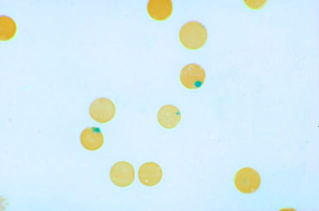
A 22-year-old male presents with shortness of breath and fatigue. He is planning a trip to an area in Africa in which chloroquine-resistant malaria is endemic, and he was started on a prophylactic antimalarial drug regimen several days ago. A blood smear is performed, and stained with a supravital stain. What are the round inclusions within the red cells?
Pappenheimer bodies
Howell-Jolly bodies
Heinz bodies
Hemoglobin H bodies
Warthin-Finkeldey bodies
The inclusions in these red cells are Heinz bodies, which are characteristic of G6PD deficiency. In this disease, the red cells are less able to handle oxidant exposure (common precipitating factors include infection, drug ingestion, and exposure to certain foods, such as fava beans). Following oxidant exposure, hemoglobin denatures and precipitates, forming so-called Heinz bodies which stick to the inside of the red cell membrane, leading to intravascular hemolysis and splenic destruction. Patients with G6PD deficiency typically develop symptoms of hemolysis after a period of two to three days following oxidant exposure. The anemia is usually short-lived and self-limiting.
-Kristine Krafts, MD, is an Assistant Professor of Pathology at the University of Minnesota School of Medicine and School of Dentistry and the founder of the educational website Pathology Student.
A 2-year-old male with no past medical history presented to the emergency department with fever and 2 days of bloody diarrhea. Stool cultures were sent to the laboratory. A Gram stain of the specimen showed the morphology seen in Figure 1. On the 5% sheep blood agar plate, the predominant organism had colonies that appeared flattened and spreading (Figure 2A). On MacConkey agar the colonies were noted to be non-lactose fermenting (Figure 2B). A Hektoen enteric (HE) agar was used as a differential and selective media to differentiate Salmonella from Shigella. On the HE agar the colonies were clear with a green appearance due to the color of the agar (Figure 2C).
Gram stain showing Gram-negative rodsIsolate growing on (A) 5% sheep blood, (B) MacConkey, and (C) Hectoen Enteric agars
Identification
Shigella is a bacterium in the Enterobacteriaceae family and is a Gram-negative rod that is facultatively anaerobic. It is non-motile, a non-spore former, and does not ferment lactose. There are four species of Shigella that are associated with subgroups A-D. Our isolate was identified as Shigella sonnei, which is the most common species in the U.S. and comprises subgroup D. The other subgroup/species correlations are listed in Table 1. The slide agglutination antisera test is used to aid in serogrouping. The suspected colony is mixed on a slide with antisera that contains specific antibodies to Shigella. If clumping (agglutination) occurs, it is considered a positive result for the specific subgroup. The organism was identified as Shigella sonnei by slide agglutination antisera testing. In addition, Shigella has certain biochemical properties that aid in further identification and confirmation.
Table 1: Shigella sp. determination by serogroup
Serogroup
Organism
A
Shigella dysenteriae
B
Shigella flexneri
C
Shigella boydii
D
Shigella sonnei
Clinical Significance
Shigella is one of the most common causes of bacterial gastroenteritis and is often associated with poor sanitation and overcrowded conditions. Transmission occurs through routes such as: fecal-oral and person to person contact. Of note, only a small amount of the bacteria (as low as 10 organisms) is required to cause disease. Hemolytic-uremic syndrome is a complication that may occur with shiga-toxin producing Shigella (the most commonly associated is S. dysenteriae). Shigella has demonstrated antibiotic resistance and therefore does undergo susceptibility testing.
References:
Nataro JP, Bopp CA, Fields, PI, Kaper JB, Strockbine, NA. 2015. Escherichia, Shigella, and Salmonella, p 603-626. In Jorgensen J, Pfaller M, Carroll K, Funke G, Landry M, Richter S, Warnock D (ed), Manual of Clinical Microbiology, Eleventh Edition. ASM Press, Washington, DC.
-Valerie Juarez, M.D., 3rd year Anatomic and Clinical Pathology resident, UT Southwestern Medical Center
-Erin McElvania TeKippe, Ph.D., D(ABMM), is the Director of Clinical Microbiology at Children’s Medical Center in Dallas Texas and an Assistant Professor of Pathology and Pediatrics at University of Texas Southwestern Medical Center.
The purpose of real-time PCR is to perform efficient amplification of a target sequence and quantify the PCR products in “real time” by employing the use of a fluorescent reporter. Fluorescent reporters can found in the form of DNA-binding dyes or fluorescently labeled primers or probes. It is extremely important to understand the difference between DNA-binding dyes, and the various fluorescent primer and probe based chemistries. The best way to grasp these theories is often to have a visual illustration of each of the different chemistries.
DNA-binding Dyes
SYBR Green Dye – SYBR Green I is a fluorescent DNA binding dye that is commonly used as it binds to all double-stranded DNA.
SYBR Green is detected by quantifying the increase in fluorescence during PCR.
Advantages to using SYBR Green are that it is inexpensive, easy to use, and easily incorporated into the PCR reaction.
Disadvantages of using SYBR Green are that there is usually an increase in background and non-specific binding that can lead to detection of false positive results.
Fluorescent PCR Primer and Probe Based Chemistries
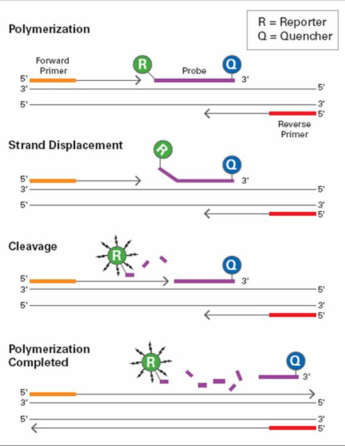
Taqman Chemistry – Utilizes 5’ – 3’ exonuclease activity of Taq Polymerase (enzyme that copies DNA and necessary for PCR) to generate a signal.
The probe is composed of a single stranded DNA oligonucleotide which is complementary to the specific target sequence of the PCR template.
The probe has a modification to the 3’ end so that the polymerase cannot extend the sequence.
The 5’ end has the fluorescent dye and the 3’ end contains the quencher
During DNA synthesis, the exonuclease activity of the Taq Polymerase will degrade the probe, thus resulting in release of the reporter from the quencher.
Molecular Beacon – This type of chemistry measures the accumulation of product during the annealing phase of PCR.
Signal is detected only when probes are bound to the template before displacement by the polymerase.
A chemical modification prevents degradation during the extension step of PCR.
The 5’ end contains the reporter fluorophore and the 3’ end contains the quencher.
The amount of fluorescence is directly related to the amount of initial template available for binding and inversely proportional to the cycle threshold (CT) value.
During extension, the probe is displaced by Taq Polymerase and the hair-pin (non-fluorescent) structure is restored.
Unbound molecular beacon probe à reporter is too close to quencher à no signal is generated.
Beacon probe binds to target à reporter is separated à signal is generated.
Understanding the various primer-probe chemistries including the interactions between the reporters and quenchers will provide some basic groundwork for those interested in pursuing a career in molecular biology.
-LeAnne Noll, BS, MB(ASCP)CM is a molecular technologist in Wisconsin and was recognized as one of ASCP’s Top Five from the 40 Under Forty Program in 2015.
Cryoprecipitate, or cryo for short, is a fresh frozen plasma (FFP)-derived concentrate including fibrinogen, factors VIII and XIII, von Willebrand factor, and fibronectin. Cryo contains only 40-50% of the coagulation factors found in a unit of plasma but is concentrated into a reduced 15-20 ml volume. Cryo is prepared from FFP as it is thawed slowly at 4° C. A precipitate forms at the bottom of the bag, which is then separated from the supernatant plasma. Cryo is stored frozen at at least 18° C and must be transfused within 6 hours of thawing or 4 hours of pooling. Each unit from a separate donor is suspended in 15 mL plasma prior to pooling.
Dose per unit
Half-life
Fibrinogen
150-250 mg
100-150 hours
Von Willebrand factor
100-150 U
24 hours
Factor VIII
80-150 U
12 hours
Factor XIII
50-75 U
150-300 hours
Cryo is used most commonly for replacement of fibrinogen in patients that are bleeding or at increased risk of bleeding. Fibrinogen replacement may be indicated for hypofibrinogenemia (fibrinogen < 100 mg/dL) or dysfibrinogenemia. The target increase in fibrinogen level is 30-60 mg/dL in adults and 60-100 mg/dL in pediatric patients. Many institutions transfuse cryo prior to administration of factor VIIa concentrate to ensure adequate fibrinogen for clot formation given the cost and short half-life of factor VIIa of about 4 hours. Fibrinogen replacement can be monitored with a fibrinogen level assay and clinical response.
Cryo may be used to treat von Willebrand disease, Hemophilia A (factor VIII deficiency), or Factor XIII deficiency only when the appropriate plasma-derived or recombinant factor concentrates are unavailable and/or desmopressin (DDAVP) is ineffective or contraindicated. Cryo is sometimes useful if platelet dysfunction associated with renal failure does not respond to dialysis or DDAVP. Cryo also contains fibronectin; however there are no clear indications for fibronectin replacement.
Topical application of cryo in combination with thrombin as a “fibrin glue” has been used as a surgical hemostatic agent. This application is being discontinued due to the preferred commercially available virus-inactivated fibrin sealants with higher fibrinogen concentrations.
Historically, the dosing was a 10-unit pool for adults and 1-2 units/10kg for pediatric patients based on fibrinogen content. However, Blood Bank and Transfusion services should check with their blood supplier on actual fibrinogen content in individual and pre-pooled units as the fibrinogen content has likely increased (~325 mg) due to improved preparation. Therefore Blood Bank and Transfusion services can probably decrease the standard dose to 4-5 pooled units for adults and 1 unit/10 kg for kids.
A previous version of this post said that cryo is frozen at 1-6°C; this is incorrect. The correct temperature is 18°C, and has been corrected in the text. Thank you, astute readers, for correcting our errors! –Lablogatory editors
-Thomas S. Rogers, DO is a second-year resident at the University of Vermont Medical Center, a clinical instructor at the University of Vermont College of Medicine, and the assistant medical director of the Blood Bank and Transfusion Medicine service.
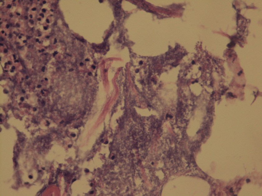
A 72 year old man had been complaining of many months of constipation followed by several months of severe diarrhea prior to admission. He did not receive medical care for these symptoms and had no medical history on file. He presented to an outside hospital unresponsive, and an emergent CT showed subcutaneous emphysema and stranding of the lower abdominal tissue extending to the left hemiscrotum. Also noted was irregular wall thickening in the distal rectum. Of note, his family reported that the patient had hit his left scrotum with a power cord recoil a few days prior to presentation. He was transferred to our hospital, where the surgical team reported severe cellulitis and necrosis of the scrotum, perineum, anterior abdominal wall, and upper thighs. Due to his poor prognosis, the family decided to transition his care to comfort measures only and he passed away several hours after presentation. An autopsy was performed, which revealed liquefaction of the subcutaneous tissue in the lower anterior abdominal wall, dusky gray connective tissue of the left testis as well as a small abscess at the superior pole, and a circumferential distal rectal mass.
Laboratory Investigation:
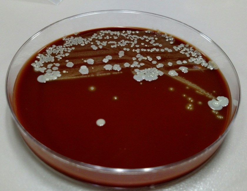
A gram stain of the scrotal abscess and tissue of the anterior abdominal wall both showed mixed gram positive and gram negative organisms. Cultures of these specimens both grew mixed gram positives including alpha hemolytic Streptococcus and coagulase negative Staphylococcus, as well as mixed anaerobes including Bacteroides fragilis group.
Gram stain of the scrotal abscess showing mixed gram negative and gram positive organisms.Histologic section of anterior abdominal wall (400X) showing many polys and many bacterial organisms.Chocolate agar plate showing mixed growth.
Discussion:
The case above represents an example of Fournier’s gangrene, a necrotizing infection of the external genitalia and/or perineum. These infections are most often polymicrobial and often include organisms that would not be particularly aggressive by themselves, suggesting a synergy between anaerobic and aerobic organisms. In a large literature review of over 4,000 cases of Fournier’s gangrene, the most common pathogens involved were Escherichia coli, Streptococcus, Bacteroides, Enterobacter, and Staphylococcus. Our case fits well with this profile, consisting of a mixture of gram positives and gram negatives including Streptococcus, Staphylococcus, and Bacteroides fragilis group. Antibiotic coverage in these cases must be broad spectrum and include coverage for aerobes, anaerobes, gram negative and gram positive organisms.
Risk factors for Fournier’s gangrene include diabetes mellitus, smoking, alcoholism, renal failure, hypertension, and coronary artery disease. It is unclear whether the patient in this case had any of these risk factors as he did not routinely seek medical care.
The rectal mass in this case proved to be adenocarcinoma. There have been multiple case reports of Fournier’s gangrene in association with rectal cancer. The theory behind the mechanism of this association is that perforation of the rectal cancer occurs with tumor infiltration of surrounding tissues spreading infectious organisms. It is possible that our case was caused by the patient’s rectal cancer, but it is also possible that the rectal cancer was simply coincidental. The recent trauma to the patient’s scrotum could also have acted as a vector for infection.
The patient in our case presented too late in the course of his disease to receive appropriate treatment, which would consist of broad spectrum antibiotic administration and aggressive debridement. Even with such treatment, the mortality rate of Fournier’s gangrene is quite high, ranging from 5% to 40% in different case series.
References:
Bjurlin MA et al. Causative pathogens, antibiotic sensitivity, resistance patterns, and severity in a contemporary series of Fournier’s gangrene. Urology 2013;81(4):752-8.
Bruketa T et al. Rectal cancer and Fournier’s gangrene – current knowledge and therapeutic options. World J Gastroenterol 2015;21(30):9002-9020.
-Laurie Griesinger is a Pathology Student Fellow at University of Vermont Medical Center.
-Christi Wojewoda, MD, is the Director of Clinical Microbiology at the University of Vermont Medical Center and an Assistant Professor at the University of Vermont.


