The purpose of real-time PCR is to perform efficient amplification of a target sequence and quantify the PCR products in “real time” by employing the use of a fluorescent reporter. Fluorescent reporters can found in the form of DNA-binding dyes or fluorescently labeled primers or probes. It is extremely important to understand the difference between DNA-binding dyes, and the various fluorescent primer and probe based chemistries. The best way to grasp these theories is often to have a visual illustration of each of the different chemistries.
DNA-binding Dyes
- SYBR Green Dye – SYBR Green I is a fluorescent DNA binding dye that is commonly used as it binds to all double-stranded DNA.
- SYBR Green is detected by quantifying the increase in fluorescence during PCR.
- Advantages to using SYBR Green are that it is inexpensive, easy to use, and easily incorporated into the PCR reaction.
- Disadvantages of using SYBR Green are that there is usually an increase in background and non-specific binding that can lead to detection of false positive results.

Fluorescent PCR Primer and Probe Based Chemistries
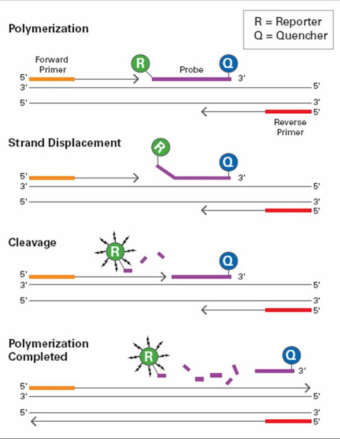
- Taqman Chemistry – Utilizes 5’ – 3’ exonuclease activity of Taq Polymerase (enzyme that copies DNA and necessary for PCR) to generate a signal.
- The probe is composed of a single stranded DNA oligonucleotide which is complementary to the specific target sequence of the PCR template.
- The probe has a modification to the 3’ end so that the polymerase cannot extend the sequence.
- The 5’ end has the fluorescent dye and the 3’ end contains the quencher
- During DNA synthesis, the exonuclease activity of the Taq Polymerase will degrade the probe, thus resulting in release of the reporter from the quencher.
- Fluorescent Resonance Energy Transfer (FRET) – Energy is transferred between two light sensitive molecules.
- Increase in target à More probes bind à Increase in fluorescence
- The 5’ end is the donor (catalyst) and the 3’ end is the acceptor (fluorophore)
- The energy is detected in the form of heat or fluorescence emission.
- If probes bind, energy is transferred from donor to acceptor and generates the signal.

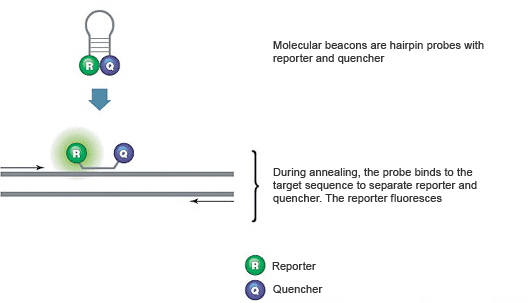
- Molecular Beacon – This type of chemistry measures the accumulation of product during the annealing phase of PCR.
- Signal is detected only when probes are bound to the template before displacement by the polymerase.
- A chemical modification prevents degradation during the extension step of PCR.
- The 5’ end contains the reporter fluorophore and the 3’ end contains the quencher.
- The amount of fluorescence is directly related to the amount of initial template available for binding and inversely proportional to the cycle threshold (CT) value.
- During extension, the probe is displaced by Taq Polymerase and the hair-pin (non-fluorescent) structure is restored.
- Unbound molecular beacon probe à reporter is too close to quencher à no signal is generated.
- Beacon probe binds to target à reporter is separated à signal is generated.

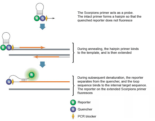
- Scorpion – Scorpion probes use two PCR primers, where one serves as a probe and once contains a stem-loop structure.
- The stem-loop structure contains a 5’ fluorescent reporter and a 3’ quencher.
- The loop of the scorpion probe contains complementary sequence to the internal portion of the target sequence.
- If the primer binds and extends, the reporter is separated from the quencher and a signal is given off.

Understanding the various primer-probe chemistries including the interactions between the reporters and quenchers will provide some basic groundwork for those interested in pursuing a career in molecular biology.

-LeAnne Noll, BS, MB(ASCP)CM is a molecular technologist in Wisconsin and was recognized as one of ASCP’s Top Five from the 40 Under Forty Program in 2015.

Very informative and important post. Thank you, it helped me in my study.
thank a lot. I learn some useful things form the post.