Microbiology Case Study: A 43 Year Old Woman with Headaches and Stiff Neck

Case history

A 43 year old woman with no significant medical history arrived at the emergency room complaining of several days of right-sided headaches, and worsening neck stiffness. Two days prior to coming into the emergency room, she had noticed some “bumps” on her posterior scalp, which her hairdresser looked at for her and confirmed the presence of a rash. Physical exam revealed a low-grade temperature of 100.6F. A small rash on the right side of the head was seen, consisting of a few erythematous patches and vesicles. A lumbar puncture was performed revealing clear and colorless cerebrospinal fluid, and the patient was given doses of ceftriaxone, vancomycin, and acyclovir.

Laboratory diagnosis

Analysis of the CSF was as follows:

  • Glucose: 45
  • Total Protein: 148 (H)
  • RBC Count: 15
  • Nucleated cell Count: 314
  • Neutrophils: 5%
  • Lymphocytes: 72%
  • Monocytes: 20%
  • Eosinophils: 3%

PCR of the CSF was positive for varicella-zoster virus (Figure 1).

vzv1

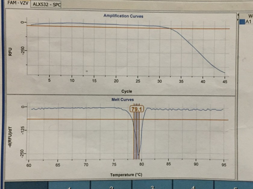
Figure 1. Real-time PCR amplification curves and melting curves for Varicella Zoster Virus in patient’s CSF sample. The amplification curve demonstrates decrease of Relative Fluorescence Units (RFUs) corresponding with detection of viral DNA. The melting point is determined by the composition of nucleic acids, and is unique to VZV, confirming the identity of the virus detected.


Discussion

Primary infection with VZV causes the classic chickenpox illness characterized by a widespread vesicular rash, with lesions of varying ages. Herpes zoster (shingles) occurs when latent varicella-zoster virus (VZV) in a sensory ganglion becomes reactivated, resulting in a painful vesicular rash typically in a dermatomal distribution. By 7-10 days after eruption, the vesicles of the rash will crust over and are not considered infectious. However, in immunocompromised patients, new vesicles may continue to erupt. The predominant complaint is pain along the site of the rash, in 75% of patients.

Even in immunocompetent patients, there is a risk of aseptic meningitis and even encephalitis with VZV reactivation. This can occur from the virus spreading centrally, to the CNS, rather than distally down a spinal nerve. Some studies have even indicated that subclinical meningitis (defined as CSF pleocytosis) occurs in up to 50% of individuals with herpes zoster. In one study, 0.5% of patients with herpes zoster developed meningitis. The typical findings of zoster meningitis on lumbar puncture include elevated protein as well as lymphocytosis.

Antiviral therapy (either with valacyclovir, famciclovir, or acyclovir) is often advised for the treatment of shingles if patients present within 3 days of symptom onset; it has the benefits of shortening the duration of skin lesions and acute neuritis, though its effects on post-herpetic neuralgia are less clear. After three days, the clinical benefit of antiviral treatment is debatable; however, it is recommended for patients with neurologic complications or with compromised immune systems.

The patient had chickenpox when in college. Although she had her zoster outbreak for 4 days by the time of presentation, because of the meningitis a course of oral Valtrex was prescribed. She was discharged home as she was clinically stable.


-Alison Krywanczyk, MD is a 3rd year anatomic and clinical pathology resident at the University of Vermont Medical Center.

Wojewoda-small

-Christi Wojewoda, MD, is the Director of Clinical Microbiology at the University of Vermont Medical Center and an Assistant Professor at the University of Vermont.


Microbiology Case Study: A 50 Year Old Male with High Fevers and Chronic Cough

Case History

A 50 year old male of Indian descent presents to the pulmonary clinic with complaints of high fevers and chronic cough. The cough has persisted for the past month and recently became productive with green sputum.  His fevers are cyclic in nature and reach 104°F.  He denies hemoptysis, unintentional weight loss or chest pain.  He has tried over the counter decongestants and cough suppressants as well as a course of levofloxacin, with minimal improvement. His past medical history is significant for rheumatoid arthritis, which is currently treated with methotrexate and prednisone. He works as a long distance truck driver and is a non-smoker.  A recent chest x-ray demonstrated a left hilar opacity with a nodular appearance. A computed tomography scan of the chest shows focal consolidation of the left lower lobe along with mediastinal and hilar adenopathy. Also, there are innumerable non-calcified nodules seen throughout bilateral lung fields. A bronchoscopy with bronchoalveolar lavage (BAL), transbronchial biopsy, and fine needle aspiration (FNA) of the enlarged lymph nodes were performed. BAL fluid was transported to the microbiology lab for bacterial, fungal and mycobacterial cultures.

Laboratory Identification

cocimm1

Figure 1. Histologic evaluation of the lung biopsy showed diffuse necrotizing granulomas which contained large yeast-like forms (red arrow) (H&E, 100x).

cocimm2

Figure 2. The large yeast-like forms measured between 10-25 µm in size and demonstrated a thick walled capsule (Grocott’s methenamine silver (GMS), 600x).

cocimm3

Figure 3. White mycelium with a downy texture and faint brown reverse grew on Mycosel agar after 28 days of incubation at 25°C.

cocimm4

Figure 4. Numerous coarse, septate hyphae producing thick walled, “barrel shaped” arthroconidia (lactophenol cotton blue stain, 1000x oil immerson).

Histology of the lung biopsy specimen showed necrotizing granulomas with occasional large, yeast-like spherules which measured between 10-25 µm in diameter (Figures 1 & 2). The spherules had a thickened capsule and endospores were not visualized. The fungal BAL cultures grew a white mycelium with a downy texture and light brown reverse after incubation for 28 days at 25°C on Mycosel agar (Figure 3). Microscopic morphology of a lactophenol cotton blue prep illustrated alternating thick walled arthroconidia suggestive of Coccidioides immitis/posadasii (Figure 4). The dimorphic mold was confirmed by DNA probe testing. Due to the findings on histology and the unusually slow growth of this particular isolate, Coccidioides IgM and IgG antibodies were performed by ELISA in the interim. They were found to be 5.4 and 4.4, respectively, suggestive of a current or recent infection. Laboratory studies for Aspergillus galactomannan, Fungitell, Cryptococcus antigen, and Histoplasma & Blastomyces urinary antigens were all negative. A Quantiferon Gold for Mycobacterium tuberculosis and all other cultures were also negative.

Discussion

Coccidioides immitis is often considered a thermally dimorphic mold geographically distributed to the arid climate of the southwestern United States and Mexico. It is morphologically identical to the C. posadasii, a species which is more widespread and endemic in South America. The two species can only be differentiated by molecular methods, although it is not routinely necessary as there is no difference in symptoms and treatment between the two.

Inhalation of infectious arthroconidia occurs as a result of environmental exposure to dust, sand and soil that has been disturbed. While many immunocompetent individuals who are exposed to C. immitis will show mild flu-like symptoms which resolve with no treatment, a portion of patients will go on to have pulmonary disease. A severe disseminated infection can occur in individuals with underlying immune system disorders, including rheumatologic diseases, HIV and transplant recipients on immunosuppression. C. immitis can have direct invasion of adjacent structures and can cause eruptive chronic granulomatous cutaneous disease. Women who are diagnosed with Coccidiomycosis during pregnancy are also at high risk for disseminated disease due to the presence of estrogen-like receptors in the fungus.

In the environment and when cultured in the laboratory at 25°C, C. immitis grows as a hyphal mold with alternating barrel-shaped arthroconidia (3-6 µm) separated by disjunctor cells. The arthroconidia are highly infectious and cultures in the laboratory must be worked up in a biological safety cabinet to minimize the risk of accidental exposure. The yeast-like phase occurs in tissue at temperatures above 35°C and is characterized as the transformation of arthroconidia in to large spherules (10-80 µm) which become filled with endospores. The cell wall of the spherule ruptures and the endospores are released into the tissue to become additional spherules.

Most patients with primary Coccidiomycosis do not require specific therapy as the disease will resolve on its own. For those patients who are immunocompromised, or whom exhibit severe disease, treatment is amphotericin B followed by fluconazole or itraconazole as maintenance therapy. In the case of our patient, he was placed on oral fluconazole twice daily for at least 3 months.


ka

-Kristen Adams, MD, is a fourth year Anatomic and Clinical Pathology resident at the University of Mississippi Medical Center. 

Stempak

-Lisa Stempak, MD, is an Assistant Professor of Pathology at the University of Mississippi Medical Center in Jackson, MS. She is certified by the American Board of Pathology in Anatomic and Clinical Pathology as well as Medical Microbiology. She is the director of the Microbiology and Serology Laboratories.  Her interests include infectious disease histology, process and quality improvement and resident education.  

Microbiology Case Study: A 60 Year Old Woman with a Skin Papule

Case history

A 60-year old woman residing in Vermont presented to the dermatology clinic for a routine annual skin exam. She had no complaints. On physical exam, a pink papule was seen on the patient’s back, with a centrally embedded tick (Figure 1). The tick was removed and sent for identification, with the plan to give a single prophylactic dose of doxycycline if identified as Ixodes scapularis.

Laboratory Identification

The tick was examined and noted to be less than 1 mm in size, with six legs (Figure 2). This was identified as the larval stage of Ixodes scapularis.

ixo1

Figure 1. Photograph of Ixodes scapularis larva embedded in the patient’s back. Six legs are visible.

ixo2

Figure 2. Photograph of the tick larva received in the laboratory, demonstrating a light tan-brown color.


Discussion

I scapularis, also known as the blacklegged tick, deer tick, or bear tick, is most clinically significant for its ability to transmit the pathogens Borrelia burgdorferi, Babesia spp., and Anaplasma phagocytophilum. It has four separate life stages (egg, larva, nymph, and then adult), spanning approximately 2 years. Each of these stages feeds on different preferred host animals.

Eggs are deposited on the ground by blood-engorged females in the late spring, where they subsequently hatch into 6-legged larvae. Because they have not yet fed, larva forms generally do not carry or transmit B. burgdorferi or other tick-borne pathogens. Trans-ovarial transmission of Borrelia, Anaplasma, or Babesia from adult I. scapularis females to eggs of is not a significant mode of pathogen transmission; however, in a similar tick species, I. ricinus (prominent in Europe), trans-ovarial transmission of Babesia divergens does occur, and so infection may be transmitted by larvae. The I. scapularis larvae will take their first blood meal from small mammals and birds, and then when engorged fall to the ground and molt into nymphs.

The nymph forms, which have already taken a blood meal, can carry pathogens and in fact are more likely to transmit pathogens to humans than the adult form of the tick. This is because the nymph form is much smaller (<2mm in size) than the adult form, and therefore is likely to go undetected when it attaches to a host. The nymphs are dormant over the winter, and re-activate the following spring to take their second meal. By fall, nymph forms have molted into adult ticks, which prefer to feed on white-tailed deer. However, while these deer support the tick population, they are not a large reservoir for Lyme disease. Rather, it is the white-footed mice preferentially fed upon by larvae and nymph forms that act as the main reservoir for B. burgdorferi, B. microti, and A. phagocytophilum. The female adults of I. scapularis are red to orange and larger than males, around 1/8 of an inch long, with a dark brown to black dorsal shield. If females do not feed in the fall, they can remain dormant over the winter and may emerge if the weather gets temporarily warmer (so the onset of cold weather does not necessarily mean the risk of tick exposure is over). Male adults do not take blood meals, and so do not transmit blood-borne pathogens.

To be considered in the differential diagnosis is Dermacentor variabilis, or the American dog tick. This tick species is larger than Ixodes spp., and adult forms have a white-to-gray collar on their backs. D. variabilis have more rectangular-shaped head and mouth parts than the deer tick. Both nymph and larvae forms are yellow-brown in color before feeding, and then turn gray once engorged. It is extremely uncommon for nymph and larval forms of D. variabilis to feed on humans, in contrast to I. scapularis. D. variabilis does not transmit Lyme disease, though in endemic areas it may transmit Rickettsia rickettsii or Francisella tularensis.

Because the tick in the presented case was identified as an I. scapularis larva, the patient was not treated with antibiotics as there was an exceedingly low risk of pathogen transmission.


-Alison Krywanczyk, MD is a 3rd year anatomic and clinical pathology resident at the University of Vermont Medical Center.

Wojewoda-small
-Christi Wojewoda, MD, is the Director of Clinical Microbiology at the University of Vermont Medical Center and an Assistant Professor at the University of Vermont.

Microbiology Case Study: A 21 Month Old Boy with Diaper Rash and Diarrhea

Case history

The patient was in his otherwise healthy state of being until 3 days prior when he developed non-bloody diarrhea. On the morning of presentation the stool had become bloody. The patient was afebrile, had some reduced intake of food, but drinking fine. Most notably, he periodically stops walking and bends over as if he is in pain. This happened 3-4 times the previous day and these episodes tended to last for about one minute, after which the boy would continue to play. He does not attend daycare and his immunizations are up to date.

Stool culture was sent and a predominant organism was an oxidase negative, lactose-fermenting, Gram-negative rod (Figure 1). The organism was non-sorbitol fermenting based on growth on Sorbitol-MacConkey agar, and grew as mauve colonies on E. coli 0157 screening agar. MUG testing was negative.

O157.png
Figure 1: Subculture of the disease causing organism on (A) MacConkey, (B) Sorbiol-MacConkey, (C) and E. coli O157 screening agars.

Discussion

The isolate was E. coli O157. Isolates of E. coli O157 commonly produce shiga toxins (sxt1 and sxt2) which are responsible for diarrhea, hemorrhagic colitis, and most famously hemolytic-uremic syndrome (HUS). Typical illness starts with non-bloody diarrhea which becomes bloody after 2-3 days due to onset of hemorrhagic colitis. Often severe abdominal pain and low grade fever are present as well. HUS is a serious complication of E. coli O157 infection which results in acute renal dysfunction. HUS most often occurs in children < 5 years of age, of which 15% of those with laboratory confirmation of E. coli O157 developing this complication, compared to 6% in the general population. It is possible for other E. coli to produce shiga toxins, with 1% of HUS is caused by non-E. coli O157 infection.

It is recommended that all patients with suspected HUS should have stool cultured on selective and differential media for detection of E. coli O157 and direct shiga toxin detection should be performed to identify non-E. coli O157 isolates that are producing toxin. E. coli 0157 isolates look exactly the same as non-E. coli O157 normal fecal flora on 5% sheep blood, chocolate, and MacConkey agars. All E. coli ferment lactose on MacConkey agar (Figure 1A). E. coli O157 can be differentiated from other E. coli strains by growth on Sorbitol-MacConkey (SMAC) agar; E. coli O157 is a non-sorbitol fermenter while most other E. coli will ferment sorbitol (Figure 1B). Chromagenic agar for E. coli O157 is another option to screen stool specimens for E. coli O157. E. coli O157 grow mauve colored colonies on this particular agar (BBL CHROMagar O157 , Becton Dickinson) (Figure 1C). A summary of this data can be found in Table 1.

Growth of organisms suspicious for E. coli O157 on any media requires confirmation prior to reporting. Biochemical confirmation tests include E. coli O157 antiserum or latex agglutination and 4-methylumbelliferyl-beta-D-glucuronide (MUG) testing. For latex agglutination or antisera testing, it is essential to test the isolate of interest with the E. coli O157-specific reagent as well as a non-specific control to exclude non-specific binding. Unlike most E. coli strains, E. coli-O157 does not express beta-glucuronidase and is therefore MUG test negative (Table 1).

Table 1. Characteristics of E. coli O157 in comparison to other E. coli strains

Test Non-E. coli O157 E. coli O157
Appearance on MacConkey agar Lactose fermenter Lactose fermenter
Appearance on Sorbitol-MacConky agar Sorbitol fermenter Non-sorbitol fermenter
MUG testing Positive Negative

For direct detection of shiga toxin, there are several commercially available immunoassays available for detection of shiga toxin protein. New on the market are multiplex gastrointestinal panels that can be used for molecular based detection of shiga toxin genes sxt-1 and sxt-2 among a host of other agents of gastrointestional disease.

E. coli O157 is spread via fecal oral route. It can be acquired directly from person to person or indirectly through food and water sources contaminated with fecal matter from infected humans and animals. Classic scenarios are undercooked ground beef, leafy greens, unpasteurized milk and juice, petting zoos, and contaminated drinking water. The incubation period prior to symptoms is 3-4 days (range 1-8 days).

Treatment for E. coli O157 is largely supportive consisting of fluids to prevent dehydration. The role of antibiotics is controversial with some studies suggesting antibiotics increase the risk of developing HUS while others found no association between the their use and increased HUS.

Following our patient’s stool culture result for E. coli O157, he was recalled to the Emergency Department for evaluation. He was still having diarrhea and vomiting, but it was reduced compared to the previous day. The patient was given fluids and sent home without antibiotic treatment and via phone conversation with his mother, his symptoms resolved a few days later.

-Erin McElvania TeKippe, PhD, D(ABMM), is the Director of Clinical Microbiology at Children’s Medical Center in Dallas Texas and an Assistant Professor of Pathology and Pediatrics at University of Texas Southwestern Medical Center.

Microbiology Case Study: A 73 Year Old Man with Altered Mental Status and Fever

Case History

A 73 year old man was brought to the emergency room with altered mental status and fever, which developed a few days following a 1-2 day illness characterized by myalgia and diarrhea. He was admitted to the hospital and blood cultures were drawn.

Laboratory Identification

The bottles flagged positive after 12 hours and Gram stain showed small, Gram positive rods (Figure 1). Growth of white, smooth translucent colonies was seen on the blood and chocolate plates, with a small rim of beta-hemolysis on the blood plate (Figure 2). MALDI-TOF confirmed the identification as Listeria monocytogenes.

list2

Figure 1. Gram stain morphology of the colonies growing, demonstrating short Gram positive bacilli.

 list1

Figure 2. Smooth white colonies growing on the blood and chocolate plates, with a soft rim of beta-hemolysis visible on the blood plate.

Discussion

Listeria monocytogenes is prevalent throughout the environment, and can also colonize the human gastrointestinal tract. Humans are exposed by consumption of contaminated food, particularly soft cheeses, deli meats, and fruit. Listeria can grow at 4C which means it can multiply in refrigerated foods, making even low-level contamination a potential hazard. On gram stain, it is a short gram positive rod which may form chains. In some cases, the rods may be so short as to resemble chains of Streptococci, and with the soft surrounding beta hemolysis, could potentially be confused for Group B Streptococcus. However, Listeria is catalase positive, while Group B Strep is negative. Another characteristic feature of Listeria is the “tumbling motility” on wet prep at 20-25C, or “umbrella motility” in tube agar. Listeria also has the unique feature of manipulating the host cells’ intracellular actin framework, using it to facilitate direct cell-to-cell spread of the bacteria. The main virulence factor is the listeriolysin toxin, which is postulated to permit survival of the organism within macrophages via cytotoxic activity.

Listeria can cause a self-limited febrile gastroenteritis in previously healthy individuals, but typically only if they consume a large inoculum. However, in neonates, the elderly, or the immunosuppressed, it can invade and cause sepsis, meningitis, or meningoencephalitis. In pregnant women, Listeria can cross the placenta and lead to intrauterine fetal demise, premature labor, or neonatal meningitis, as well as the typically fatal condition granulomatosis infantiseptica in which the newborn develops widespread abscesses throughout multiple organ systems. Infection during pregnancy usually happens during the 3rd trimester, though the effects seem to be more severe with earlier infection.

 Listeria has been cultured from the stool of up to 3.4% of healthy, asymptomatic humans, and so there is little utility in stool cultures for Listeria except for epidemiologic purposes during an outbreak. Infections due to outbreaks of Listeria are far less common than sporadic infections, which comprise 95% of Listeria infections. Additionally, traditional stool cultures are poor at detecting Listeria and selective media is usually required. Blood and cerebrospinal fluid are the preferred sites of culture if there is suspicion for disseminated infection. Meningitis caused by Listeria is unique in that is can cause a lymphocyte-predominant CSF pleocytosis, which may result in confusion for viral meningitis. Additionally, gram stains of the CSF are only positive in approximately 1/3 of patients, so a high index of suspicion needs to be maintained while awaiting final culture results. While antibiotic treatment is not recommended for otherwise healthy patients with febrile gastroenteritis, it is recommended for those with disseminated infection or at high risk of dissemination (i.e. extremes of age, immunocompromised, or pregnant).


-Alison Krywanczyk, MD is a 3rd year anatomic and clinical pathology resident at the University of Vermont Medical Center.

Wojewoda-small

-Christi Wojewoda, MD, is the Director of Clinical Microbiology at the University of Vermont Medical Center and an Assistant Professor at the University of Vermont.

Microbiology Case Study: A 57 Year Old Woman with Recurrent Fever

Case History

A 57 year old female presents to the hospital with complaints of a recurrent fever for the past few days. Her past medical history is significant for acute myeloid leukemia (AML). On physical examination, she has multiple, warm, erythematous, non-painful papules and nodules involving her extremities. One of the lesions, located on the dorsal aspect of her finger, had ulcerated. She is found to be neutropenic with a white blood cell count of 0.36 TH/cm2 (reference range 4.0-10 TH/cm2). A chest CT scan is performed and reveals multiple, small hyperdense pulmonary nodules.  As part of the work up for febrile neutropenia, blood cultures are collected. Dermatology was also consulted and a skin biopsy at the advancing edge of the lesion was performed.

Laboratory Identification

fus1

Figure 1. Review of the deep portion of the punch biopsy demonstrates panniculus with a deep, dilated vessel containing fibrin and possible fungal organisms (H&E, 40x).

fus2

Figure 2. Deep dilated blood vessel with fibrin and fungal forms consistent with a hyaline septate mold were identified (H&E, 400x). Mycotic organisms were confirmed by a Grocott’s methenamine silver (GMS) stain.

fus3

Figure 3. Gram stain from a positive blood culture vial highlighting narrow hyphal elements with acute angle branching consistent with a hyaline septate mold (400x).

fus4.png

Figure 4. Lactophenol cotton blue preparation highlighting septate hyphae and numerous macroconidia that can be described as canoe or banana shaped (400x).


All of the specimens were collected on the same day and the results from the skin biopsy which showed fungal elements consistent with a hyaline septate mold were reported first (Figure 1). After 2 days of incubation, multiple blood culture bottles were positive for a mold similar to what was seen on tissue biopsy, indicating a disseminated fungal infection in this severely immunocompromised patient (Figure 2). Fungal cultures were set up from the blood and after 5 days of incubation at 30°C, lavender cottony colonies with lighter periphery grew on Sabouraud’s dextrose agar (SDA). The lactophenol cotton blue preparation showed many macroconidia with three to five septa (Figure 4). All of the findings are consistent with Fusarium spp.

Discussion

Fusarium spp. are filamentous fungi which are classified as a rapidly growing, hyaline septate mold. This opportunistic mold can be found all over the United States in the soil and on plants. In immunocompetent individuals, Fusarium spp. can cause localized infections, most commonly as the result of traumatic inoculation. Frequently, the eye is the site of infection, leading to keratitis. This can be due to trauma, contamination of contact lenses or solution, or corticosteroid drops. Fusarium has been reported as the infectious pathogen and has many clinical manifestations such as pneumonia, sinusitis, and wound infections. However, in immunocompromised patients, Fusarium spp. pose a greater threat for invasive and disseminated infections. Of the fungal organisms routinely implicated in fungemia (in addition to Candida, Cryptococcus and Histoplasma), Fusarium spp. have a high frequency of positive blood cultures.

Blood cultures positive for a septate mold and the presence of characteristic skin lesions are highly indicative of a disseminated Fusarium infection, especially in a severely neutropenic patient. To support the diagnosis of fusariosis, it is reassuring to have two different specimens each growing the same pathogen. In the case of our patient, both blood and skin tissue cultures grew Fusarium spp. with the same colony and microscopic morphology. The patient was unable to produce sufficient sputum for respiratory cultures, but the lung abnormalities were attributed to the fungal process as well.

In the laboratory, Fusarium spp. grow relatively rapidly on Sabouraud’s dextrose agar and can usually be identified within 3 to 5 days. Colonies are typically cottony in appearance and develop a pink to lavender color as they mature. The reverse of the plate is usually light. Fusarium spp. produce both macroconidia and microconidia. The characteristic macroconidia have been described as canoe, banana or sickle shaped and are separated by 3 to 5 transverse septa. The microconidia arise from short conidiophores and are more oval in shape, containing zero to one septa and can be single or arranged in clusters.

In cases of invasive fusariosis, anti-fungal agents such as voriconazole or high-dose amphotericin B are therapies of choice. In the case of neutropenic patients, growth factors (G-CSF or GM-CSF) or granulocyte transfusions are potential treatment options as well. Surgical debridement of necrotic tissue has shown benefit in patients with large abscesses. Additionally, if the cause of the fungemia is thought to the result of an infected line, the catheter should be removed.

Disseminated involvement of Fusarium spp. has a high rate of mortality associated with the infection.  Often times the prognosis is related to the extent of infection and the degree immunosuppression.  It is important to suspect Fusarium infections in the clinical setting of a severely neutropenic patient with skin lesions and having a low threshold for beginning anti-fungal therapy. An accurate and prompt diagnosis will lead to appropriate treatment and improved outcomes.


kt

-Katie Tumminello, MD, is a fourth year Anatomic and Clinical Pathology resident at the University of Mississippi Medical Center. 

Stempak

-Lisa Stempak, MD, is an Assistant Professor of Pathology at the University of Mississippi Medical Center in Jackson, MS. She is certified by the American Board of Pathology in Anatomic and Clinical Pathology as well as Medical Microbiology. She is the director of the Microbiology and Serology Laboratories.  Her interests include infectious disease histology, process and quality improvement and resident education. 


Microbiology Case Study: A 5 Month Old with Redness in her Left Eye

Case History

A 5 month old girl was brought to her pediatrician by her mother for redness of the medial canthus of her left eye. There was some associated thick discharge as well. The mother mentioned that her daughter had excessive tearing from the left eye since birth. The pediatrician diagnosed dacryocystitis, and prescribed a course of oral cefdinir. However, the swelling and redness continued to worsen, and the infant began to have low-grade fevers. She was sent to pediatric ophthalmology for a consult, and admitted to the hospital on IV clindamycin. The following day, she was taken to the operating room, where they opened the obstructed nasolacrimal duct and sent the contents for culture.

Laboratory Work-Up

Gram stain and smear: No neutrophils, no bacteria seen.

Routine bacterial culture showed growth of small, smooth and translucent colonies on the chocolate agar only. The colonies had a distinct wet-mouse odor.

Repeat review of the gram stain showed moderate neutrophils, with gram negative coccibacilli both extracellular and intracellular.

Hflu1
Chocolate plate demonstrating growth of smooth, translucent colonies.
Hflu2
Thick, uninterpretable region of gram stain.
Hflu3
Repeat review of gram stain showed gram negative coccobacilli, with intraleukocytic organisms.

Discussion

The gram stain and colony morphology described above are consistent with Haemophilus influenzae. Identification of the organism was confirmed by MALDI-TOF.

Congenital nasolacrimal duct obstruction is a common problem, affecting up to 6% of newborns. The vast majority of cases will resolve without treatment by the time the child is 6 months old. However, complications can arise, include acute or chronic dacryocystitis. Acute dacryocystitis causes swelling, warmth, and erythema with or without purulent discharge. The organisms most commonly implicated are alpha-hemolytic streptococci, Staphylococcus epidermidis, and Staphylococcus aureus. In chronic dacryocystitis, there is purulent drainage from the eye but no other signs or symptoms. The most common organisms isolated in these cases are Streptococcus pneumonia, Haemophilus influenzae, Pseudomonas aeruginosa, and viridans group Streptococci.  Acute dacryocystitis usually requires systemic antibiotics, while chronic can be treated with topical antibiotics. In this case, the infant had clinical features of acute dacryocystitis, but infection with an organism more typically associated with chronic dacryocystitis.

H. influenzae is a gram negative coccobacillus, which grows only on chocolate agar due to its requirement for factors V and X. However, H. influenzae can grow on blood agar if it is growing around an organism that hemolyzes the red blood cells in the media, releasing factor V (i.e. Staphylococcus aureus), a phenomenon known as satelliting.

As illustrated by this case, reviewing gram smears inconsistent with the final culture is an important aspect of quality assurance in the microbiology laboratory. This allows the opportunity to provide continuous feedback to technologists on their technique, and lets us keep track of any trends or common mistakes that may be occurring. In this instance, on review of the gram smear it appears the original reader examined only the very thick portions of the smear, which were uninterpretable. However, by moving out to the edges, neutrophils and bacteria were clearly visible. Feedback was provided to the original reader. The provider was immediately called and notified that a corrected report would be issued; the patient was switched to oral Augmentin based on these results.

-Alison Krywanczyk, MD is a 3rd year anatomic and clinical pathology resident at the University of Vermont Medical Center.

Wojewoda-small

-Christi Wojewoda, MD, is the Director of Clinical Microbiology at the University of Vermont Medical Center and an Assistant Professor at the University of Vermont.

Microbiology Case Study: 15 Year Old with Bacteremia

Case History

A 15-year-old male patient with acute myeloid leukemia (AML) had a central line placed for chemotherapy and subsequently developed symptoms of fever, abdominal pain, and diarrhea a few weeks later. He was treated with metronidazole for intra-abdominal infection and experienced improvement in diarrhea and abdominal pain, however his fever remained. Blood cultures were drawn from his central line and were positive.

gran1

Figure 1. Gram stain of the positive blood culture showing Gram-variable cocci in pairs and chains

gran2

Figure 2. Growth profile on organism on 5% sheep blood and chocolate agars

gran3

Figure 3. Colony Gram stain demonstrating pleomorphic, Gram-variable cocci in pairs and short chains

Laboratory diagnosis

Gram stain of the blood specimen revealed Gram-variable cocci in pairs and chains (Figure 1). The specimen was cultured on 5% sheep blood, chocolate blood, MacConkey, and Columbia colistin nalidix agar (CNA) agars. The next day, growth of small, round, grey colonies was seen only on the chocolate agar (Figure 2). Catalase test was negative. MALDI-TOF (Matrix Assisted Laser Desorption/Ionization, Time-of-Flight) identified Granulicatella adiacens with a 2.267 match score.

Discussion

The Granulicatella genus is a former member of the Abiotrophia genus, previously known as nutritionally variant or satelliting streptococci. The Granulicatella genera consists of G. adiacens and G. elegans (formerly A. adiacens and A. elegans respectively); of the Abiotrophia genus, only A. defectiva remains. These organisms are normal flora of the oral cavity, upper respiratory tract, gastrointestinal tract, and genitourinary tract. Granulicatella endocarditis is an uncommon, but well-documented phenomenon of both native and prosthetic valves and accounts for 5-6% of all streptococcal endocarditis. Other potential complications include ocular, central nervous system, musculoskeletal infections, bacteremia, pneumonia, scrotal abscess, septic arthritis, peritoneal dialysis-associated peritonitis, and breast-implant associated infections.

The Granulicatella spp. are catalase-negative, oxidase-negative, facultative anaerobic, gram-positive coccobacillus arranged in pairs and chains; although pleomorphism may occur (Figure 3, colony Gram stain). Notably, Granulicatella will grow on chocolate blood agar, but not 5% sheep blood agar or CNA agar because it requires Pyridoxine or vitamin B6 for growth. In addition to chocolate agar, Granulicatella can grow on Brucella agar with 5% horse blood and in thioglyconate broth. Granulicatella and other nutritionally variant streptococci exhibit satelliting behavior.  Satellite testing is performed on a media that supports no or little growth of Granulicatella (e.g. sheep blood agar). A single streak of Staphyloccous aureus across an area of the media inoculated with Granulicatella is incubated at 35°C in a high CO2 atmosphere. Granulicatella will grow only in the vicinity of S. aureus growth.  An alternate test for satelliting involves supplementation of media with pyridoxine hydrochloride, to allow growth of Granulicatella.

Biochemical reactions include: pyrrolidonyl arylamidase production (PYR) positive, leucine aminopeptidase production (LAP) positive, 6.5% NaCl negative, and bile esculin negative. Species identification is accomplished by arginine hydrolysis (ARG) and beta-glucuronidase (BGUR) activity testing. G. elegans is ARG-positive, while G. adiacens and Abitrophia are both negative. G. adiacens is further identified by a positive BGUR analysis, and Abiotrophia is again negative.

Abiotrophia and Granulicatella have shown variable susceptibility to both penicillin and aminoglycoside antibiotics. There is documented resistance to clindamycin, tetracycline, erythromycin, and ciprofloxacin, but not to rifampin or vancomycin. Current recommendations are to treat similarly as for enterococcal endocarditis using a combination therapy of a beta-lactam antibiotic with an aminoglycoside antibiotic, such as penicillin plus gentamycin. Unfortunately, relapse rates appear high despite appropriate treatment.

The likely source of this patient’s Granulicatella bacteremia is bacterial translocation from the gut in the setting of an immunocompromised state. The work up for a central line source of the bacteremia is still currently in progress, and echocardiogram was negative for vegetation.  He is currently being treated with vancomycin for bacteremia, and cefepime and metronidazole for intra-abdominal infection.

References

  1. Ruoff, K. Aerococcus, Abiotrophia, and other aerobic catalase-negative, gram-positive cocci. Manual of Clinical Microbiology, 10th Edition(pp. 365–376). American Society of Microbiology.
  2. Procop, G. W., Church, D. L., Hall, G. S., Janda, W. M., Koneman, E. W., Schreckenberger, P. C., & Woods, G. L. (2016). Koneman’s Color Atlas and Textbook of Diagnostic Microbiology(7th ed.). Philadelphia: Wolters Kluwer.
  3. Cargill, J., Scott, K., Gascoyne-Binzi, D., Sandoe, J. “Granulicatella infections: diagnosis and management.” Journal of Medical Microbiology 16 (2012): 755-761.


-Melinda Flores, MD, is a 1st year clinical and anatomic pathology resident at the University of Texas Southwestern Medical Center, Dallas, Texas.

-Erin McElvania TeKippe, PhD, D(ABMM), is the Director of Clinical Microbiology at Children’s Medical Center in Dallas Texas and an Assistant Professor of Pathology and Pediatrics at University of Texas Southwestern Medical Center.


Candida auris: An Emerging Pathogen

Clinical laboratory professionals, microbiologists, and pathologists need to be aware of an emerging fungal pathogen. According to the CDC, Candida auris is a threat because:

  • It’s multi-drug resistant
  • It’s often incorrectly identified by common laboratory techniques and analyzers
  • It causes outbreaks in the healthcare setting.

Here are a few resources if you’d like to read more.


Microbiology Case Study: A Newborn Baby in Respiratory Distress

Case History

A 29 year old G2P1 woman presented in labor at 39+2 weeks gestational age. Her pregnancy had been previously uncomplicated. Prenatal infectious disease testing showed that she was negative for HIV and Hepatitis C, but that she was positive for Group B Streptococcus. No test results were available for rubella, VZV, toxoplasmosis, or syphilis.

A term male infant was born shortly afterwards by spontaneous vaginal delivery; the mother received less than 4 hours of antibiotics.  The baby was noted to be covered in petechiae, and in a moderate amount of respiratory distress. A CBC showed thrombocytopenia to 23 K/cmm. The baby was transported emergently to the neonatal intensive care unit, where platelet transfusions were given. Blood cultures were drawn. The baby was started empirically on ampicillin/gentamycin, and the following day, once platelet counts were improved, a lumbar puncture was performed. The cell counts in the CSF were unremarkable. A cranial ultrasound showed scattered bilateral parenchymal calcifications, mineralized vasculature of the lenticulostriate arteries, and a subependymal cyst. Urine PCR testing was positive for CMV.

Laboratory Work-up

  • Bacterial Culture and Smear, CSF: No neutrophils, no bacteria. No growth.
  • CSF Viral PCR: Negative for HSV and VZV.
  • Urine CMV PCR: Positive.
  • The placenta was not sent for pathologic examination.

CMV1

Cranial ultrasound demonstrating scattered parenchymal calcifications.

CMV2
Photomicrograph of a lung from a 20 week gestation fetus demonstrating the characteristic “Owl’s Eye” inclusion of CMV.
CMV3
Photomicrograph of the placenta from the same case as B. The chronic villitis with plasma cells seen here is a sign of CMV infection.


Discussion

Cytomegalovirus is one of the classic “TORCH” infections. TORCH is an acronym for a group of pathogens that can cause in-utero or intrapartum infections:

  •                 T= Toxoplasmosis
  •                 O= other (syphilis, VZV, parvovirus)
  •                 R= rubella
  •                 C= CMV
  •                 H= HSV

Although these infections share several common signs and symptoms, there are clinically suggestive findings that can help target testing. The combination of thrombocytopenia and intracranial calcifications in this infant raised strong suspicion for congenital CMV. CMV is a member of the herpesvirus family.  It is a double-stranded DNA virus with both a viral capsid and envelope. While most babies born with congenital CMV are asymptomatic (~90%), congenital CMV infection is the main etiology of non-hereditary sensorineural hearing loss. This occurs in up to 50% of symptomatic infants and in 10-15% of asymptomatic infants. Symptomatic infants may be small for gestational age, and can be afflicted by thrombocytopenia, petechiae, intracranial calcifications, chorioretinitis, hepatosplenomegaly, microcephaly, and jaundice. While toxoplasmosis can also cause intracranial calcifications, it does not typically cause thrombocytopenia. Congenital HSV can cause thrombocytopenia, but is not associated with intracranial calcifications.

CMV infection during pregnancy is most often acquired by contact with young children. CMV has the ability to remain latent in the host, and become reactivated at a later time, so pregnancies can be affected by either primary infection or by reactivation of the virus. The risk of vertical transmission is much higher with primary CMV infection (32%) than with recurrent infection (1.4%). Although the rate of vertical transmission increases if the infection occurs later in pregnancy, infections acquired in early pregnancy are more likely to cause symptomatic disease. Treatment for the baby is generally supportive, with antivirals generally used only in symptomatic disease (their utility in asymptomatic infection is debated).

An audiology screen and ophthalmologic exam were both normal in the infant presented here. Oral valgancyclovir was started in addition to other supportive measures.


-Alison Krywanczyk, MD is a 3rd year anatomic and clinical pathology resident at the University of Vermont Medical Center.

Wojewoda-small

-Christi Wojewoda, MD, is the Director of Clinical Microbiology at the University of Vermont Medical Center and an Assistant Professor at the University of Vermont.