Microbiology Case Study: A 10 Year Old Girl with New Onset Fever

A 10 year old girl presented to the ED with several month history of worsening CNS symptoms, including gait instability, left-sided weakness, nausea, vomiting, several witnessed seizure events and loss of consciousness. MRI revealed right frontal mass measuring 7.0 cm in greatest dimension with midline shift. The patient underwent a right-sided craniotomy with tumor resection and was closely followed in the PICU. On post-op day 3, the patient appeared pale and lethargic with a temperature of 39.9 C. Blood cultures were collected and the patient empirically started on ceftriaxone and vancomycin for meningitis coverage. Due to worsening fever and altered mental status, a lumbar puncture was performed with the following results:

  • Opening pressure: 10.5 cm
  • Appearance: slightly cloudy
  • Cells: 91% PMN
  • Gram negative rods were identified on gram smear


OLYMPUS DIGITAL CAMERA
Gram stain shows gram negative rods. Photo credit: Dr. Gary Kaiser.

pseudostut2

Photo credit: University Medical Center of Rotterdam, Dept. of Microbiology and Infectious Disease

Discussion

Pseudomonas stutzeri is an aerobic gram negative bacilli (GNB) with a single polar flagellum that is both catalase and oxidase positive. Several defining features help distinguish P. stutzeri from other Pseudomonas species, including its nutritional versatility (utilizes starch, maltose, and ethylene glycol), its inability to produce fluorescent pigment and its “coral-like” appearance on growth medium.  There are no known virulence factors for P. stutzeri. Hospital distribution rates of P. stutzeri are approximately 1-2% of Pseudomonas spp. Nosocomial infection via medical devices or solutions is the most common route of infection. P. stutzeri isolates are sensitive to beta lactam antibiotics—meropenem is commonly used.


-Christina Litsakos is a Pathology Student Fellow at University of Vermont Medical Center.

Wojewoda-small

-Christi Wojewoda, MD, is the Director of Clinical Microbiology at the University of Vermont Medical Center and an Assistant Professor at the University of Vermont.

Microbiology Case Study: A 25 Year Old Male with Fevers, Headache, and Neck Pain

Case History

A 25 year old Caucasian male with no significant past medical history presented to the emergency department (ED) with a several day history of persistent fevers, headache (pain 10/10), dizziness and neck pain. He also reported facial and hand numbness and difficulty focusing at work.  His laboratory values at that time showed a normal white blood cell count (5.3 TH/cm2), normal hemoglobin (13.8 g/dL), slightly decreased platelet count (142,000 TH/cm2) and slightly elevated liver enzymes. A computed tomography (CT) of his head showed no abnormalities, and a lumbar puncture was performed that was suggestive of viral meningitis (92% lymphocytes). After obtaining blood cultures, the patient received a dose of vancomycin and ceftriaxone and was discharged home.

Two days later he returned to the ED with complaints of worsening neck pain, photophobia, decreased appetite, and fevers reaching 105°F. He reported fevers of such intensity that he resorted to soaking himself in ice baths. On further questioning, he reported working in a microbiology lab that handles cytomegalovirus (CMV) and attenuated mycobacterium, but was unaware of any exposures or sick contacts. He has 2 dogs he rescued (one with a history of heartworm), 3 cats (one with a history of tapeworms) and a mouse. Infectious disease was consulted and a thorough workup was initiated, which included repeat blood cultures and testing for hepatitis, human immunodeficiency virus (HIV), respiratory pathogens and syphilis. He was started on cefepime initially and then later changed to meropenem and levofloxacin.

Laboratory identification

brucan1

Figure 1. Gram stain from a positive blood culture illustrating small Gram negative coccobacilli (100x, oil immersion).

The microbiology laboratory reported the blood cultures collected during his second hospital visit were positive with small gram negative coccobacilli after approximately 60 hours on the automated instrument (Figure 1). No growth was noted after 24 hours incubation at 35°C in 5% CO2 on standard media. After 48 hours, small white colonies grew on sheep blood and chocolate agars but failed to grow on MacConkey agar. Biochemical tests revealed the organism was positive for catalase, oxidase and urease. In accordance with the suspected agents of bioterrorism manual, the culture was sent to the State Department of Health for further classification. The organism was identified by PCR as Brucella spp.  Subsequently, the Centers for Disease Control and Prevention (CDC) performed species level PCR and identified the isolate as B. canis.

On further questioning, the patient denied consuming unpasteurized milk products but reported recently adopting a pregnant dog from a local shelter, who had subsequently delivered stillborn puppies of which the patient had been in close proximity. At this point, the patient’s antibiotics were switched to a 6 week course of oral doxycycline and rifampin. On follow up visits, he was doing well and symptom free. Unfortunately, the dog also tested positive for B. canis and had to be euthanized.

Discussion

Brucella spp. are common zoonoses among wildlife and domestic animals including cattle (B. abortus), pigs (B. suis), goats (B. melitensis) and dogs (B. canis) who are usually asymptomatic carriers. While rare in the United States due to vaccination of livestock, Brucella spp. is considered endemic in areas of the Middle East, Central and South America and the Indian subcontinent. Symptoms of infection generally occur during an infectious abortion in which the placenta, fetal tissues and secretions contain high levels of the bacteria which can survive in the environment under various conditions for long periods of time. Humans are usually infected due to consumption of unpasteurized milk and cheeses. High risk professions such as veterinarians and slaughterhouse workers can also be infected by direct contract with contaminated materials or inhalation of aerosolized particles. Symptoms generally appear 1 to 2 weeks after infection with remittent/undulant fever the characteristic feature of the illness, in addition to arthralgias, fatigue, weight loss and hepatomegaly.

Laboratory identification of Brucella spp. is the gold standard but can be challenging as it is a slow growing organism and can infect personnel leading to laboratory acquired infections (LAI). When small Gram negative coccobacilli are identified that fail to grow on MacConkey agar, this should alert the laboratory worker of a potential agent of bioterrorism and work up should be performed in appropriate biosafety cabinets. Brucella spp. grows as small, smooth white colonies that appear after 24 to 48 hours incubation. It is catalase, oxidase and urease positive. Automated systems and MALDI-TOF mass spectrometry are not terribly reliable or recommended for identification of this organism due potential aerosolization events. When Brucella spp. is suspected, the level A clinical laboratory (a sentinel lab) should notify and send samples to a Level B/C lab (state health department) for confirmation.  Subsequently, confirmed isolates can be forwarded to a level D lab (CDC) for speciation.

While overall, the mortality for Brucella spp. is very low, significant morbidity can result with long term non-specific symptoms and cardiac and osteoarticular complications. Good outcomes result when acute presentations are treated with combined regimens of antibiotics. The World Health Organization (WHO) recommends the use of oral doxycycline and rifampin for 6 to 8 weeks. Susceptibility testing is not recommended as resistance is rare and the concern for laboratory safety. In the case of laboratory exposures, prophylaxis with doxycycline and rifampin for 3 weeks is recommended for high risk workers. In the case of low risk employees, temperature monitoring for 6 months and serologic testing at defined time points is standard as the incubation period for Brucella can be this long.


mb

-Melissa Brents, MD, is a 4rd year Anatomic and Clinical Pathology resident at the University of Mississippi Medical Center.

Stempak

-Lisa Stempak, MD, is an Assistant Professor of Pathology at the University of Mississippi Medical Center in Jackson, MS. She is certified by the American Board of Pathology in Anatomic and Clinical Pathology as well as Medical Microbiology. She is the director of the Microbiology and Serology Laboratories.  Her interests include infectious disease histology, process and quality improvement and resident education. 


Microbiology Case Study: A 71 Year Old Man with a dehised Corneal Wound

Case History

A 71 year old man presented with a dehisced corneal wound status post corneal transplant.

Laboratory Diagnosis

Corneal scrapings from the ulcer were submitted for interpretation and two separate organisms were isolated from the blood agar plates. The first was a non-motile gram negative rod that grew on 5% horse blood agar and MaConkeys agar. The organism was oxidase negative and spot indole negative and was identified as Klebsiella pneumoniae.

The second organism grew in bright yellow-pigmented colonies on 5% horse blood agar (Image 1) but did not grow on MacConkeys. The organism was oxidase positive and spot indole positive and was identified as Chryseobacterium indologenes by MALDI.

chryseo1
Image 1.

Discussion

Chryseobacterium indologenes, previously known as Flavobacterium indologenes is a yellow pigmented, gram-negative filamentous, non-motile rod that is a non-glucose fermenter and can be found in soil, plants, foodstuffs and water sources including those found in hospitals. It produces a water-insoluble pigment, flexirubin which gives it its characteristic color. It was first isolated from a clinical specimen in 1983 however there have been more recent reports of bacteremia related to C. indologenes related to use of indwelling devices, such as a catheters.

  1. indologenes typically exhibits resistance to multiple antibiotics, however, a case series of 16 patients with C. indologenes infections, all nosocomial and in patients with comorbidities, showed no clear relationship between antibiotic susceptibility and response to treatment (1).


Reference:

  1. Lin Y-T, Jeng Y-Y, Lin M-L, Yu K-W, Wang F-D, Liu C-L. 2010. Clinical and Microbiological Characteristics of Chryseobacterium indologenes Bacteremia. J. Microbiol. Immunol. Infection.43:498-505.


-Agnes Balla, MD is a 3rd year anatomic and clinical pathology resident at the University of Vermont Medical Center.

Wojewoda-small

-Christi Wojewoda, MD, is the Director of Clinical Microbiology at the University of Vermont Medical Center and an Assistant Professor at the University of Vermont.


Microbiology Case Study: A 14 Year Old Boy with Cystic Fibrosis

Case History

A 14 year-old-boy with Cystic Fibrosis had a respiratory culture collected at his routine clinic visit. It grew abundant mixed respiratory flora, and rare Gram-negative coccobacilli. This organism grew as non-lactose fermenting colonies on MacConkey agar (Figure 1) in approximately 36 hours and was oxidase and catalase positive. The isolate was identified by MALDI-TOF MS with a score of 2.39, which is acceptable for species-level identification.

bord1

Discussion

Our isolate was identified as Bordetella bronchiseptica. Bordetella spp. are small Gram-negative rods that often appear as coccobacilli. Like other Bordetella spp., our isolate was catalase positive. Oxidase results vary across the genus, but B. bronchiseptica is oxidase positive. Some Bordetella spp. including B. pertussis and B. parapertussis, are very sensitive to metabolites and toxic substances found in many types of microbiological media. For the best chance of recovering these fastidious Bordetella spp. in culture, specialized agar such as Regan-Lowe, Bordet-Gengou, or Stainer-Scholte medium along with extended incubation periods are used. Due to the difficulty of culturing B. pertussis and B. parapertussis, these days culture is only performed at large reference laboratories or public health facilities. Testing by PCR is the current clinical practice and has greatly improved the sensitivity of detection for these fastidious organisms. In contrast, other Bordetella spp. including B. bronchiseptica are routinely recovered in culture using standard laboratory methods.

The most clinically relevant Bordetella spp. in humans are B. pertussis, the infectious agent of whooping cough, and B. parapertussis, which causes a similar illness to B. pertussis but generally symptoms are less severe. B. bronchiseptica causes respiratory infections in many animals including cats and dogs, and is the infectious agent of kennel cough.  B. bronchiseptica can cause respiratory infection in humans who acquire the bacterium primarily from their infected pets. Human infection is rare, and most likely to occur in immunocompromised patients such as those with poorly controlled HIV or Cystic Fibrosis.

B. bronchiseptica produces a β-lactamase making the organism resistant to penicillin and many cephalosporins. Most strains are resistant to trimethoprim-sulfamethoxazole as well. In contrast, strains of B. bronchiseptica are generally susceptible to β-lactam/ β-lactamase inhibitor combinations, quinolones, aminoglycosides, and tetracycline.

Our patient was not having an exacerbation at the time of specimen collection, so he continues to do well. We expect to find B. bronchiseptica in his future sputum specimens, but the pathogenicity of B. bronchiseptica in such a low amount compared to respiratory flora is unclear.

Erin McElvania TeKippe, PhD, D(ABMM), is the Director of Clinical Microbiology at Children’s Medical Center in Dallas Texas and an Assistant Professor of Pathology and Pediatrics at University of Texas Southwestern Medical Center.

Microbiology Case Study: A 3 Year Old Girl with Abdominal Pain and Fever

Case History

A 3 year old girl initially presented with abdominal pain and fevers. Ultrasound identified a left kidney mass, leading to a left radical nephrectomy and excision of retroperitoneal mass. Pathology showed a Wilms’ tumor, diffuse anaplasia type. Staging uncovered multiple pulmonary metastases and involvement of a supraclavicular lymph node. She received chemotherapy and radiation without regression of disease.  She then presented to the ED and was admitted for neutropenic fever. She was started on broad-spectrum antibiotics. She continued to spike fevers so an antifungal, micafungin, was added.  While admitted, she developed scattered erythematous papules. Infectious disease was consulted and a skin biopsy from the left forearm was obtained.

Laboratory Results

  • Bacterial cultures, blood: negative
  • Fungal cultures, blood: negative
  • Blastomyces urine antigen: negative
  • Skin biopsy: slight epidermal hyperplasia with follicular dilatation, mild vascular ectasia, and focal erythrocyte extravasation. Negative for organisms.
  • Bacterial culture, tissue: no growth.
  • Gram stain: rare budding yeast forms seen
  • Fungal culture, tissue: no growth to date
blasto-1
The gram smear made from skin biopsy tissue for bacterial culture displayed rare broad-based budding yeast forms, consistent with Blastomyces dermatiditis.

blasto-2

The gram smear made from skin biopsy tissue for bacterial culture displayed rare broad-based budding yeast forms, consistent with Blastomyces dermatiditis.


Discussion

Blastomyces dermatitidis is a dimorphic fungus found in moist soil and decomposing matter. It is endemic within the Mississippi and Ohio River valleys as well as the Great Lakes region and Southern United States.  There are reports of infection in Africa and India. The fungus is transmitted by inhalation of as few as 10-100 conidia. Once in the lungs, the spores convert to yeast and multiply. Infection usually results in a flu-like illness with pulmonary involvement 3-15 weeks post-exposure. Hematogenous spread can further result in involvement of the skin, bone, genitourinary tract, and central nervous system.

The gold standard for diagnosis is culture or cytopathology/histology. However, the organism is a slow grower, which can take 2-4 weeks, and may fail to grow in one-third of cases. On culture at room temperatures (25-30°C), the mold form appears wrinkled and waxy and is cream to tan in color. Microscopically, they form septate hyphae with short or long conidiophores bearing small round to pear-shaped conidia (2-10 microns). This arrangement of the mold is described as a “lollipop” appearance. At 35-37°C, the fungus is a yeast (8-10 microns) with classic broad-based budding and double contoured walls.

Antigen testing is available on urine, serum, bronchoalveolar lavage fluid, and CSF. Antigen testing is more rapid, utilizing enzyme immunoassay, but has a lower sensitivity. Antigen testing is most sensitive in patients with isolated pulmonary disease. Serial urine antigen testing can be used to indicate disease regression or relapse.

A real-time PCR assay is available for confirmation of B. dermatitidis. The probe targets the promoter region of the BAD1 gene, which encodes an adhesin molecule and virulence factor. This method can be performed in five hours, but is only available at reference laboratories.

Mild to moderate pulmonary and extrapulmonary blastomycosis can be treated with oral itraconazole. Severe cases, CNS involvement, or infection of immunosuppressed patients, pregnant women, or children require amphotericin B followed by step-down therapy with itraconazole for 6-12 months.

Upon report of the mold on gram smear, micafungin was discontinued and amphotericin B treatment initiated. Her fever and rash resolved. The patient was transitioned to oral itraconazole prior to discharge and will remain on therapy for 12 months.

REFERENCES

  1. https://www.cdc.gov/fungal/diseases/blastomycosis/index.html
  2. Frost HM, Novicki TJ. Blastomyces Antigen Detection for Diagnosis and Management of Blastomycosis. Journal of Clinical Microbiology. 2015;53(11):3660-3662. doi:10.1128/jcm.02352-15.
  3. Sidamonidze K, Peck MK, Perez M, et al. Real-Time PCR Assay for Identification of Blastomyces dermatitidis in Culture and in Tissue. Journal of Clinical Microbiology. 2012;50(5):1783-1786. doi:10.1128/jcm.00310-12.
  4. Chapman SCAW, Dismukes WE, Proia LA, et al. Clinical Practice Guidelines for the Management of Blastomycosis: 2008 Update by the Infectious Diseases Society of America. Clinical Infectious Diseases. 2008;46(12):1801-1812. doi:10.1086/588300.


-Prajesh Adhikari, MD is a 2nd year anatomic and clinical pathology resident at the University of Vermont Medical Center.

Wojewoda-small

-Christi Wojewoda, MD, is the Director of Clinical Microbiology at the University of Vermont Medical Center and an Assistant Professor at the University of Vermont.

Microbiology Case Study: 81 Year Old Man with Prior History of Bladder Cancer

Case history

An 81 year old man with prior history of bladder cancer treated with Bacillus Calmette–Guérin  (BCG) presented with dysuria. Three urine samples were sent for AFB culture.

Laboratory Workup

The urine samples were plated on 7H11 solid media and Middlebrook liquid media. Colonies grew in the liquid media in five days and the solid media in 6 days (Figure 1). The colonies were subcultured to 7H11 solid media and Lowenstein-Jensen (LJ) media.  At this time, the colonies were probed for Mycobacterium tuberculosis complex, M. avium complex and M. gordonae, all of which were negative.  The organism grew on all of the subcultured media within one day. This fulfilled the criteria for rapidly- growing Mycobacterium species given the organism had grown in less than 7 days on subculture from solid media to solid media.  The specimen was sent out to our reference laboratory for further speciation and was identified as Mycobacterium chelonae.

chel1

Figure 1: 7H11 media with non-pigmented white colonies.


Discussion

Rapidly growing mycobacteria include many species, but the main clinically relevant species are M. fortuitum, M. chelonae, and M. abscessus. These organisms are widely distributed in nature and can survive nutritional deprivation and extreme temperatures. They have been isolated from soil, dust, natural surfaces, water, wild animals and domestic animals.  Risk factors for infection include patients with immunosuppression, organ transplant and autoimmune disorders. Immunocompetent patients are also at risk if they have had trauma or invasive medical procedures.  M. chelonae may cause a spectrum of human disease. The most common manifestations are cutaneous infection, osteomyelitis and catheter infections. Nosocomial outbreaks of M. chelonae have been reported and linked to various water sources, including water-based solutions, distilled water, tap water and ice.  Rapidly growing mycobacterium are generally resistant to the classic antituberculosis drugs (rifampin, ethambutol and isoniazid). M. chelonae is usually sensitive to aminoglycosides, however treatment should be determined by antibiotic susceptibility testing.  In our patient, we had expected the colonies to be M. bovis because of the patient’s history of BCG treatment which is a live attenuated strain of M. bovis. Cystitis induce by M. chelonae is a rare clinical manifestation. We believed this is a true infection, as opposed a contaminated patient sample, given the patient’s symptoms in conjunction with all three urine samples being positive for M. chelonae.


-Jill Miller, MD is a 4th year anatomic and clinical pathology resident at the University of Vermont Medical Center.

Wojewoda-small

-Christi Wojewoda, MD, is the Director of Clinical Microbiology at the University of Vermont Medical Center and an Assistant Professor at the University of Vermont.

Microbiology Case Study: A 22 Year Old Female with Recent Travel

Case History

A 22 year old female with recent travel to Nicaragua noted passage of a 10-12 cm long worm in her stool. She also noted some intermittent hematochezia over the past several days and had developed an itchy eczematous rash on her extremities.

Laboratory Diagnosis

Stool sample was submitted for ova and parasite exam. Stool sediment exam showed the presence of multiple fertilized eggs measuring 50 microns (Image 1). Based on the size of the egg, and the presence of the thick and yellow mammelated coat, she was diagnosed with an Ascaris lumbricoides infection.


ascaris-1
Image 1.

Discussion

Ascaris is the largest of the common nematode parasites of humans with females measuring 20-35 cm long and males measuring 15-31 cm. Notably, males have a curved posterior end. Infection is acquired through ingestion of the embryonated eggs from contaminated soil. In the larval migration phase of infection, diagnosis can be made by finding the larvae in sputum or in gastric washings. One female worm can lay up to 20,000 eggs, therefore enumeration of eggs does not correlate with worm burden. Both fertilized and unfertilized eggs can be easily be recovered using the sedimentation concentration from a fecal sample. It is estimated that 25% of the world population is infected with Ascaris and since transmission depends on fecal contamination of the soil, in areas where infection rates are high, mass population treatment plans with Abendazole have been successful.


-Agnes Balla, MD is a 3rd year anatomic and clinical pathology resident at the University of Vermont Medical Center.

Wojewoda-small

Christi Wojewoda, MD, is the Director of Clinical Microbiology at the University of Vermont Medical Center and an Assistant Professor at the University of Vermont.

Microbiology Case Study: A 72 Year Old Man with Extreme Fatigue

Patient History

A 72 year old Caucasian man with diabetes presented to his primary care physician in late September complaining of recent extreme fatigue while baling hay on his farm in southern New England. A thorough interview revealed recent anorexia with a 15 pound weight loss, dyspnea on exertion, joint pain and easy bruising. Blood work demonstrated pancytopenia with 17% blasts on peripheral smear. Further work-up established a diagnosis of acute myelomonocytic leukemia (AML) and the patient was started on chemotherapy.  During his admission, he spiked a fever (101°F) and based on a chest x-ray showing a left basilar consolidation was consistent with pneumonia and he treated with vancomycin and Zosyn. Symptoms persisted despite the addition of acyclovir and anidulafungin. Given an infectious etiology was continually suspected based on a chest CT showing right upper and left lower lobe opacities, infectious disease (ID) was consulted.

The detailed ID work-up noted an exposure history including interaction with chickens and cows, though the veterinarian reported that the livestock were avian flu negative and vaccinated against brucellosis, respectively. Further, it was revealed that the farmer’s hay had recently been infested with voles and that mold had been found growing in his home and barn. ID recommended a bronchoscopy and a CT-guided lung biopsy to characterize the patient’s pathology more completely. The patient remained febrile (102.9°F) and his condition deteriorated to the point of requiring MICU admission. A new chest CT showed rapid progression of airspace opacities.

Gross findings from the bronchoscopy raised concern for an invasive fungal infection but all specimens obtained for cytology and culture were negative for a fungal process.  The patient continued to decline with multisystem organ failure, the development of new hypoechoic liver lesions on ultrasound and a brainstem mass without evidence of herniation on head CT. His fever peaked at 106.2°F at which time he was transitioned to comfort measures only and passed away shortly after.

Pathology Identification

Post-mortem, a limited autopsy was performed and gross, histologic and special stains finding are shown in Figure 1.

lichthem1

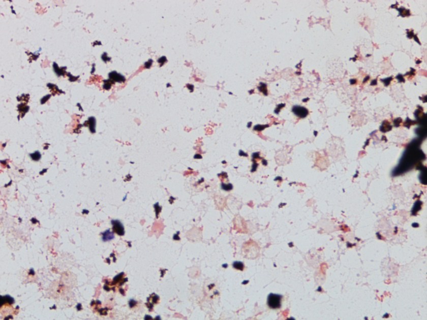
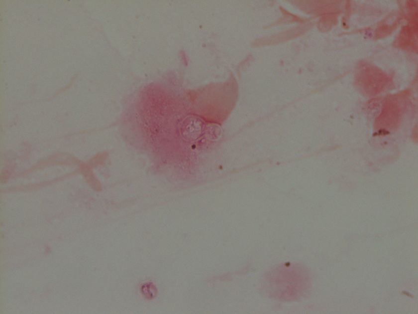
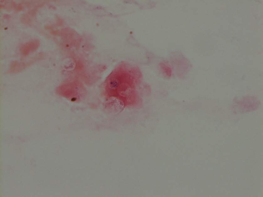
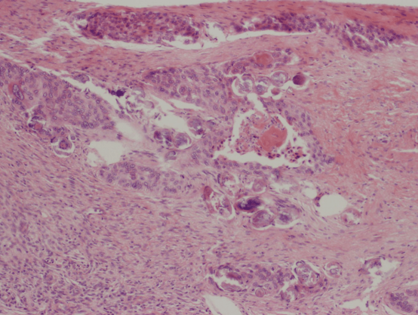
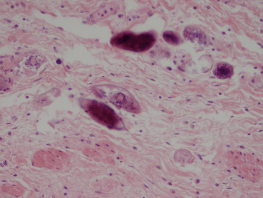
Figure 1. Gross and histologic lung images from autopsy. Target shaped lesions on the lung surface can be seen in this gross photograph of the upper and middle lobes of the right lung in situ (A). A sample of lung parenchyma was sectioned and H&E staining revealed broad, irregularly branched, pauci-septate fungal elements admixed with a brisk inflammatory infiltrate within damaged alveoli (B). Depiction of angioinvasion as the fungal organisms penetrate the walls of pulmonary vessels; GMS and PAS fungal stains, respectively (C and D).

Grossly, both lobes were abnormal. Specifically, as seen in Figure 1A, the right upper lobe exhibited multiple target shaped lesions on its surface in addition to a thin fibrinous coat involving the visceral and parietal pleurae. Moreover, the right upper lobe was adhered to the chest wall. Histologic examination of the lesions demonstrated abundant inflammatory cells and board fungal hyphae that were irregular and “ribbon like” with occasional septations within the disrupted lung parenchyma and invading into blood vessels as seen in Figure 1B-D.  Tissue from the lung lesions were cultured at autopsy and grew Lichtheimia spp.

A battery of additional tests collected premortem for Streptococcus pneumoniae, Cryptococcus neoformans, Mycoplasma, Histoplasma, Blastomyces and Toxoplasma gondii were all negative.  The AFB culture from the BAL specimen grew Mycobacterium avium after 8 weeks of incubation.

Discussion

Lichtheimia is a fungal genre, which shares the order Mucorales with variety of other clinically relevant organisms including Rhizopus, Rhizomucor and Mucor. It was formerly referred to as Absidia and many resources still use this term. As a saprophyte, Mucorales species live freely in the environment and can often be found indoors as well as outdoors. In the healthcare setting, infections are most frequently encountered in diabetic patients. In fact, along with its other Mucorales relatives, Lichtheimia spp. demonstrate a particular tropism for high glucose environments. Immunosuppressed individuals, especially those with hematologic malignancy, are also commonly infected with the fungi of this order – an observation, which speaks both to the ubiquity of these organisms in the human environment as well as the crucial role played by the immune system in their control. Today, the infection caused by Lichtheimia and its relatives is referred to as mucormycosis, though the related term zygomycosis remains deeply ingrained in the medical lexicon. The most striking presentations of mucormycosis are those involving the rhino-orbital-cerebral tissues. However, Mucorales organisms can also colonize many other organ systems. Relevant to this case, pulmonary mucormycosis is a particularly severe form of the infection with a mortality rate nearing 90%. Further, in a patient with pulmonary mucormycosis, the likelihood of concomitant disseminated mucormycosis is also very high (nearing 50%). Though in the present case the post-mortem examination was limited to only particular lung lobes, clinicoradiographic findings preceding the patient’s death strongly suggested that the infection also involved the liver and brain.

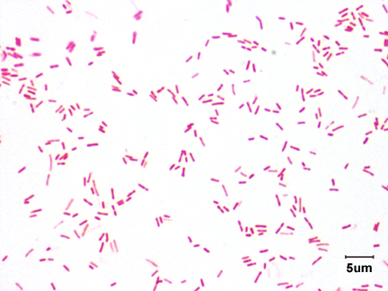
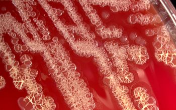
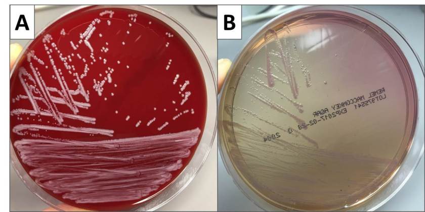
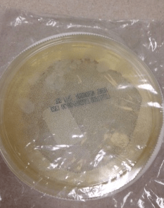
Diagnosis of pulmonary mucormycosis can be tricky and often hinges upon histopathologic findings and culture results. As in the present case, Mucorales organisms are differentiated from other common fungi with similar presentations based on their broad hyphae with limited septations and irregular branching. By contrast, Aspergillus spp. exhibits more narrow hyphae with septations and regular, acute-angle branching patterns. Additionally, Mucorales organisms, like Lichtheimia, are often angioinvasive and histologic examination may demonstrate fungal elements entering vascular lumina and even inducing thrombotic infarctions (both noted in the current case). In the laboratory, if a Mucorales is in the differential diagnosis, the tissue specimen should be minced instead of ground in order to preserve viability of the organisms. Mucorales grow rapidly as non-descript, whitish-gray molds within 4 days and are described as “lid lifters” due to their predilection to completely fill the plate. Due to its highly infectious nature, plates should be wrapped in parafilm and only examined in certified biosafety cabinets. On a lactophenol cotton blue prep, the sporangiophores of Lichtheimia and Rhizomucor spp. arise internodally between the rhizoids. This is in contrast to Rhizopus spp. in which the sporangiosphores arise directly over the rhizoids and Mucor spp. where rhizoids are not produced.

While there is little doubt that the patient in the above case was particularly susceptible to environmental Lichtheimia spores as a consequence of his immunosuppressed condition and status as a diabetic, the contribution played by his occupational exposures is less clear. Though provocative, it would be challenging to establish a link between the patient’s terminal infection and his agricultural encounters with decaying vegetable matter and the associated molds.

lavik

-JP Lavik, MD/PhD, is a 3rd year Anatomic and Clinical Pathology Resident at Yale New Haven Hospital.

Stempak

-Lisa Stempak, MD, is an Assistant Professor of Pathology at the University of Mississippi Medical Center in Jackson, MS. She is certified by the American Board of Pathology in Anatomic and Clinical Pathology as well as Medical Microbiology. She is the director of the Microbiology and Serology Laboratories.  Her interests include infectious disease histology, process and quality improvement and resident education. 

Microbiology Case Study: A 50 year Old Man with Dysuria and Hematuria

Case History

A 50 year old healthy man presented with dysuria and hematuria for 4 months. He had briefly lived in the Middle East 3 years ago. The patient underwent cystoscopy which demonstrated a solid mass in the lateral wall of the bladder. Bladder biopsies were performed and showed invasive squamous cell carcinoma associated with ova consistent with Schistosoma haematobium (Figures 1&2).

schisto1

Figure 1. H&E of bladder biopsy showing invasive squamous cell carcinoma and ova of Schistosoma haematobium.

schisto2

Figure 2. High power view of Schistosoma haematobium ova.

Discussion

Schistosomiasis is caused by blood flukes of the genus Schistosoma. There are three major species related to human disease: S. haematobium, S. japonicum, and S. mansoni. Clinical presentation of schistosomiasis depends on the species. S. haematobium infection causes urinary schistosomiasis. Urinary schistosomiasis can range from asymptomatic to gross hematuria and possible obstruction resulting in renal failure.  S. haematobium is geographically distributed primarily in Africa and the Middle East. Transmission to humans requires direct contact with water harboring snails infected with S. haematobium. The cercaria that are released from infected snails penetrate human skin and then migrate to venules of the bladder and ureters. The cercaria develop into adult male and female flukes.  The adult schistosomes reside in the bloodstream and lay eggs that pass through the urine. The eggs are highly immunogenic and produce an intense inflammatory response resulting in hematuria and dysuria. Progression to fibrosis, renal failure and carcinoma may occur as in our patient with squamous cell carcinoma of the bladder. In addition to detection in surgical specimens, S. haematobium may be detected by identification of ova in urine. The ova of S. haematobium are oval and 112-170 µm x 40-70 µm in size with a characteristic terminal spine.  In patients with a high clinical suspicion of S. haematobium, serology may be useful when ova are not identified in urine or surgical specimens. The recommended treatment for schistosomiasis is praziquantel.  The timing of treatment is important because praziquantel is most effective against the adult worm and requires a mature antibody response to the parasite. The Centers for Disease Control and Prevention recommend starting treatment for infected travelers at least 6-8 weeks after the last exposure to contaminated water.


-Jill Miller, MD is a 4th year anatomic and clinical pathology resident at the University of Vermont Medical Center.

Wojewoda-small

-Christi Wojewoda, MD, is the Director of Clinical Microbiology at the University of Vermont Medical Center and an Assistant Professor at the University of Vermont.

Microbiology Case Study: A 4 Day Old with Eye Drainage

A 4 day old infant presented to the ED with copious amounts of left eye drainage. He was born at 38 weeks via vaginal delivery and was discharged home on day 2 of life. On day 3 of life the baby had acute onset of left eye drainage. On day 4 of life the discharge had become so profuse that the mother was cleaning the baby’s eye several times per hour and his eye was red. In the ED, the discharge was collected and sent to the lab for culture. The specimen Gram stain was reported as many polymorphonuclear leukocytes (pmns) and rare gram negative diplococci. Two days later the following growth was noted on chocolate, 5% sheep blood, and Thayer Martin agars (Figure 1).

ngono1

Figure 1. Growth of organism on (A) chocolate, (B) 5% sheep blood, and (C) Thayer Martin agars

Discussion

The organism was identified as Neisseria gonorrhoeae. N. gonorrhoeae are gram negative diplococcic that grow on solid media in 24-48 hours when incubated under aerobic conditions at 35-37°C with 5% CO2. They exhibit more robust growth on chocolate agar, but can grow on 5% sheep blood agar as well (Figure 1A and B). Like most other Neisseria spp., N. gonorrhoeae are oxidase and catalase positive. Species-level identification can be made by testing carbohydrate utilization; N. gonorrhoeae can only utilize glucose while N. meningitidis can utilize glucose and maltose.

To increase the sensitivity of N. gonorrhoeae detection from mucosal (non-sterile) sites, selective media such as Thayer Martin agar is commonly used (Figure 1C). Selective media for N. gonorrhoeae contains colistin, vancomycin, and antifungal agents to suppress gram negative bacteria, gram positive bacteria, and yeast, respectively, along with other inhibitory agents. N. gonorrhoeae are fastidious bacteria that are at great risk for dying in transport. For maximal recovery of N. gonorrhoeae, special transport packs have been developed. Figure 2 is an image of a Thayer Martin transport pack including agar plate, CO2 generating tablet, and plastic bag for transport back to the lab.

ngono2

Figure 2. Components of the Thayer Martin transport system for bedside plating of specimen.

Neisseria spp. reside in the mucosal membranes of animals including humans and most species are considered normal flora of the upper respiratory tract. N. gonorrhoeae is an exception and is always considered a pathogen no matter what amount or from what location it is isolated. N. gonorrhoeae is spread by sexual transmission where it infections the mucosal surfaces of the urethra, cervix, rectum, and pharynx. Infection presents as acute urethritis in men with symptoms of urethral discharge and sometimes dysuria, but infection is asymptomatic in up to 10% of cases. In women, N. gonorrhoeae infection is most often asymptomic. If symptoms are present, they are generally mild and non-specific including increased vaginal discharge, dysuria, and intermenstrual bleeding. Delay in treatment due to lack of recognition of infection can lead to assentation of the bacteria resulting in pelvic inflammatory disease. Rectal and pharyngeal infections occur in both men and women and are generally asymptomatic. N. gonorrhoeae can cause conjunctivitis which leads to corneal ulceration. In adults conjunctivitis is caused by autoinoculation of the eye. In newborns, such as our case patient, it is transmitted from infected mother to child as the baby moves through the birth canal. In the olden days N. gonorrhoeae infection of newborns was a cause of blindness, but now it is standard to administer 1% aqueous solution of silver nitrate or antibiotic ointment containing erythromycin to newborns just after birth to prevent infection.

These days, molecular detection of N. gonorrhoeae from urine is the most common method used to identify N. gonorrhoeae infection in adult males and females. One of the benefits of molecular detection is that it does not require live bacteria for detection. Many reference laboratories and public health clinics have validated their molecular assays for detection of N. gonorrhoeae from rectal and pharyngeal swabs. Traditionally for sites such as eyes, molecular detection was not available and culture was the only means of N. gonorrhoeae detection. This is a rapidly changing area of microbiology as we were able to get our patient’s eye drainage tested by transcription-mediated amplification (APTIMA) at our reference lab and the specimen was positive.

Our patient was treated with cefotaxime and is being followed outpatient by his pediatrician and ophthalmology clinic.

References

  1. MCM, 10th edition
  2. CDC (https://www.cdc.gov/std)


-Erin McElvania TeKippe, PhD, D(ABMM), is the Director of Clinical Microbiology at Children’s Medical Center in Dallas Texas and an Assistant Professor of Pathology and Pediatrics at University of Texas Southwestern Medical Center.