Microbiology Case Study: A 16 Year Old with Rhinosinusitis

Case

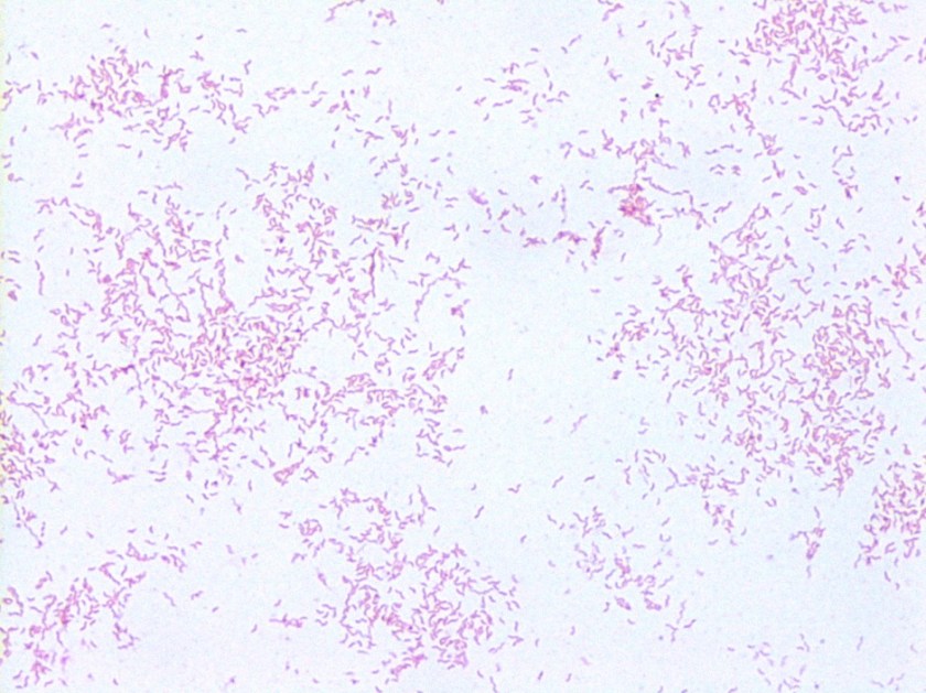
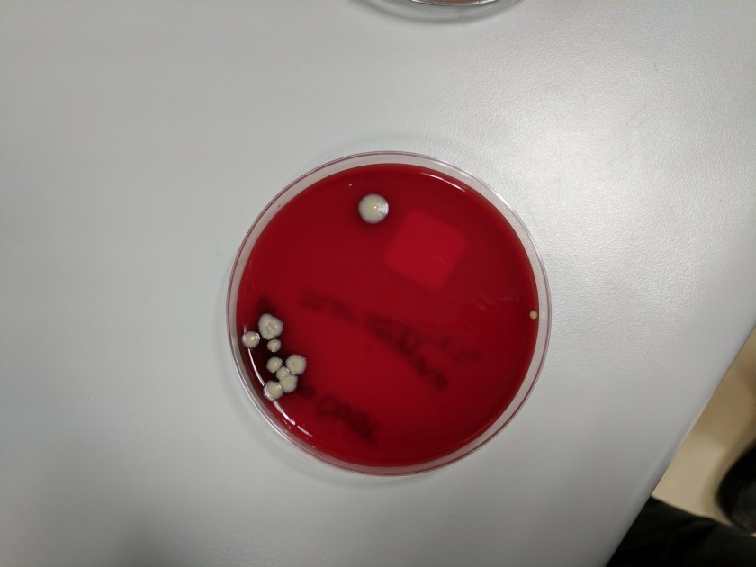
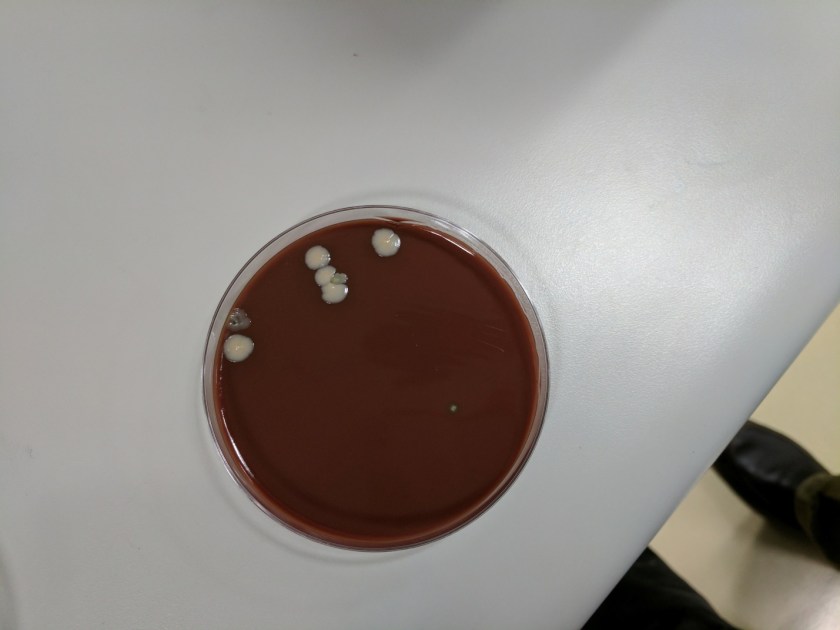
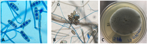
A 16-year-old male presented with recurrent sinusitis and rhinitis. He had a history of left sinus surgery two years ago, and at that time pathologic examination of the tissue demonstrated eosinophilia and fungal culture grew Curvularia, consistent allergic fungal sinusitis. The patient was doing well without allergy or immunotherapy management until three months ago when he could not breathe out of the right nostril and began snoring loudly. He underwent bilateral endoscopic frontal sinusotomy with tissue removal of the ethmoid and sphenoid sinuses. Tissue was sent to the laboratory for fungal culture. After five days, fungal cultures grew mold on inhibitory mold agar with gentamicin. The surface was a gray speckled color (Image 1C). The reverse color of the mold colony was dark brown to black. The microscopic appearance can be seen in Image 1A-B.

Bipolaris figure
Image 1. A) Ellipsoid conidia with the common number of 3-5 septations stained with lactophenol cotton blue counterstain, B) a cluster of conidia surrounded by less lactophenol cotton blue stain better demonstrating brown melanin pigment in the cell wall, and C) a dark gray speckled fungal colony.


Discussion

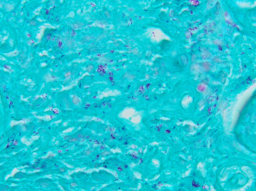
These features are consistent with the identification of Bipolaris. Microscopic examination using lactophenol cotton blue tape prep demonstrated oblong conidia characteristic of Bipolaris (Image 1 A-B). The conidia are ellipsoidal with pale brown pseudoseptations that contain three to five septa. Four septa are the most common. Bipolaris is a dematiaceous fungus, meaning the cell walls contain dark brown melanin pigment. This can be seen by microscopic observation of the fungal cell wall which contains pigment (Image 1B) and is also demonstrated by the dark reverse color of the fungal colony.

To distinguish Bipolaris from Drechslera and Exserohilum, the Germ tube test can be utilized. When conidia are incubated with a drop of water on a glass slide for 8-24 hours, they will begin to form Germ tubes. Bipolaris species germinate from both poles of the oblong conidium at a 180 degree angles (hence the name “Bipolaris”), whereas Exserohilum germinate from just one pole at a 180 degree angle and Dreschslera species germinate at a 90 degree angle from the central cells of the conidium. Dreschslera can be confused for Bipolaris based on colony appearance and microscopic appearance, but unlike Bipolaris, Dreschslera is not associated with human disease.1

Pathogenic strains of Bipolaris include Bipolaris australiensis, Bipolaris hawaiiensis, Bipolaris maydis, Bipolaris melanidis, Bipolaris speicifera, and Bipolaris sorokiniana.2 The most common cause of infection is Bipolaris spifcifera. Bipolaris species are the most common cause of fungal sinusitis in immunocompetent individuals which often presents as allergic rhinitis. Allergic rhinitis could also be a risk factor for acquiring Bipolaris. Treatment often consists of prompt surgical excision to prevent expansion, superficial deformity and dissemination. If fungal chemotherapy is pursued, itraconazole and amphoterin B have been reported as effective agents.3

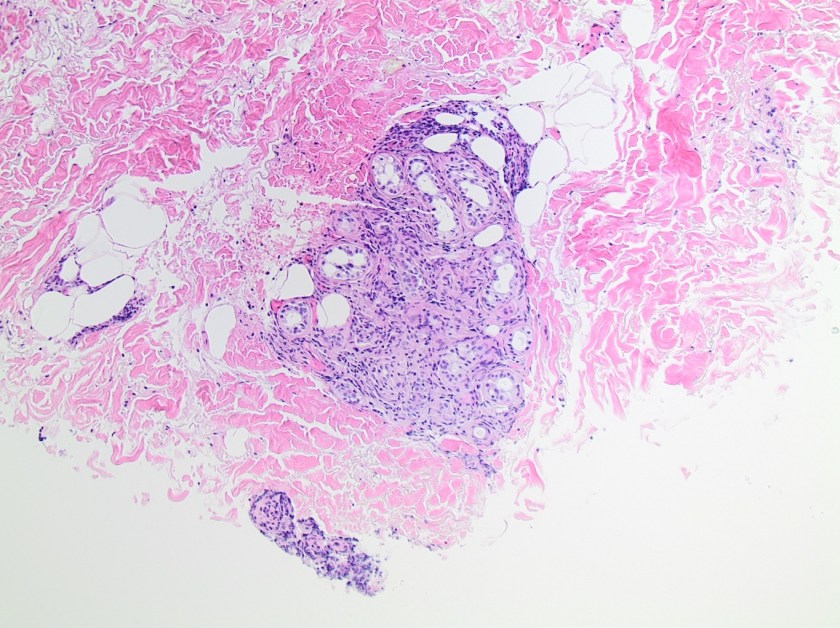
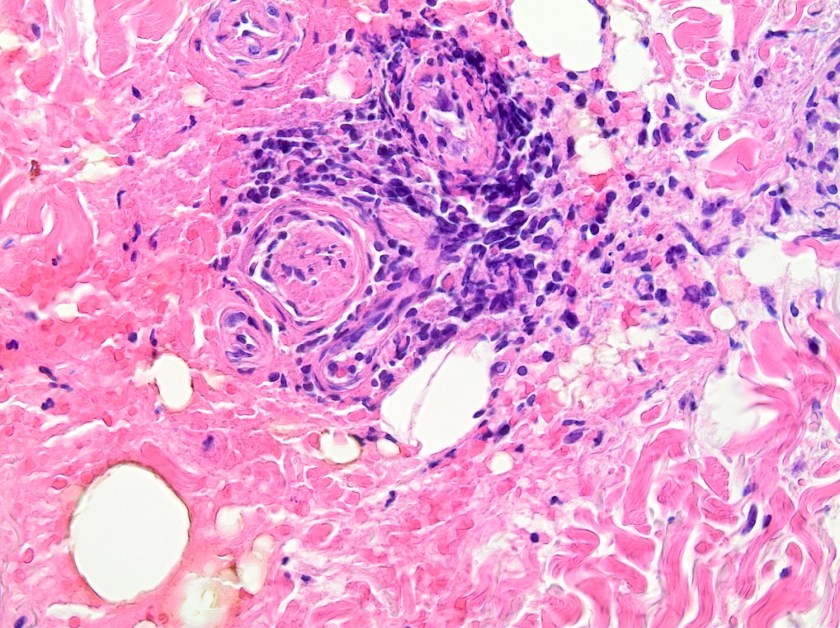
Bipolaris is one of the most common causes of allergic fungal sinusitis, typified by nasal polyps and mucus plugs consisting of eosinophils, fungal hyphae and Charcot-Leyden crystals. It is a type 1 and 3 hypersensitivity reaction mediated process due to high levels of mold-specific IgE.4 Skin prick testing is also positive in patients with allergic fungal rhinosinusitis (AFRS) which further indicates that the pathophysiology is an immunologic versus infectious process.4 While the exact process of fungal allergic sensitization has not been codified, chitin, a structural fungal protein has been shown to elicit a Th2 immune response.5 It will be interesting to see how this research evolves so that we might one day see why fungi can cause both erosive infections and allergies within human patients.


References

  1. Fothergill AW. Identification of Dematiaceous Fungi and Their Role in Human Disease. Clin Infect Dis. 1996; 22 (S2): S179-84.
  2. Shafili SM, Donate G, Mannari RJ, Payne WG, Robson MC. Diagnostic Dilemmas: Cutaneous Fungal Bipolaris Infection. Wounds. 2006; 18(1):19-24.
  3. Saenz RE, Brown WD, Sanders CV. Allergic Bronchopulmonary Disease Caused by Bipolaris hawaiiensisPresenting as a Necrotizing Pneumonia: Case Report and Review of Literature. The American Journal of Medical Sciences. 2001; 321(3):209-12.
  4. Manning SC, Holman M. Further evidence for allergic pathophysiology in allergic fungal sinusitis. Laryngoscope. 1998;108(10):1485–1496.
  5. Reese TA, Liang HETager AMLuster ADVan Rooijen NVoehringer DLocksley RM. Chitin induces accumulation in tissue of innate immune cells associated with allergy. Nature 2007; 3;447(7140):92-6.


 JS

-Jeffrey SoRelle, MD, is a 1st year Clinical Pathology Resident at UT Southwestern Medical Center.

Erin McElvania TeKippe, PhD, D(ABMM), is the Director of Clinical Microbiology at Children’s Medical Center in Dallas Texas and an Assistant Professor of Pathology and Pediatrics at University of Texas Southwestern Medical Center.

Microbiology Case Study: Elderly Woman with Signs of Shock

Case History

The lab received two sets of blood cultures drawn at the time of admission from an elderly woman presenting with escalating signs of shock in the setting of suspected ischemic bowel. An exploratory laparotomy led to resection of a gangrenous segment of large bowel without signs of perforation or abscess. The woman’s medical history in the previous three months included two episodes of GI bleed, including one associated with signs of ischemic bowel, treated conservatively, and a hospitalization for health care acquired pneumonia treated with levofloxacin and steroids.

Lab Identification

Bacterial growth was detected in two bottles from one site. The anaerobic bottle demonstrated gram negative bacilli. The aerobic bottle demonstrated gram positive cocci that were identified by the Verigene microarray as non-VRE Enterococcus faecium. However the Verigene system could not identify the gram negative rod.

The gram negative rod grew on blood, chocolate, and McConkey agar. It was a lactose non-fermentor. Additionally, the gram negative colonies were indole positive and oxidase negative.

escfer1

Image 1. Gram stain of anaerobic blood culture bottle.

escfer2

escfer3
Image 2. Blood and MacConkey agars with growth of the organism.


These colonies were identified by the MALDI-TOF mass spectrometer as Escherichia fergusonii.

Discussion

Originally described as a unique species in 1985, Escherichia fergusonii is the closest relative of the much more common and well-known Escherichia coli.1 It is differentiated from E. coli by being lactose and sorbitol negative but adonitol, amygdalin, and cellobiose fermentation positive. Due to its molecular and structural similarities to E. coli, platforms such as the MALDI-TOF or Vitek 2 may have difficulty discriminating between the two species.2, 3 It has been identified as normal intestinal flora of warm blooded animals, though not specifically humans, and has been implicated in a salmonellosis-like disease in sheep and cattle.3

Descriptions in the literature regarding the clinical relevance of E. fergusonii to human disease are limited. There have been reports of E. fegusonii acquiring known virulence factors from E. coli and Salmonella, however, the prevalence and significance of such transformation is not well understood. In one report, E. fergusonii was found to have acquired the E. coli O157 antigen, but did not acquire the associated virulence factors or Shiga toxin.4 It has been identified as the pathogenic agent in cases of urinary tract infection, wound infection, diarrhea, bacteremia, cholangiosepsis, pleuritis, and endopthalmitis. These reports are geographically widespread with E. fergusonii identified in patients from the US, Nicaragua, Italy, India, and Taiwan. There is also variable information on antimicrobial susceptibility. The original description by Farmer et al. reported that all strains were susceptible to colistin, gentamicin, and chloramphenicol, and resistant to penicillin.1 An analysis of 600 specimens from eastern India revealed high rates of resistance to gentamicin and chloramphenicol and moderate resistance to ampicillin.5 An analysis of urinary tract infection specimens from Nicaragua reported resistance to nitrofuratoin, ciprofloxacin, ceftriaxone, and amoxicillin-calvulanate.6

Interestingly, E. fergusonii has been identified as the most quickly evolved member of the Escherichia genus.7 Though it is unclear whether that rapidity of differentiation translates to an ability to acquire clinical relevant features, it can at least be concluded E. fergusonii is a widely distributed and possibly under-identified gram negative facultative bacillus with the potential to acquire virulence factors and antibiotic resistance that could make it a significant pathogenic organism in humans.

References

  1. Farmer JJ, Fanning GR, Davis BR, O’Hara C, Riddle C, Hickman-Brenner FW, Asbury MA, Lowery VA, Brenner DJ. Escherichia fergusonii and Enterobacter taylorae, two new species of Enterobacteriaceae from clinical specimens. J. Clin. Microbiol., 21 (1985): 77–81.
  2. Crawford-Miksza LK, Himathongkam S, Dodd ML, Badoiu AS, Badoiu OM, Guthertz LS. Misidentification of a variant biotype of Escherichia coli O157:H7 as Escherichia fergusonii by Vitek 2 compact. J. Clin. Microbiol., 47 (2009): 872–873.
  1. Gastra W, Kusters JG, van Duijkern E, Lipman LJA. Escherichia fergusonii. Vet. Microbiol., 172 (2014): 7-12.
  1. Fegan N, Barlow RS, Gobius KS. Escherichia coli O157 somatic antigen is present in an isolate of E. fergusonii. Curr. Microbiol., 52 (2006): 482–486.
  1. Mahapatra A, Mahapatra S, Mahapatra A. Escherichia fergusonii: an emerging pathogen in South Orissa. Indian J. Med. Microbiol., 23 (2005): 204–208.
  1. Bours PHA, Polak R, Hoepelman AIM, Delgado E, Jarquin A, Matute AJ. Increasing resistance in community-acquired urinary tract infections in Latin America, five years after the implementation of national therapeutic guidelines. Int. J. Infect. Dis., 14 (2010): e770–e774.
  2. Walk ST, Alm EW, Gordon DM, Ram JL, Toranzos GA, Tiedje JM, Whittam TS. Cryptic lineages of the genus Escherichia. Appl. Environ. Microbiol., 75 (2009): 6534–6544.


-Taylor Goller is a Pathology Student Fellow at University of Vermont Medical Center.

Wojewoda-small

-Christi Wojewoda, MD, is the Director of Clinical Microbiology at the University of Vermont Medical Center and an Assistant Professor at the University of Vermont.

Microbiology Case Study: A 34 Year Old Female with Nausea, Vomiting, Diarrhea and Tender Extremities

Case History

A 34 year old female presented to the emergency department with a chief complaint of nausea, vomiting and diarrhea as well as tenderness in her extremities. These symptoms had been present for the previous 4 days with multiple episodes of diarrhea, associated low grade fevers & chills and she had poor oral intake as a result. Her past medical history was significant for human immunodeficiency virus (HIV) and chronic kidney disease. She has not be compliant with her anti-retroviral therapy and infectious disease prophylactic medications. Her vitals were within normal range and her physical exam elicited tenderness to palpation of her extremities and torso. No rashes and no erythema are seen. Routine laboratory tests as well as infectious disease work up, which included blood, stool & urine cultures, C. difficile and ova & parasite exam, were ordered. Notable findings included a slightly elevated white blood count (11.3 TH/cm2), creatinine of 7.1 mg/dL, HIV RNA viral load of 671 VC/mL and an absolute CD4 count of 7 cells/cm2. Two days after collection, her blood cultures were signaled as positive by the automated instrument.

Laboratory Identification

ngono1
Image 1. Gram stain from the blood culture bottles showed Gram negative cocci arranged in pairs (1000x oil immersion).
ngono2.png
Image 2. Small, whitish glistening colonies grew on blood and chocolate agars after 48 hours incubation in a 35°C incubator with 5% CO2.

The pathogen of interest grew from two sets of blood cultures and the direct Gram stain showed Gram negative cocci arranged in pairs (Image 1). After 48 hours incubation, small, whitish colonies were observed on blood and chocolate agars. No growth was seen on the MacConkey plate (Image 2). The isolate was positive for both catalase and oxidase. It was identified as Neisseria gonorrhoeae by both MALDI-TOF MS and a Vitek NH card.

Discussion

N. gonorrhoeae is the second most common sexually transmitted infection (STI) in the United States, only surpassed by Chlamydia trachomatis and they are often acquired together as a co-infection. Uncomplicated infections with N. gonorrhoeae typically present as acute urethritis with discharge. Asymptomatic infection occurs in 10% of males and upwards of 50% of females. As a result, females are at risk for the development of ascending infections and pelvic inflammatory disease leading to further reproductive issues. Disseminated gonococcal infection is uncommon (less than 1% of all gonococcal infections) but can occur and manifests as purulent arthritis with or without an accompanying dermatitis. In the case of our patient, her tenderness to palpation of the extremities could be a symptom of this disseminated septic arthritis.

In the laboratory, N. gonorrhoeae can be fastidious and requires special media such as chocolate, Martin-Lewis, modified Thayer-Martin or New York City agars as well as an enhanced CO2 environment in order to grow. The Gram stain of N. gonorrhoeae is described as Gram negative cocci with adjacent flattened sides and helpful biochemicals include catalase and oxidase (both positive).  Traditionally, in order to further speciate members of the Neisseria genus, sugar fermentation was necessary. N. gonorrhoeae only ferments glucose, while another notable member, N. meningitides, ferments both glucose and maltose.  Additionally, N. lactamica ferments glucose, maltose and lactose. Currently, commonly used methods of identification include API NH strips and automated instruments such as Vitek and MALDI-TOF MS.

Susceptibility testing for N. gonorrhoeae is usually limited to testing for beta-lactamase activity, although CLSI guidelines are available if deemed necessary. Current therapeutic guidelines recommend empiric treatment of uncomplicated infections with intramuscular ceftriaxone and oral azithromycin.


ka

-Kristen Adams, MD, is a fourth year Anatomic and Clinical Pathology resident at the University of Mississippi Medical Center. 

Stempak

-Lisa Stempak, MD, is an Assistant Professor of Pathology at the University of Mississippi Medical Center in Jackson, MS. She is certified by the American Board of Pathology in Anatomic and Clinical Pathology as well as Medical Microbiology. She is the director of the Microbiology and Serology Laboratories.  Her interests include infectious disease histology, process and quality improvement and resident education. 

Microbiology Case Study: A 50 Year Old Male with Fever and Diarrhea

Case History

A 50 year old male initially presented with cold symptoms. He was seen and evaluated at urgent care, with suspicion for bronchitis, but with no improvement with albuterol. Physical exam raised a suspicion for bacterial sinusitis. The patient was treated with amoxicillin/clavulanic acid with little improvement, and he was admitted to the hospital a week later for fever and diarrhea. Blood cultures were obtained. He was initially treated with cefepime prior to the speciation of the culture, and then switched to erythromycin for a 7 day course.

Laboratory Identification

Blood cultures were positive for gram negative curved/spiral rods. Gram stain and colony morphology were consistent with Campylobacter which was confirmed as C. jejuni by MALDI-TOF.

campy1

Image 1. Gram stain showing gram negative curved/spiral rods.

Discussion

C. jejuni are gram negative curved or spiral rods. Campy CVA agar is used for stool cultures because it is selective for Campylobacter and contains cefoperazone, vancomycin, and amphotericin B (CVA) which inhibit normal fecal flora. The media is incubated at 42°C under microaerophilic conditions, supporting the growth of Campylobacter jejuni and C. coli. C. jejuni is thermophilic, with growth on blood agar at 37°C and 42°C. Growth does not occur at 25°C. The colonies on blood agar are non-hemolytic, gray and smooth. Our isolate grew, albeit not happily, on blood and chocolate at 37°C with 5-10% CO2.

Infection is often transmitted by contaminated foods such as undercooked chicken. C. jejuni are most commonly associated with human infections however, C. coli have also been implicated. Guillain-Barre syndrome has been associated with patients following an infection with C. jejuni. It is not known how our patient was exposed. Macrolides are effective treatment modalities for C. jejuni, as well as fluoroquinolones, however, resistance to fluoroquinolones is increasing.


-Mustafa Mohammad, MD is a 3rd year anatomic and clinical pathology resident at the University of Vermont Medical Center.

Wojewoda-small

-Christi Wojewoda, MD, is the Director of Clinical Microbiology at the University of Vermont Medical Center and an Assistant Professor at the University of Vermont.

Microbiology Case Study: An 18 Year Old with Vaginal Discharge

Case Presentation

An 18 year old girl presents to her pediatrician with her mother for her pre-college check-up. She has no past medical history. After her mother leaves the room for the social history component, the girl admits to having sex with her boyfriend for the first time two weeks ago and complains of a yellow green malodorous vaginal discharge that started a week ago. She endorses mild pelvic pain. A pelvic exam is performed and mild cervical tenderness is noted. The cervix is pink, nulliparous, inflamed and is covered by small red punctate spots. A thin yellow green frothy discharge of fishy odor is also detected. A wet prep is made and reveals squamous cells and numerous motile organisms.

trich1
Figure 1.  Trichomonas vaginalis in a Pap test. The protozoa are often found next to squamous cells. (ThinPrep)

trich2
Figure 2.  Collection of Trichomonas vaginalis parasites eating at a squamous cell in a Pap (ThinPrep)

Discussion

Our patient was diagnosed with Trichomonas vaginalis (TV). TV is a flagellated parasitic protozoan for which humans are the only known host. It is 10-20 um long and 2-14 um wide with multiple flagella projecting from the anterior and posterior sides. It has a single trophozoite stage and does not survive well outside of its host. TV is a predatory obligate parasite that eats bacteria, vaginal epithelial cells, and red blood cells. It uses fermentative metabolism to produce the carbohydrates needed for fuel. TV is a sexually transmitted disease; however, because it is not reportable to local health departments, the true epidemiologic incidence rate is unknown. Its prevalence is highly variable by population and location. For example, some studies cite a prevalence of 3.1% of American pre-menopausal women (2.3% of adolescents) [1], while in certain high-risk populations the rate might be as high as 47% [2]. Most affected patients are asymptomatic; about a third of females become symptomatic within six months of infection. Symptoms for females include vulvar and vaginal irritation and itching, pain with urination and a diffuse, malodorous, yellow-green vaginal discharge. The cervix becomes reddened in a punctuated fashion causing the well-known strawberry cervix seen on colposcopy. In males, urethritis can develop. TV is often diagnosed via wet mount microscopy, where the protozoa can be seen moving around (Video 1). However, the sensitivity is relatively low, especially among males. Detection by nucleic acid probe from urine, endocervical, and vaginal swabs are considered more sensitive. TV can also be incidentally discovered on Pap tests (Figures 1 and 2). Treatment typically consists of a single dose of metronidazole [1,2]. It is critical that partners be treated as well, because otherwise reinfection may occur.


References

  1. Kissinger P. Trichomonas vaginalis: a review of epidemiologic, clinical and treatment issues. BMC Infectious Diseases. 2015; 15(307): 1-8.
  2. Meites E et al. A review of evidence-based care of symptomatic trichomoniasis and asymptomatic Trichomonas vaginalis infections. Clinical Infectious Diseases. 2015; 61(S8): S837-48.


AM

-Amanda Strickland, MD, is a 2nd year Anatomic and Clinical Pathology Resident at UT Southwestern Medical Center.

Erin McElvania TeKippe, PhD, D(ABMM), is the Director of Clinical Microbiology at Children’s Medical Center in Dallas Texas and an Assistant Professor of Pathology and Pediatrics at University of Texas Southwestern Medical Center.


Microbiology Case Study: A 58 Year Old Female with Lung Nodules

Case History

A 58 year old female with past medical history significant for Type II diabetes, hypertension, hyperlipidemia, chronic diastolic heart failure, and hypothyroidism was hospitalized for following a fall and was found to have compression fractures. The hospitalization was complicated by flash pulmonary edema requiring intubation. CT chest obtained during this hospitalization demonstrated lung nodules, which were biopsied and cultured.

Laboratory Identification

The bacterial and mycobacterial cultures grew gram positive bacilli which were positive on Modified Kinyoun stain. They were negative for Auramine/Rhodamine. The organism grew on several media, including 7H11, Chocolate, and Buffered Charcoal Yeast Extract (BCYE). They formed chalky, white-pink colonies. The organism was confirmed as Nocardia nova by a reference laboratory.

nocard1

Image 1. Modified acid fast bacilli on Modified Kinyoun stain.

nocard2

Image 2. Chalky white-pink colonies on BCYE agar.

Discussion

Nocardia nova is a weakly acid fast, aerobic filamentous, beaded, gram positive bacilli with right-angled branching. It is identified by a Modified Kinyoun stain. Nocardia grows best on BCYE agar; however it also can grow within 3-5 days on blood and chocolate agar. It forms chalky white-pink colonies. Molecular testing is performed to speciate Nocardia, primarily 16S ribosomal RNA gene sequencing, as well as mass spectrometry. Most infections can be treated with sulfonamides for 6-12 months, however, the CDC recommends performing speciation and susceptibility testing on every isolate due to specific susceptibility profiles and drug resistant strains. Our patient was treated with high dose sulfamethoxazole/trimethoprim and meropenem.

Nocardia nova is commonly found in soil and is one of several pathogenic Nocardia species. Nocardia is often inhaled and presents as a chronic pulmonary infection with cough, shortness of breath, and fever. Nocardia can also cause pleural effusions, empyema, pericarditis, abscesses, or dissemination to deep organs, especially the brain. Nocardia can also be contracted though trauma, causing cutaneous diseases such as a mycetoma or cellulitis. Because of its low virulence, Nocardia generally affects immunocompromised patients, however those with preexisting pulmonary disease can also be susceptible to infection.


-Mustafa Mohammad, MD is a 3rd year anatomic and clinical pathology resident at the University of Vermont Medical Center.

Wojewoda-small

-Christi Wojewoda, MD, is the Director of Clinical Microbiology at the University of Vermont Medical Center and an Assistant Professor at the University of Vermont.

Microbiology Case Study: A 60 Year Old Male with Longstanding Skin Lesions

Case History

A 60 year old male from Louisiana presents to his family doctor with a chief complaint of longstanding skin lesions for approximately the last two years. On physical exam, there are several sharply defined reddish-brown plaques on his upper back and extremities. He reports sensory loss involving his chest, back and upper extremities. The lesions have not responded to conventional topical anti-fungal treatments. Punch biopsies along the margin of the most active lesion were obtained and sent to the Microbiology laboratory for bacterial, fungal and mycobacterial cultures and to the Pathology Department for histologic diagnosis.

Tissue sections

mycolep1
Image 1. Section from the right upper extremity skin punch biopsy demonstrates a normal basket-weave stratum corneum and normal epidermis with nodular superficial and deep granulomatous inflammatory infiltrate. A Grenz zone, a narrow layer beneath the epidermis that is not infiltrated or involved in the same way as are the lower layers of the dermis, is noted (H&E, 40x).
mycolep2
Image 2. Inflammation engulfing eccrine glands in the deep portion of the dermis (H&E, 100x).
mycolep3
Image 3. Portion of punch biopsy demonstrating perineural inflammation consisting predominantly of mononuclear cells (H&E, 400x).
mycolep4
Image 4. Fite stain highlighting numerous acid fast bacilli within macrophages surrounding the eccrine glands (1000x oil immersion).


On histologic examination of the skin biopsy, nodular, superficial and deep granulomatous inflammation was noted surrounding eccrine glands and engulfing nerves (Images 1-3). Fite staining illustrated numerous acid fast bacilli (Image 4) and, given the geographic location of the patient and clinical symptoms, was felt to be highly suggestive of Mycobacterium leprae. The case was sent for confirmatory testing by polymerase chain reaction (PCR). All cultures collected were negative.

Discussion

Mycobacterium leprae is a chronic, granulomatous disease which presents as anesthetic skin lesions and peripheral neuropathy with nerve thickening. While rare in the United States (US) today, historically it was one of most prominent pathogens in Mycobacterium genus apart from M. tuberculosis. In the past, leprosy (also known as Hansen’s disease) was prevalent throughout Europe, but due to systematic control programs aimed at underserved and rural locations, the number of cases drastically decreased and countries with the majority of recent cases include India, Brazil and Indonesia. According to National Hansen’s Disease Registry, a total of 178 cases were reported in the US in 2015. Of these, 72% (129) of cases were reported in Arkansas, California, Florida, Hawaii, Louisiana, New York and Texas. Transmission to those who are in prolonged and close contact with an infected person is thought to occur via shedding from the nose. While humans are the only known reservoir of leprosy, infections with organisms indistinguishable from M. leprae have been detected among wild armadillos in parts of the southern US.

The diagnosis of M. leprae is largely a clinical one as the organism is not able to be grown on artificial media, but histology and confirmatory PCR are useful adjuncts. Skin biopsies should be full thickness and include the deep dermis. Ideally, the most active edge of the most active lesion should be biopsied. There is a spectrum of M. leprae which ranges from few lesions and a paucity of bacilli (tuberculoid leprosy) to widespread skin involvement with numerous bacilli (lepromatous leprosy).  Histologically, there are granulomatous aggregates of epithelioid cells, multinucleate giant cells and lymphocytes and inflammation often engulfs sweat glands and nerves. Small lesions that have poorly defined borders and are found on the elbows, knees or ears are where bacilli tend to be located. A Fite stain is useful to highlight the acid fast bacilli located in the macrophages within the inflammatory nodules. M. leprae PCR can also be performed on blood, urine, nasal cavity specimens and skin biopsies as a sensitive diagnostic technique. PCR can also be used to detect certain genes that confer resistance to common treatment drugs such as rifampin, ofloxacin and dapsone.

As with other mycobacterial diseases, the treatment for M. leprae infections consists of a long term multidrug regimen. The six most commonly used medications include rifampin, dapsone, clofazimine, minocycline, ofloxacin, and clarithromycin.


kt

-Katie Tumminello, MD, is a fourth year Anatomic and Clinical Pathology resident at the University of Mississippi Medical Center. 

Stempak

-Lisa Stempak, MD, is an Assistant Professor of Pathology at the University of Mississippi Medical Center in Jackson, MS. She is certified by the American Board of Pathology in Anatomic and Clinical Pathology as well as Medical Microbiology. She is the director of the Microbiology and Serology Laboratories.  Her interests include infectious disease histology, process and quality improvement and resident education. 

Microbiology Case Study: A 51-Year-Old Woman with a Tick Bite

Case History

A 51-year-old female with a past medical history of chronic lower back pain, depression, and anxiety presented to an outpatient dermatology clinic for a lesion on her left cheek that was present for years but has recently grown and become irritated.  Additionally, she reported a “skin taggy thing” that she first noticed on her posterior neck about two days ago.  Upon physical examination, a tick was observed latched onto the right posterior neck.  After being alerted to the presence of a tick, the patient did disclose that she was in the woods three days prior.  The bite site was locally anaesthetized and the tick was removed and sent to the laboratory for arthropod identification.  Furthermore, the patient received a single prophylactic dose of doxycycline 200 mg.

deertick1
Image 1. Ixodes scapularis, adult female, dorsal surface: an anterior capitulum (A) that contains mouth parts (paired palpi (B) and a median (partially intact) hypostome (C)) and the basis capituli (D) with two porose areas (E). The scutum (F) is inornate. 
deertick2
Image 2. Ixodes scapularis, adult female, ventral surface: eight coxa (G) of paired legs I-IV, a genital aperture (H), two spiracular plates (I), and an anal groove (J) that is anterior to the anus (K).


Discussion

Ixodes scapularis (black-legged ticks), also known as deer ticks (their preferred hosts are white-tailed deer), are small arachnids.  As obligate ectoparasites of vertebral hosts, I. scapularis are also willing to feed on humans.  Importantly, infected arthropods can be vectors of multiple pathogens including: the spirochete, Borrelia burgdorferi, that causes Lyme disease; the intracellular gram-negative bacterium, Anaplasma phagocytophilum, that causes human granulocytic anaplasmosis; the Apicomplexan parasites, Babesia spp, that cause babesiosis; and the flavivirus, Powassan virus, that causes encephalitis.

The I. scapularis life cycle, ranging from one to two years in length, is composed of four developmental stages: egg, larva, nymph, and sexually dimorphic adult.  Compared to nymphs and adults that have eight legs, larvae are smaller and have six legs.  The term “three-host cycle” implies that during each of the three motile stages, I. scapularis takes a blood meal from a different host animal, at which time the tick’s saliva is injected and transmission of pathogens can occur.

Hard ticks possess an anterior capitulum (Image 1, A) whereas soft ticks lack a capitulum.  The capitulum is made up of mouth parts that are attached to the basis capituli.  The mouth parts refer to paired appendages called palpi (Image 1, B) that are parallel to a median hypostome (Image 1, C).  The hypostome holds teeth-like structures, called denticles, arranged in a specific formula useful for identification.  The mouth parts (palpi and hypostome) are longer than the width of the basis capituli (Image 1, D) and this ratio is also useful in identification.  The dorsal surface of the basis capituli has two porose areas (Image 1, E) in adult females that secrete wax to waterproof eggs.  The dorsal shield, called a scutum (Image 1, F), is absent in soft ticks and inornate compared to other hard ticks.  In adult males, the scutum covers nearly the entire dorsum.  Other hard ticks have eyes (lateral markings on the scutum) and festoons (grooved bulges on the posterior margin) that are both absent in I. scapularis.  The ventral surface demonstrates coxa (Image 2, G) that are the basal segments of paired legs, numbered I-IV from anterior to posterior.  Posterior to coxa IV are paired spiracular plates (Image 2, I), external openings of the respiratory system.  A median genital aperture (Image 2, H) is present in adults.  The distinct anal groove (Image 2, J) is an inverted U-shaped curve located anterior to the anus (Image 2, K) in all Ixodes species, as opposed to posterior or indistinct anal grooves of other genera of hard ticks.

Following arthropod identification in parasitology, microbiology laboratory reports include: the genus (Ixodes), species (scapularis), developmental stage (adult female), level of engorgement (unengorged), and status of mouth parts (partially intact).  The genus and species are of medical importance because of their characteristic associations with various human pathogens.  Of the developmental stages, nymphs and adults are most frequently associated with human transmission of A. phagocytophilum.  Also, larvae are unable to transmit B. burgdorferi because the spirochete is not transmitted vertically to eggs; as such, a blood meal from a reservoir host is required.  Nymphs, being smaller in size than adults, are more likely to complete feeding undetected and thereby transmit B. burgdorferi.  Feeding is necessary for adult females to achieve fertility; therefore males are less likely to be discovered on hosts.  The level of engorgement, an estimate of feeding time, is relevant because approximately 36 hours are required for B. burgdorferi to multiply in the tick’s midgut and migrate to salivary glands for transmission to a host, by which time ticks are visibly engorged.  Lastly, if the mouth parts (palpi and hypostome) are intact, this suggests that there was only brief host contact and it is less likely that pathogen transmission occurred.

Reference

-Patterson FC and Winn WC. Practical identification of hard ticks in the parasitology laboratory. Pathology Case Reviews 2003; 8(4):187-198.


-Adina Bodolan, MD is a 1st year anatomic and clinical pathology resident at the University of Vermont Medical Center.

Wojewoda-small

-Christi Wojewoda, MD, is the Director of Clinical Microbiology at the University of Vermont Medical Center and an Assistant Professor at the University of Vermont.

Microbiology Case Study: A 65 year Old Male with Decubitus Ulcer

Case History

A 65 year old male with a history of T7-8 paraplegia and lengthy history of a decubitus ulcer presented for surgical debridement of the wound. An ischial bone biopsy for culture was performed.

Lab Identification

The primary gram stain demonstrated mixed gram positive and gram negative organisms, and the tissue sample from the wound grew Corynebacterium striatum and Bacteroides fragilis. The bone sample grew Enterococcus faecalis and anaerobic gram positive cocci. He was initially treated with ertapenem alone but daptomycin was added to cover enterococcus and he continued this dual regimen. Daptomycin was discontinued due to elevated creatinine kinase levels and he was transitioned to intravenous vancomycin. He completed 42 days of ertapenem and vancomycin. He was transitioned to oral amoxicillin/clavulanic acid and he has not developed new fever, chills, sweats, fatigue or increased drainage from his wound.

corstri1

Image 1. Blood agar plate with gray-white, moist, smooth, non-hemolytic bacterial colonies.

corstri2

Image 2. Chocolate agar plate with gray-white, moist, smooth, bacterial colonies.

The blood and chocolate agar plates grew bacterial colonies while the MacConkey agar had no growth. The gram stain was consistent with Corynebacterium and mass spectrometry identified the organism as Corynebacterium striatum.

Discussion

Corynebacterium striatum is a gram positive bacilli that is part of normal skin and mucosal flora. In immunocompromised patients or through direct inoculation of a sterile site, C. striatum can cause infectious endocarditis, bacteremia, pneumonia, lung abscess, arthritis and chorioamnionitis. Studies have shown that C. striatum also can cause wound infections in patients with underlying disease and previous antibiotic use. Foreign medical devices can also be infected by C. striatum, and removal of the device may be necessary. Vancomycin is used to treat C. striatum due to the variable susceptibility to other antibiotics.


-Mustafa Mohammad, MD is a 3rd year anatomic and clinical pathology resident at the University of Vermont Medical Center.

Wojewoda-small

-Christi Wojewoda, MD, is the Director of Clinical Microbiology at the University of Vermont Medical Center and an Assistant Professor at the University of Vermont.

Microbiology Case Study: A 14 Year Old Female with Neck Swelling

Case History

A previously healthy 14-year-old female presents to the emergency department with three days of progressive facial and neck swelling. The swelling started on the left side. Two days ago she visited her primary care physician where she had negative monospot and mumps IgM testing.  She is fully vaccinated, but was exposed to a mumps outbreak at school.

Discussion

Our patient was diagnosed with mumps by positive RT-PCR from a buccal swab. The mumps virus is a member of the Paramyxoviridae family which includes notable human pathogens parainfluenza, Hendra, and Nipah viruses. Members of this family are enveloped, helical viruses with single-stranded, non-segmented RNA genomes with negative polarity. Mumps is an obligate human pathogen that replicates in the epithelial cells of the upper respiratory tract and subsequently moves to regional lymph nodes. It is spread from person to person via direct contact with respiratory secretions or contact with contaminated fomites. Mumps is a highly contagious disease with as high as 85% of naïve individuals becoming infected after contact with a mumps infected individual. It spreads most efficiently in areas where there is close contact among individuals for prolonged periods of time such as college campuses and close-knit religious communities.

Prior to vaccination for mumps in the 1960s, greater than 150,000 cases of mumps occurred each year in the US. The incubation period for infection is 16-18 days, with the majority of infected persons being asymptomatic or having mild respiratory symptoms. Orchitis causing sterility in post-pubescent males is the main concern of mumps infection but other rare but serious complications include mastitis and oophoritis in females, meningoencephalitis, pancreatitis, and deafness.

Due to sporadic outbreaks of measles since the introduction of the vaccine, the vaccine schedule has been revised from one dose of the MMR (measles, mumps, and rubella) vaccine at age 12-15 months to include another MMR booster at age 4-6 years. We are currently in the middle of yet another outbreak with nearly 6,000 cases of mumps reported to the CDC in 2016 and a high rate of infections reported thus far in 2017 (Figures 1 and 2).

mumps1
Figure 1. Number of cases identified by the CDC in 2017 by state. (Figure courtesy of the CDC Mumps website at https://www.cdc.gov/mumps/outbreaks.html. Content source: National Center for Immunization and Respiratory Diseases [NCIRD], Division of Viral Diseases)
mumps2
Figure 2. Number of cases of mumps per year identified by the CDC.
(Figure courtesy of the CDC Mumps website at https://www.cdc.gov/mumps/outbreaks.html. Content source: National Center for Immunization and Respiratory Diseases [NCIRD], Division of Viral Diseases)


Diagnostic Testing for Mumps

Serological testing for IgM and RT-PCR from a buccal swabs are the mainstay of mumps diagnosis. IgM becomes positive in the first 3-4 days after symptom onset and will remain positive for 8-12 weeks. IgG becomes positive 7-10 days following symptom onset and will remain at high levels for many years and detectable for life. In a vaccinated individuals, IgM testing has less utility as it may be non-reactive or weakly positive following a secondary immune response.

RT-PCR from a buccal swab specimen is the most sensitive test for diagnosis of mumps. It should be performed as soon as a patient is symptomatic, as testing by this method is the most sensitive in the first few days following symptom onset and becomes less sensitive as time goes on.

Urine specimens can be used to isolate mumps in viral culture. Urine is not positive for mumps until greater than 4 days post symptom onset and is less sensitive than PCR performed on the bucal swab. For these reasons, viral isolation from urine is no longer a commonly used test for diagnosis of mumps, although viral culture is still considered the gold standard for mumps conformation.

Resolution

The patient and her family were counseled on the infectious nature of mumps. She was instructed to remain in isolation at home for 6 days after resolution of swelling.


References

  1. Manual of Clinical Microbiology, 11th edition
  2. CDC Mumps Website (www.cdc.gov/mumps/index.html)


Erin McElvania TeKippe, PhD, D(ABMM), is the Director of Clinical Microbiology at Children’s Medical Center in Dallas Texas and an Assistant Professor of Pathology and Pediatrics at University of Texas Southwestern Medical Center.