Microbiology Case Study: A 55 Year Old Female with Respiratory Failure

Case History

A 55 year old female with a history of chronic obstructive pulmonary disease, alpha-1 antitrypsin deficiency, and current tobacco use was transferred to our hospital due to acute hypoxemic respiratory failure. She had a gradual six day onset of cough, fever, malaise, weakness, dizziness and wheezing. At the outside facility, she was hypoxic with an oxygen saturation of 67% at room air, hypotensive with a blood pressure of 80/50. She was intubated en route to our facility.

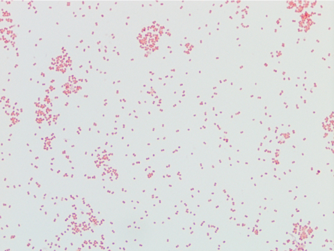
Labs were significant for a positive influenza B swab, leukopenia (WBC 1.2) with 59% bands, and acute kidney injury with a creatinine of 1.4 mg/dl and hyponatremia with a sodium level of 129 mEq/L. Blood cultures grew Streptococcus pneumoniae, sensitive to ceftriaxone. At our facility, she was started on ceftriaxone and azithromycin. She completed 14 days of ceftriaxone; however, she continued to have intermittent fevers above 38 degrees Celsius. Due to the patient’s continued fever, infectious work up was initiated and showed Candida in her urine and HSV lesions on her lips. She was started on a 14 day course of fluconazole and valacyclovir.

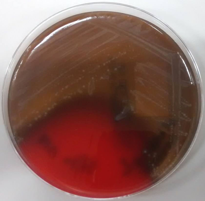
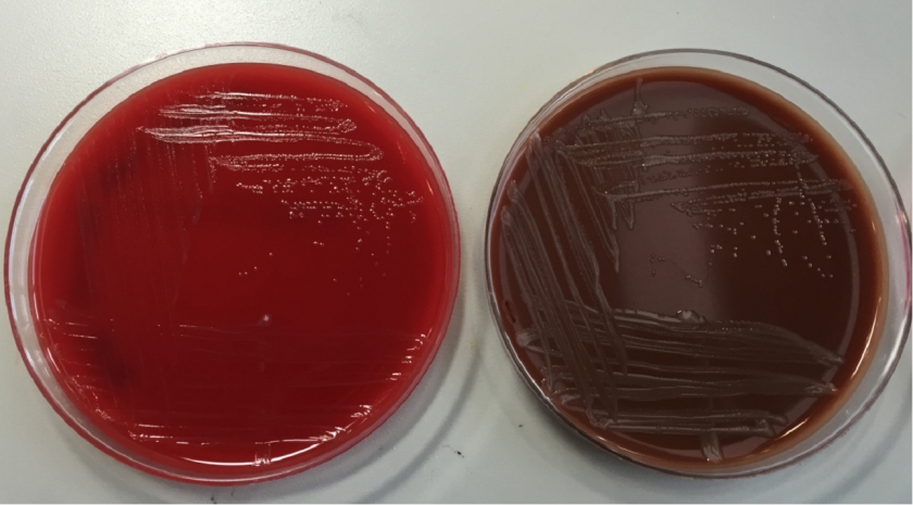
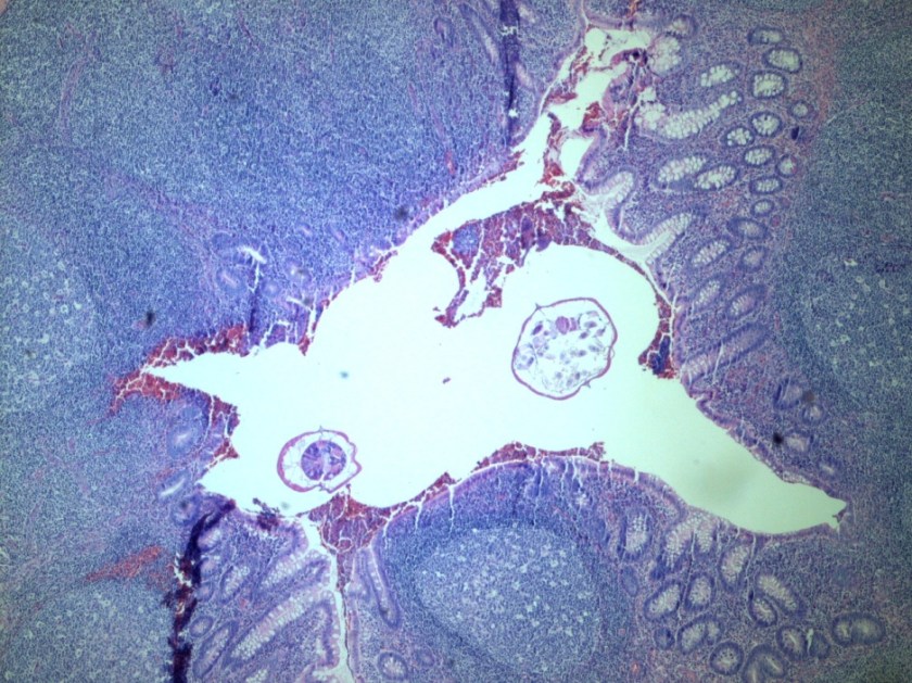
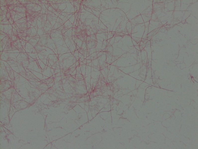
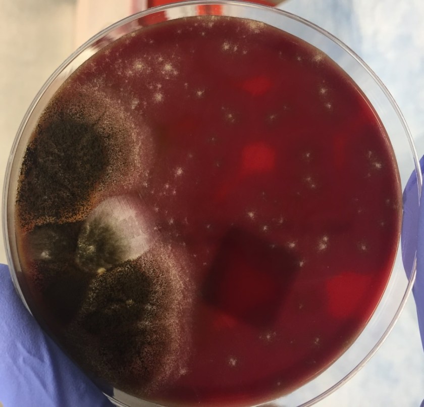
Tracheal aspirates on two occasions were also cultured and grew mixed gram positive and negative organisms as well as Syncephalastrum species. Four weeks after being admitted to our facility, she developed a right-sided hydropneumothorax in which 500 mL of exudative fluid was drawn and subsequently cultured. These cultures also grew Syncephalastrum species as well as Staphylococcus epidermis.

synrac1
Image 1: Syncephalastrum growing on a blood agar plate from the patient’s pleural fluid.
synrac2
Image 2: Lactophenol cotton blue stain of Syncephalastrum demonstrating the sporangiophore with tubular sporangia on the large round vesicle. The sporangia contain chains of round spores.

Discussion

Syncephalastrum racemosum is thought to be the only species out of the two Syncephalastrum species known to cause mucormycoses in humans (1). The only proven reported cases of infection have been due to percutaneous inoculation after trauma, however whether this is due to low pathogenicity, no case reports, or interpretation as a contaminant remains a mystery (1).

Syncephalastrum is a saprophytic fungus isolated throughout the world particularly in environments with decaying organic matter (1, 2). It is found in low levels in the air and has been reported to colonize both immunocompromised and healthy individuals after natural disasters (3).

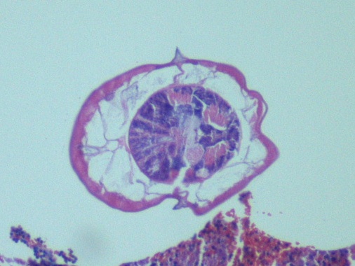
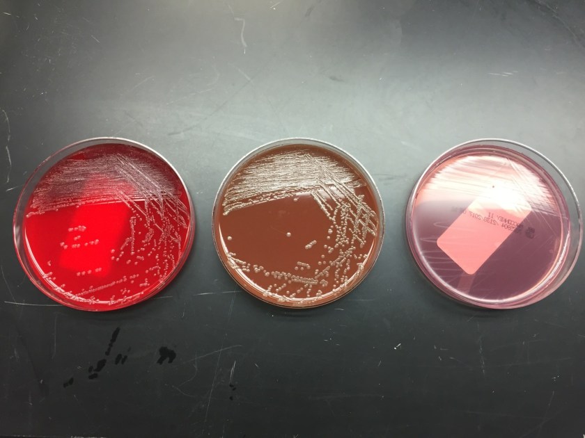
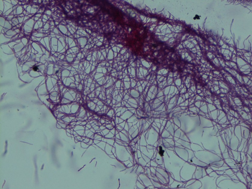
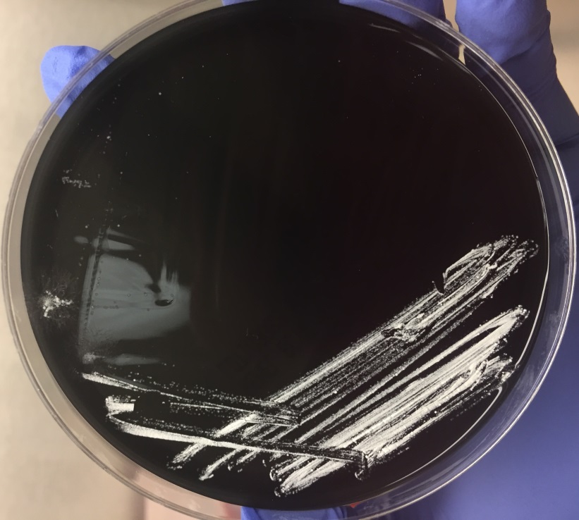
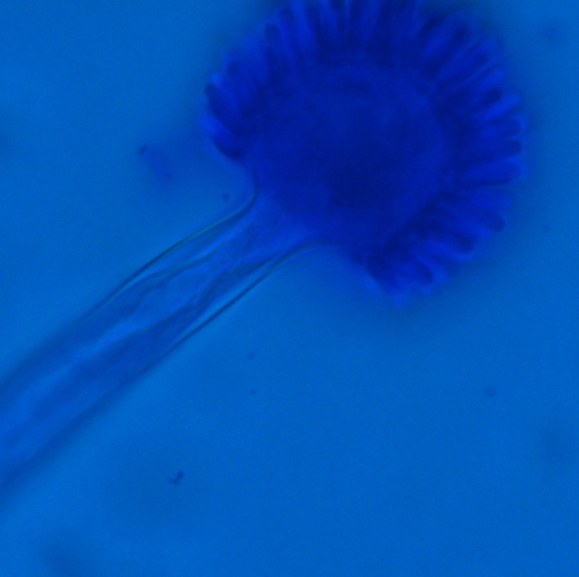
Diagnosis of Syncephalastrum can be made by visualizing pauci-septate, ribbon-like mycelium and a merosporangial sack surrounding sporangiospores from the cultures using a lactophenol cotton blue mount preparation (1). Caution should be used in distinguishing Aspergillus niger from Syncephalastrum using a direct KOH mount due to the similarities in their fruiting bodies (1). On a petri plate, it begins as fast growing white fluff and then turns dark gray to almost black with the reverse side being white (4).


References

  1. Gomes MZ, Lewis RE, Kontoyiannis DP. Mucormycosis caused by unusual mucormycetes, non-Rhizopus, -Mucor, and -Lichtheimia species. Clin Microbiol Rev. 2011;24(2):411-45.
  2. Ribes JA, Vanover-sams CL, Baker DJ. Zygomycetes in human disease. Clin Microbiol Rev. 2000;13(2):236-301.
  3. Rao CY, Kurukularatne C, Garcia-diaz JB, et al. Implications of detecting the mold Syncephalastrum in clinical specimens of New Orleans residents after Hurricanes Katrina and Rita. J Occup Environ Med. 2007;49(4):411-6.
  4. Larone DH. Medically Important Fungi, A Guide to Identification. Amer Society for Microbiology; 2011.


-Angela Theiss is a pathology resident at the University of Vermont Medical Center.

Wojewoda-small

-Christi Wojewoda, MD, is the Director of Clinical Microbiology at the University of Vermont Medical Center and an Associate Professor at the University of Vermont.

Microbiology Case Study: A 75-Year-Old Man with Polymicrobial Bacteremia After Hemicolectomy

Case History

A 75-year-old male with a past medical history of hypertension, hyperlipidemia, and benign prostatic hyperplasia underwent an elective right hemicolectomy at an outside hospital after a cecal polypectomy demonstrated an intramucosal adenocarcinoma (in situ) arising in a background of a sessile serrated adenoma. On post-op day 6, he was transferred to our institution for management of an ST-elevation myocardial infarction that was treated with placement of a drug-eluting stent to the right coronary artery. After the cardiac catheterization, he complained of acute-onset abdominal pain and was tachypneic (49/min), hypotensive (72/48 mmHg), and febrile (39.4°C). He was emergently intubated, given vasopressors, and started on vancomycin and piperacillin/tazobactam empirically for septic shock. A chest X-ray showed atelectasis but no pulmonary consolidation. An abdominal X-ray did not show definitive evidence of pneumoperitoneum and abdominal CT showed some free fluid but no acute abdominal pathology. The WBC count was 3,640/cm3 with an absolute neutrophil count (2,880/cm3) within normal limits. The anaerobic bottle in one of two blood culture sets drawn on post-op day 7 became positive at 27 hours and Gram staining (Image 1) demonstrated gram negative bacilli. Subsequently, the bacilli detected in the anaerobic blood culture bottle were identified by MALDI-TOF as Clostridium clostridioforme, requiring a laboratory corrected report. On post-op day 8, two sets of repeat blood cultures were both positive with Clostridium tertium (Images 2 and 3) and Escherichia coli, consistent with bowel flora. Therapy for the patient’s polymicrobial bacteremia, thought to arise from an ileocolic anastomotic leak, was switched to piperacillin/tazobactam and Metronidazole. Blood cultures on post-op days 10 and 14 were negative. Meanwhile, the patient developed diarrhea, secondary to Clostridium difficile colitis, treated with oral vancomycin and oral thrush treated with micafungin. His hospital course was further complicated by formation of intra-abdominal abscesses, containing E. coli, C. tertium, and C. albicans, that required percutaneous drain placement.

clos1

Image 1. Gram stain of Clostridium clostridioforme from a positive anaerobic blood culture bottle demonstrates thin gram negative bacilli with pointed ends arranged in pairs (100x, oil immersion).

clos2

Image 2. Gram stain of Clostridium tertium from a positive anaerobic blood culture bottle demonstrates gram variable bacilli arranged in short chains (100x, oil immersion).

clos3

Image 3. Clostridium tertium colonies are β-hemolytic on an anaerobic (Schaedler) blood agar plate and appear circular with slightly irregular margins, matte, and grey-white.

Discussion

The genus Clostridium contains approximately 200 species, of which approximately 32 have been associated with human pathologies (1). These organisms are normal members of the human gastrointestinal and cervical-vaginal microflora. Clostridia are also ubiquitously present in nature within soil. Thus, human infection may occur via endogenous or exogenous means. They are classified as gram positive rods and, as such, they do not grow on media that inhibit the growth of gram positive organisms (ie. MacConkey agar). However, upon gram staining, Clostridia may appear gram positive, gram variable, or gram negative. Due to the gram stain variability, inconsistent presence of spores, and atypical colony morphologies, laboratory identification of Clostridum species is problematic.

Clostridium clostridioforme was initially detected in the anaerobic blood culture bottle at 27 hours. Gram staining (Image 1) demonstrates gram negative long, thin bacilli with pointed ends, described as “elongated football shaped” that are arranged in pairs but may also lie singly or in short chains. Oval spores may not be seen but they can be central or subterminal. As obligate anaerobes, C. clostridioforme may be cultured on anaerobic blood agar plates where the gamma-hemolytic colonies appear small, convex to slightly peaked, translucent to opaque, and grey-white. They possess peritrichous flagella that confer motility. It is believed that C. clostridioforme may represent three different species that are frequently isolated anaerobically from blood cultures, particularly in association with mixed cultures, typical of colonic flora (2).

Subsequent blood cultures one day later were positive for both Escherichia coli (detected at 18 hours) and Clostridium tertium (detected at 21 hours). The anaerobic blood culture bottle gram stain (Image 2) demonstrates C. tertium staining as gram variable bacilli arranged in short chains. Terminal spores, only produced under anaerobic conditions, are not seen in Figure 2. C. tertium is one of the aerotolerant clostridia and was cultured on an anaerobic blood agar plate (Figure 3). Colonies appear circular with slightly irregular margins, low convex, matte, and grey-white. Hemolysis can be beta, alpha, or gamma. It was likely overgrown by the E. coli on the aerobic plates. This species is generally considered a weak human pathogen but it has been implicated as a cause of bacteremia in immunocompromised patients. In non-neutropenic patients, C. tertium bacteremia can occur in the setting of gastrointestinal mucosal injury due to gastrointestinal tract pathology or surgery (3).

References

  1. Tille PM. Bailey & Scott’s Diagnostic Microbiology, 13th ed. Elsevier Health Sciences; 2014. pp458-479.
  2. Finegold SM, Song Y, Liu C, et al. Clostridium clostridioforme: a mixture of three clinically important species. Eur J Clin Microbiol Infect Dis. 2005;24(5):319-24.
  3. Miller DL, Brazer S, Murdoch D, Reller LB, Corey GR. Significance of Clostridium tertium bacteremia in neutropenic and nonneutropenic patients: review of 32 cases. Clin Infect Dis. 2001;32(6):975-8.


-Adina Bodolan, MD is a 1st year anatomic and clinical pathology resident at the University of Vermont Medical Center.

Wojewoda-small

-Christi Wojewoda, MD, is the Director of Clinical Microbiology at the University of Vermont Medical Center and an Associate Professor at the University of Vermont.


Microbiology Case Study: A 2 Week Old Female with Eye Discharge

Case History

A 2 week old African American female presented to the pediatric emergency department (ED) with erythema, swelling and copious mucopurulent discharge from the right eye. One week earlier, her Mom noted similar symptoms in the left eye which spontaneously resolved. Mom denied fever, irritability, lethargy, rash, and respiratory or urinary symptoms. The baby was born at term through a spontaneous vaginal delivery with no complications. Mom received regular prenatal care and all screening tests were negative. The baby received erythromycin eye ointment at birth prior to initial discharge. Complete blood count showed a slight leukocytosis (WBC 15.7 TH/cm2) and cerebral spinal fluid (CSF) values were unremarkable. A complete sepsis work up was performed with blood, CSF, eye swabs and urine sent for bacterial cultures. Given the high suspicion for a sexually transmitted infection, an eye swab was also collected for Neisseria gonorrhoeae and Chlamydia trachomatis polymerase chain reaction (PCR). Herpes simplex virus PCR from the CSF was also performed. The patient was started on IV ampicillin, cefotaxime and oral erythromycin in the ED.

Lab results

neiss1
Image 1. The eye swab showed growth of glistening, grey bacterial colonies on sheep blood and chocolate agars after 48 hours incubation at 35°C in 5% CO2.
neiss2
Image 2. Gram stain of the bacterial colonies showing uniform Gram negative diplococci.

The organism was positive for both catalase and oxidase and identified by matrix-assisted light desorption ionization- time of flight (MALDI-TOF) as Neisseria meningitidis. The health department also confirmed the identification. PCR of the eye swab was negative for Neisseria gonorrhoeae and Chlamydia trachomatis. Bacterial cultures from the blood, CSF and urine were all negative.

Discussion

Neisseria meningitidis is an encapsulated Gram negative diplococcus (Image 2) that is usually transmitted through large droplet secretions from the oropharynx from colonized individuals. It can cause invasive meningococcal disease, which can present as meningitis (high fever, stiff neck, and headache), acute sepsis or a combination of both. Waterhouse Friderichsen-syndrome can result in severe dissemination forms of the disease and is characterized by petechial hemorrhages, involvement of the adrenal glands, and disseminated intravascular coagulopathy (DIC). Rarely, N. meningitidis can cause acute bacterial conjunctivitis (1.5 % – 2.5% of cases). Local complications, including corneal ulcers or a more systemic disease, may occur as well.

N. meningitidis produces multiple virulence factors that help cause disease and evade human immune defense mechanisms. The polysaccharide capsule represents the major virulence factor and is also the basis of meningococcal serotyping. Twelve different capsular serotypes can be distinguished, with serotypes A, B, C, W, X, and Y accounting for most invasive disease worldwide. Other virulence factors include pili, which helps the bacteria attach to host surfaces, and IgA protease, an enzyme that cleaves IgA and allows the bacteria to escape the humoral portion of the immune system.

In the laboratory, N. meningitidis grows well on both blood and chocolate agars after 24 hours of incubation (Image 1) and it is positive for both catalase and oxidase. Traditionally, sugar fermentation was used to differentiate Neisseria species from one another. N. meningitidis ferments both glucose and maltose whereas N. gonorrhoeae is only capable of fermenting glucose. Currently, more rapid identification methods (MALDI-TOF, PCR and sequencing) are being increasingly used in most laboratories for a faster and more accurate identification of Neisseria species. The work up of suspected N. meningitidis isolates must be performed using BSL 2 standards, as aerosols created during mobilization from culture plates or performance of biochemical testing has been known to cause invasive disease in laboratory workers.

In general, N. meningitidis is susceptible to penicillin and cefotaxime, but susceptibility testing by disk or gradient diffusion is recommended. Both rifampin and ciprofloxacin can be used for chemoprophylaxis in close contacts of the patient and healthcare & laboratory workers. In addition, a number of meningococcal vaccines are available in the United States (US) and the Centers for Disease Control & Prevention (CDC) recommends vaccinating all adolescents and people at high risk for infection (college students, military recruits, those who had a splenectomy and patients with complement deficiencies). The most common vaccine is a quadrivalent polysaccharide-protein conjugate vaccine which covers serotypes A, C, W and Y. Recently in 2014, the Food and Drug Administration (FDA) approved Trumenba, a vaccine effective against serotype B, which a common serotype causing invasive disease in the US.


AS-small

-Akram Shalaby, MD, is a first year anatomical and clinical pathology resident at the University of Mississippi Medical Center.

Stempak

-Lisa Stempak, MD, is an Assistant Professor of Pathology at the University of Mississippi Medical Center in Jackson, MS. She is certified by the American Board of Pathology in Anatomic and Clinical Pathology as well as Medical Microbiology. She is the director of the Microbiology and Serology Laboratories. Her interests include infectious disease histology, process and quality improvement and resident education.

Microbiology Case Study: An 8 Year Old with Acute Appendicitis

Case History

An 8-year-old female presented to an outside hospital with appendicitis-like clinical symptoms and underwent a laparoscopic appendectomy. Gross examination of the appendix (7.2 cm in length x 0.5 cm in diameter) wall was unremarkable and the lumen contained a minimal amount of hemorrhage. The specimen was entirely submitted for microscopic evaluation.

entver1

Image 1. Cross section of appendix containing two intra-luminal helminths (H & E stain).

entver2

Image 2. Cross section of female Enterobius vermicularis containing eggs (H & E stain).

entver3

Image 3. Cross section of male Enterobius vermicularis (H & E stain).

Discussion

Enterobius vermicularis (human pinworm) is an intestinal nematode (roundworm) with a worldwide distribution that is most prevalent among school-age children. Cross sections of the nonsegmented, cylindrical worms demonstrate a well-developed digestive tract, reproductive system, and two lateral alae (Images 1-3). E. vermicularis has two sexes and Image 1 demonstrates that the male is smaller than the female. Humans are directly infected upon ingestion of E. vermicularis eggs (fecal-oral route of transmission). The eggs then hatch and immature worms undergo maturation within the human gastrointestinal tract (Image 1). Eggs are shed in stool and the typical E. vermicularis eggs (Image 2) are thick-shelled with one flattened aspect, described as “D-shaped”. Patients with the infection are commonly asymptomatic or may complain of perianal pruritus. Rarely, patients present with abdominal pain secondary to E. vermicularis-associated acute appendicitis (1).

Reference

  1. Arca MJ, Gates RL, Groner JI, Hammond S, Caniano DA. 2004. Clinical manifestations of appendiceal pinworms in children: an institutional experience and a review of the literature. Pediatr Surg Int 20(5):372-5.


-Adina Bodolan, MD is a 1st year anatomic and clinical pathology resident at the University of Vermont Medical Center.

Wojewoda-small

-Christi Wojewoda, MD, is the Director of Clinical Microbiology at the University of Vermont Medical Center and an Associate Professor at the University of Vermont.

Microbiology Case Study: Specimen Referral from a 20 Month Old Male

Case History

A 20 month old male presented to an outside hospital with symptoms unknown to our laboratory. That laboratory sent us the specimen recovered from a diaper (Image 1).

asc1
Image 1.

Discussion

The nematode Ascaris lumbricoides is one of the most common helminth infections in the United States. It can grow to be 20-35 cm long. Infection occurs when an egg is ingested, usually in a small child eating dirt contaminated with human feces. When the larvae hatch they penetrate the duodenal wall. From there, the larvae go into the blood stream and eventually end up in the pulmonary circulation where the larvae grow in the alveoli.  In about three weeks, the larvae are coughed up from the lungs and swallowed.  The worms then mature in the jejunum (primarily).  Infection most often shows no symptomatology. If symptoms are present, they can range from mild abdominal discomfort to intestinal blockage and even cough as the worms migrate to the lungs [1].

Diagnosis can be made by examining concentrated stool for knobby-coated, bile-stained eggs that are oval [2].  However, some of the adult worms can pass with the feces.

References

  1. https://www.cdc.gov/parasites/ascariasis/index.html
  2. Murray PR, Rosenthal KS, Pfaller MA. Medical Microbiology, Seventh Edition. Elsevier Health Sciences; 2012.


-Angela Theiss, MD is a 1st year anatomic and clinical pathology resident at the University of Vermont Medical Center.

Wojewoda-small

-Christi Wojewoda, MD, is the Director of Clinical Microbiology at the University of Vermont Medical Center and an Associate Professor at the University of Vermont.

Microbiology Case Study: 42 Year Old Male with Bilateral Lower Extremity Rash

Case History

A 42 year old male presented to the emergency department with a chief complaint of bilateral lower extremity rash. The rash initially began on the dorsum of the patient’s foot, but progressively worsened over the past two weeks. He denied fevers, chills or night sweats. Additionally, he denied any trauma to his legs, burns or exposure to bodies of water. He reported no sick contacts or recent travel and lives at home with his sister and pet dog. His past medical history was significant for squamous cell carcinoma of the oropharynx and alcoholic cirrhosis. On physical exam, he was noted to have extensive cellulitis with the sloughing of skin. Imaging found no acute osteomyelitis or evidence of necrotizing fasciitis. Lab work showed a white count of 17.1 TH/cm2 and elevated ESR and CRP values. He was admitted and started on broad spectrum antibiotics, vancomycin and meropenem. Wound and blood cultures were collected.

Laboratory Identification

pastmult1
Image 1. Gram stain from a positive blood bottle showing Gram negative coccobacilli in pairs (100x oil immersion).
pastmult2
Image 2. Small, grayish colonies grew on blood and chocolate agars after 48 hours incubation in a 35°C incubator with 5% CO2.

The wound culture as well as two blood culture bottles showed growth of the same organism. Gram stain revealed Gram negative coccobacilli that varied from ovoid to short rods. The organism grew on blood and chocolate agars, but not on MacConkey, despite being a Gram negative rod (fastidious pattern). The colonies were non-hemolytic and opaque in appearance. The isolate was positive for catalase, oxidase and indole. MALDI-TOF MS identified the isolates as Pasteurella multocida.

Discussion

Pasteurella spp. are Gram-negative, facultative anaerobic, coccobacilli capable of fermentation. This organism is often found as part of the normal flora of many healthy animals including cats and dogs. P. multocida and P. canis are the most frequently isolated species of the genus. Both of these species are pathogenic in humans. The majority of human infections are wound infections associated with cat (most commonly) & dog bites and scratches. These often result in localized cellulitis and lymphadenitis. Furthermore, rare infections have been reported which include septic arthritis and osteomyelitis, prosthetic joint infection, meningitis, respiratory tract infections, endocarditis, sepsis and bacteremia, and perinatal infections. Systemic infection usually occurs in immunocompromised patients, particularly those with underlying hepatic disease and cirrhosis.

P. multocida is readily recovered by standard media in the clinical microbiology laboratory, growing well on 5% sheep’s blood and chocolate agars, but poorly on MacConkey agar. After overnight incubation on blood agar, small gray colonies with a characteristic musty odor are present. This characteristic musty odor is caused by the production of indole. P. multocida is also oxidase positive and catalase positive. Susceptibility testing for P. multocida from bite wounds in not routinely recommended as these infections most like represent polymicrobial infections and empiric therapy is usually effective. Susceptibility testing should be performed on isolates from normally sterile sites. P. multocida is generally susceptible to penicillin, broad spectrum cephalosporins, tetracyclines, quinolones, trimethoprim-sulfamethoxazole and azithromycin. Resistance has been documented with oxacillin, cephalexin, erythromycin and clindamycin.

In the case of our patient, susceptibility testing was performed by disk diffusion and was susceptible to all the antibiotics listed above with the exception of erythromycin. It was thought he acquired this infection from the family dog licking his feet, with his liver cirrhosis placing him at an increased risk for bacteremia. He was received IV ampicillin/sulbactam for 10 days before being transitioned to an oral regimen for an additional 4 days.


sims

Brooke Sims, MD, is a Cytology Fellow at the University of Mississippi Medical Center.

Stempak

-Lisa Stempak, MD, is an Assistant Professor of Pathology at the University of Mississippi Medical Center in Jackson, MS. She is certified by the American Board of Pathology in Anatomic and Clinical Pathology as well as Medical Microbiology. She is the director of the Microbiology and Serology Laboratories. Her interests include infectious disease histology, process and quality improvement and resident education.

Microbiology Case Study: A Routine Sputum Culture on a 20 Year Old Cystic Fibrosis Patient

Case History

A 20 year old woman with cystic fibrosis was routinely screened with bacterial sputum cultures.  The patient reported feeling well and was compliant with her treatment regimen.  She had no history of Nocardia colonization in the past and her last hospitalization was four years prior due to a pulmonary exacerbation in which cultures grew Stenotrophomonas and Pseudomonas.

no1

Image 1: Modified acid fast stain showing two unique traits of Nocardia: long, delicate rods and weak acid fastness.

no2

Image 2: Nocardia colonies on buffered charcoal yeast extract (BCYE) agar.

Discussion

Nocardia are slow growing, aerobic gram negative rods. Nocardia are unique for being weakly acid fast and displaying aerial hyphae.  They are ubiquitous organisms that can cause a variety of infections in immunocompromised individuals. Most often in the United States, nocardiosis causes a lung infection. If left untreated, it can spread to the brain or spinal cord, where up to 44% die [1].

The above patient showed no signs of an infection, making the possibility of colonization by Nocardia more likely.  Cystic fibrosis patients can be colonized by Nocardia and the clinical approach is to treat regardless of the patient’s overall health. However, there is a lack of reporting on whether Nocardia is the cause of an infection when it happens. Host and pathogen interactions are also not well known. Thorn et al. showed that treating cystic fibrosis patients that are colonized with Nocardia with oral antibiotics did not affect their clinical outcome [2]. More studies are needed to be done to see if antibiotics are warranted in circumstances like the above patient.

References:

  1. Nocardiosis. https://www.cdc.gov/nocardiosis/transmission/index.html
  1. Pulmonary nocardiosis in cystic fibrosis. Thorn, Shannon T. et al.. Journal of Cystic Fibrosis, Volume 8 , Issue 5 , 316 – 320


-Angela Theiss is a pathology resident at the University of Vermont Medical Center.

Wojewoda-small

-Christi Wojewoda, MD, is the Director of Clinical Microbiology at the University of Vermont Medical Center and an Associate Professor at the University of Vermont.

Microbiology Case Study: An Elderly Man with Abdominal Pain

Case History

The lab received two sets of admission blood cultures from an elderly man with a history of atrial fibrillation, aortic valve replacement six years ago, and idiopathic pancytopenia who presented with abdominal pain and symptoms of decompensated heart failure.

Lab Identification

At 59 hours, one aerobic bottle showed growth of a gram negative bacillus. The organism grew pure yellow-pigmented colonies on the blood and chocolate plates with no growth on the MacConkey plate.

breves4
Gram stain from blood agar plate.

The Verigene microarray was not able to identify the organism against 12 gram negative targets. The organism was identified by MALDI-TOF mass spectrometer as Brevundimonas vesicularis. The organism could not be grown for susceptibility testing.

Discussion

B. vesicularis is an aerobic, glucose non-fermenting, spore non-forming, gram negative bacillus closely related to Pseudomonas species. It will demonstrate slow growth of yellow-pigmented colonies on chocolate and blood agar plates with variable growth on MacConkey agar plates. It has in the past been classified as Corynebacterium and Pseudomonas vesicularis. It has been identified globally in environmental sources such as soil, spring water, and healthcare related water containers. It has been identified as a community-acquired, and increasing a hospital-acquired, pathogen in all age groups, and particularly in immunocompromised patients [1, 2]. It has been most commonly reported as a cause of bacteremia, but has also been implicated as the causative agent in cases of arthritis, meningitis, endocarditis, peritonitis, and urinary tract infection [1]. Interesting in vitro studies have demonstrated that the presence of B. vescularis may facilitate the growth of Legionella [3].

Even in cases of the septicemia in immunocompromised patients the mortality rate from B. vesicularis is relatively low. Strains have demonstrated resistance to most forms of antibiotics though is most consistently susceptible to amikacin and piperacillin-tazobactam [1, 2].


References

  1. Shang ST, Chiu SK, Chan MC, Wang NC, Yang YS, Lin JC, Chang FY. Invasive Brevundimonas vesicularis bacteremia: Two case reports and review of the literature. J Microbiol Immun Inf. (2012) 45: 468-472.
  2. Zhang CC, Hsu HJ, Li CM. Brevundimonas vesicularis bacteremia resistant to trimethoprim-sulfamethoxazole and ceftazidime in a tertiary hospital in southern Taiwan. J Microbiol Immunol Infect. (2012) 45(6): 448-452.
  3. Koide M, Higa F, Tateyama M, Cash HL, Hokama A, Fujita J. Role of Brevundimonas vesicularis in supporting the growth of Legionella in nutrient-poor environments. New Microbiol. (2014) 1:33-39.


-Taylor Goller is a Pathology Student Fellow at University of Vermont Medical Center.

Wojewoda-small

-Christi Wojewoda, MD, is the Director of Clinical Microbiology at the University of Vermont Medical Center and an Associate Professor at the University of Vermont.

Microbiology Case Study: A 7 Month Old Female with Fever and Seizure-Like Episodes

Case History

A 7 month old female presented to the emergency department (ED) due to fever and seizure-like episodes. Her mother reported the child had been persistently febrile for 5 days (Tmax 103.9°F) with rhinorrhea, fussiness and decreased oral intake. The patient experienced 3 seizure-like episodes on the day of admission, which the mother described as periods of “shaking” with eyes rolling back. The child was unresponsive during these episodes, which lasted 1 to 2 minutes each. The child had been taken to her pediatrician the day prior to presentation to the ED where she was given a shot of ceftriaxone for presumed otitis media. The child received a chest x-ray, influenza testing, and blood and urine cultures were collected. She also had a lumbar puncture performed and the cerebral spinal fluid (CSF) was sent for chemistries, bacterial culture and polymerase chain reaction (PCR) testing for meningitis/encephalitis pathogens. She was started on IV ceftriaxone.

Laboratory Testing

The child’s white blood cell count from peripheral blood was 7.1 TH/cm2 and chest x-ray, urinalysis and flu testing were unremarkable. The CSF was clear and colorless with 7 WBC/cm2, glucose of 57 mg/dL and protein of 21 mg/dL. The cytospin Gram stain identified no organisms. The meningitis/encephalitis panel detected the presence of human herpesvirus 6 (HHV-6).

Discussion

Human herpesvirus 6 is a member of the Herpesviridae family and was the sixth herpes virus identified. Structurally, HHV-6 possesses a double stranded DNA genome and is enveloped. Clinically, it is the etiologic agent of roseola infantum (exanthum subitum) in infants and toddlers. Primary infection occurs in early childhood and those infected can be asymptomatic or have a non-specific febrile illness while only the minority present with the characteristic red macular rash prominent on the trunk and extremities, lymphadenopathy and high fevers. HHV-6 is highly neurotropic and as such causes viral encephalitis with 5-15% of children experiencing febrile seizures as a result of this illness. HHV-6 is highly prevalent with a greater than ninety percent seropervalence rate. HHV-6 establishes latency in T lymphocytes and can reactivate & cause disease, especially in immunocompromised patients such as those recipients of stem cell or solid organ transplants.

Traditional laboratory methods of identification for HHV-6 were challenging as viral culture, while once the gold standard for active disease, is not practical for most labs and is no longer used in routine diagnostics. PCR from serum, plasma or CSF has become the preferred test as there are now FDA-cleared, commercial platforms that are easy to use, allow for rapid turnaround time and in the case of multiplex PCR panels, the ability to target multiple pathogens from one test. Serology, while helpful in the diagnosis of primary infections, may not be provide conclusive results in a timely manner and is of limited utility in reactivation. Other less commonly used methods include immunohistochemistry, in situ hybridization and electron microscopy.

The prognosis for patients infected with HHV-6 is generally good with self-limited illness not requiring treatment. Rarely, multi-organ involvement can occur and HHV-6 infection in immunosuppressed patients can be a major cause of morbidity and mortality. There is no antiviral therapy licensed for the treatment of reactivated disease in this setting, but approaches using ganciclovir and valganciclovir have been proposed.

In the case of our patient, her blood, urine and CSF cultures were negative and her antibiotics were stopped after cultures were no growth at 24 hours. She required no treatment other than supportive care with acetaminophen for fever control. Prior to discharge, she developed a fine rash on her face, the back of her neck and trunk that was characteristic of an HHV-6 rash. This case demonstrates the utility of multiplex PCR testing in providing rapid identification of pathogenic organisms allowing for real time diagnosis and the limiting of unnecessary treatment.


ET

-Eric Tillotson, MD, is a second year Anatomic and Clinical Pathology resident at the University of Mississippi Medical Center.

Stempak

-Lisa Stempak, MD, is an Assistant Professor of Pathology at the University of Mississippi Medical Center in Jackson, MS. She is certified by the American Board of Pathology in Anatomic and Clinical Pathology as well as Medical Microbiology. She is the director of the Microbiology and Serology Laboratories. Her interests include infectious disease histology, process and quality improvement and resident education.


Microbiology Case Study: A 56 Year Old Woman with Purulent Vaginal Discharge and Dysarthria

Case History

The lab received a purulent fluid from a frontal subdural empyema in a 56 year old woman with a several month history of sporadic bloody to purulent vaginal discharge and a two week history of severe headache, facial weakness, and recent dysarthria causing her to seek treatment. Pelvic exam and CT of the abdomen revealed a fungating cervical mass, later found to be adenocarcinoma, with possible fluid collection within the uterine cavity.

Lab Identification

Initial gram stain of the subdural fluid showed moderate neutrophils and no bacteria. The aerobic plate showed no growth at 48 hours. The thioglycollate broth grew “puffballs” containing long gram negative bacilli with tapered ends.


fusnuc1
Anaerobic blood agar plate on day 9. Note: large white colonies are contamination.
fusnuc2
Gram stain from anaerobic plate on day 9.

The anaerobic plate grew few grey colonies composed of similarly-shaped gram negative bacilli. MALDI-TOF mass spectrometer identified both colony morphologies as Fusobacterium nucleatum.

Discussion

F. nucleatum is a gram negative, spore non-forming, asacchrolytic, slow-growing, obligate anaerobe that can fluoresce chartreuse under UV light. It is one of 14 species within the Fusobacterium genus and is further classified into 5 subspecies (nucleatum, animalis, fusiforme, vincentii, and polymorphum) which have different pathogenic profiles. It is commonly associated with the oral cavity, though it is unclear if it is ever a constituent of usual flora. Its main virulence factors are invasion of epithelial and endothelial cells, induction of host immune response, and adhesion to tissue through a variety of adhesins. The FadA adhesin interacts with the ubiquitous cell junctional cadherins leading to increased permeability of the epithelial and endothelial barrier. This interaction may be why F. nucleatum infections can be found in such diverse locations within the body and are often part of a polymicrobial infection [1, 2].

F. nucleatum is a constituent of oral plaque and is strongly associated with gingivitis and periodontitis. It is present in increased quantity with increasing severity of disease and in patients with a history of smoking or uncontrolled diabetes mellitus. It is a known pathogen in infections and abscesses of the head and neck, brain, lung, abdomen, blood, and pleura. It is also commonly found in placental and fetal tissues and strongly associated with a variety of obstetrical conditions including preterm birth, chorioamnionitis, and neonatal sepsis. It has been implicated in at least one case as the causative agent of stillbirth. Its prevalence in cord blood in cases of neonatal sepsis is equal to or greater than that of E. coli and Group B Strep. There is also a known association between F. nucleatum and colorectal cancer and IBD, with current research investigating whether the bacteria could play a role in pathogenesis. [1].

F. nucleatum is generally responsive to treatment with a range of antimicrobials, though there are reports of strains resistant to clindamycin and beta-lactamase-based resistance to ampicillin [2, 3].

The infectious disease clinician covering the present case suggested that the patient may have been transiently bacteremic due to her fungating gynecologic malignancy and suffered a minor head trauma causing a small subdural hemorrhage that seeded the bacteria in a sufficiently protected anaerobic environment.


References

  1. Han TW. Fusobacterium nucleatum: a commensal-turned pathogen. Curr Opin Microbiol. (2015) 23:141-147.
  2. Denes E, Barraud O. Fusobacterium nucleatum infections: clinical spectrum and bacteriological features of 78 cases. Infection (2016) 44:475-481.
  3. Veloo ACM, Seme K, Raanges E, Rurenga P, Singadji Z, Wekema-Mulder G, van Winkelhoff AJ. Antibiotic susceptibility profiles of oral pathogens. Intl J Antimicrob Agents. (2012) 40:450-454.


-Taylor Goller is a Pathology Student Fellow at University of Vermont Medical Center.

Wojewoda-small

-Christi Wojewoda, MD, is the Director of Clinical Microbiology at the University of Vermont Medical Center and an Associate Professor at the University of Vermont.