Microbiology Case Study: A 65 Year Old Man with Pneumonia

Case History

A 65 year old male with a history of systolic heart failure secondary to non-ischemic (alcohol-induced) dilated cardiomyopathy underwent cardiac transplantation on 10/11/2016. He was hospitalized between 3/1/17 and 4/15/17 for neutropenia and CMV viremia. Two days after discharge, he presented to the hospital with a gradual-onset of left-sided sharp chest pain described as “soreness” over his rib cage and exacerbated by breathing. Associated symptoms included fever, malaise, and fatigue. In the emergency department, vital signs included: BP 144/75 mmHg, T 40.2°C, RR 24/min, HR 101 bpm, SpO2 97% on room air. A CBC revealed a normal white blood cell count and a chest X-ray demonstrated a lingular opacity. The immunocompromised patient was admitted for sepsis secondary to presumed pneumonia following recent hospitalization. He was treated empirically for hospital-acquired pneumonia with vancomycin and piperacillin-tazobactam. After a urine antigen test detected the presence of Legionella pneumophila serogroup 1, antibiotic treatment was changed to levofloxacin and an induced sputum culture was obtained for Legionella surveillance.

legion1
Image 1. Sputum culture on BCYE agar with PAV shows Legionella pneumophila colonies that are circular with smooth edges, grey-white, and glistening in addition to few usual oropharyngeal flora.

Discussion

Legionnaires’ disease, caused by Legionella bacteria, is a cause of 1-9% of both community-acquired and hospital-acquired pneumonias. Symptoms of fever, chills, cough, and chest pain are similar to other causes of pneumonia; however multiple organ systems may be involved, producing additional symptoms including gastrointestinal (diarrhea, nausea, and vomiting) and central nervous system (headache and confusion) findings. Legionella was first discovered after a 1976 outbreak of pneumonia among Pennsylvania State American Legion members who attended a convention at a Philadelphia hotel that had infected water in the air conditioning system; it is reported that 29 out of 182 infected people died. At present, the mortality rate of Legionnaires’ disease ranges from less than 10% in treated community-acquired cases to approximately 30% for hospital-acquired cases.

The genus Legionella contains greater than 60 species of which approximately 20 are human pathogens. Legionella pneumophila (consisting of serogroups 1-16) is the most common cause of Legionnaires’ disease and, in particular, L. pneumophila serogroup 1 causes 70-90% of cases. The organisms are ubiquitous in nature, particularly in warm freshwater environments including lakes and streams, where they infect and multiply within single-celled host organisms. Of pathogenic concern, they can be present in high numbers in human-made complex water systems (such as cooling towers, whirlpool spas, humidifiers, and decorative fountains). After environmental aerosols are inhaled or contaminated water is aspirated into the lungs, alveolar macrophages are infected by the obligate intracellular bacteria. Host risk factors for developing Legionnaires’ disease include organ transplantation, immunocompromised state, immunosuppresion, age greater than 60 years, chronic lung disease, and smoking.

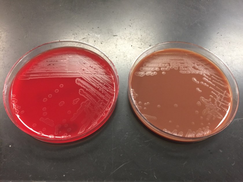
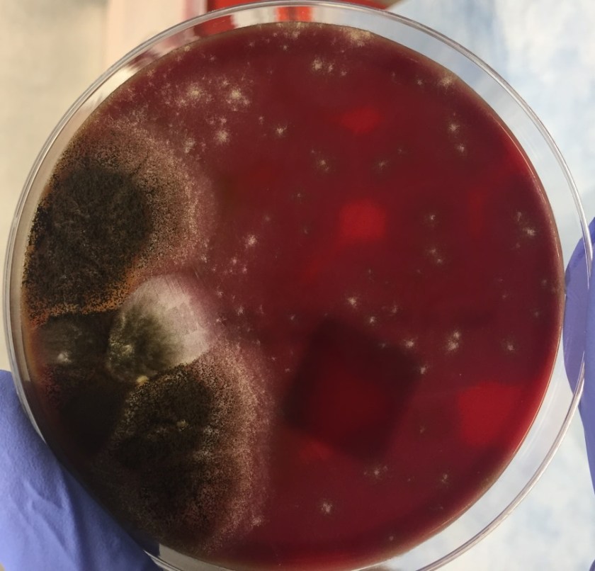
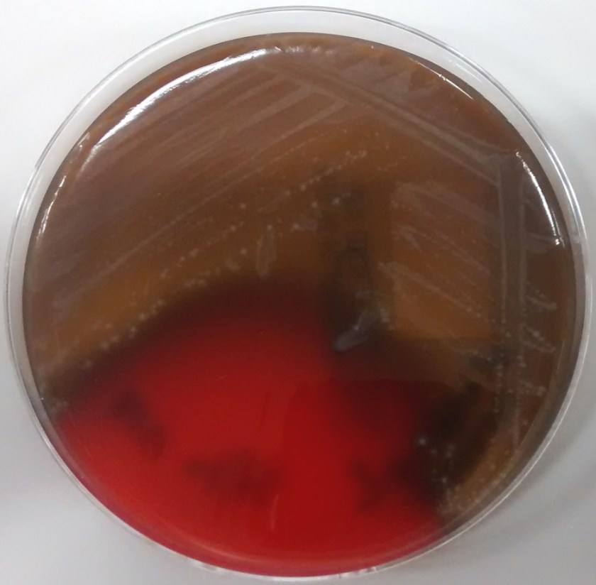
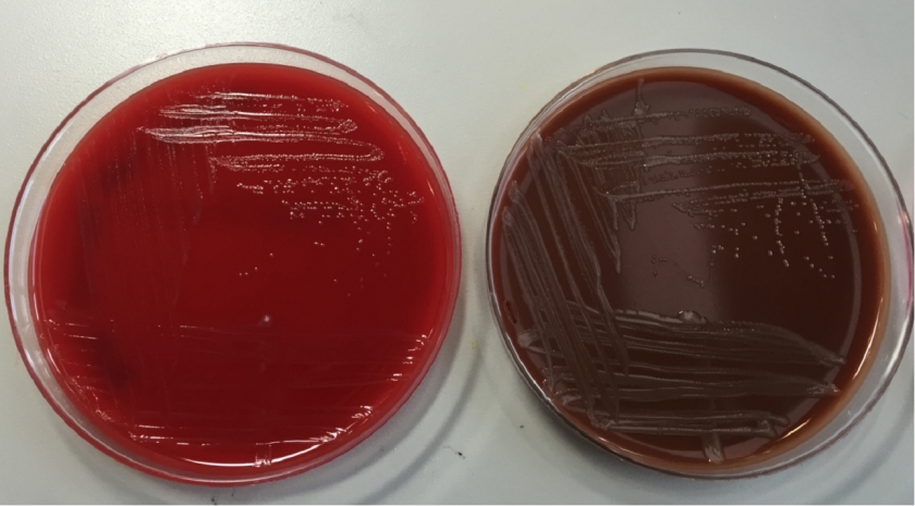
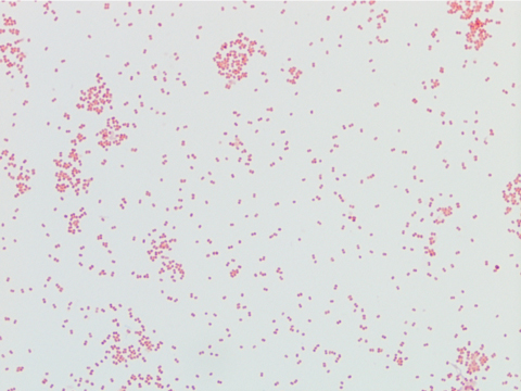
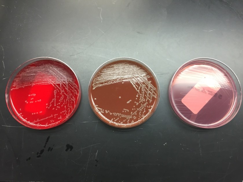
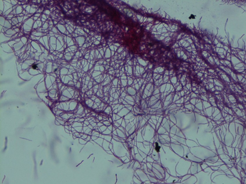
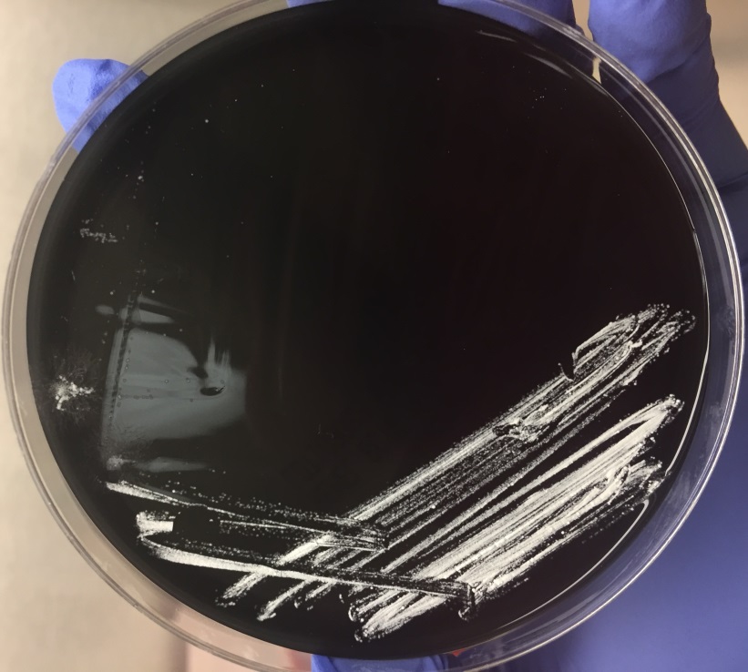
In the microbiology laboratory, Legionella are mesophilic (20-45 °C) obligate aerobes. The small, thin gram negative rods react poorly with Gram stains and are not usually stained in direct clinical samples. The patient’s Gram smear revealed moderate neutrophils, few squamous epithelial cells, and mixed gram positive and gram negative organisms present. Sensitivity for detecting the biochemically inert and fastidious bacteria is increased with culture on buffered charcoal yeast extract (BCYE) agar. For sputum samples that are likely contaminated with usual oropharyngeal flora, BCYE agar with polymyxin B, anisomycin, and vancomycin (PAV) media are used. After 3-5 days of incubation, Legionella colonies appear convex, circular, 3-4 mm in diameter, grey-white to blue-green, and glistening. This identification was confirmed by MALDI-TOF MS. Laboratory in vitro susceptibility studies are not recommended on individual isolates, as they do not correlate with clinical responses. Monotherapy with a fluoroquinolone (Levofloxacin) or macrolide (Azithromycin) is active against Legionella.


-Adina Bodolan, MD is a 1st year anatomic and clinical pathology resident at the University of Vermont Medical Center.

Wojewoda-small

-Christi Wojewoda, MD, is the Director of Clinical Microbiology at the University of Vermont Medical Center and an Associate Professor at the University of Vermont.

Microbiology Case Study: A 24 Year Old Refugee with Eye Irritation

Case History

A twenty-four year-old male Kenyan refugee had been in the United States for about a month when he received a mandatory health screen for infectious diseases. He had no complaints and stated that overall, he was generally healthy. Physical exam was significant only for bilateral red conjunctiva. He stated at times his eyes get irritated and have since birth. As part of routine work-up, an ova and parasite stool exam was ordered. Organisms were detected as seen in Image 1.

giardia1
Image 1. Trichrome stained slides of patient’s stool sample.


Discussion

The patient’s stool examination showed Giardia cysts. Two nuclei are visible in the figure above with centrally located karyosomes. Also visible are the intracytoplasmic fibrils, seen as a darker purple area.

Giardia is a flagellated protozoan that causes giardiasis, a diarrheal illness. It is the most commonly diagnosed intestinal parasitic disease in the United States. It is known as Giardia intestinalis, Giardia lamblia, or Giardia duodenalis. The most common mode of transmission is drinking water contaminated with feces from infected mammals (1).

Symptoms vary and can last 1 week to years if untreated (2). Typical symptoms of giardia are “greasy, foul-smelling, frothy stools that float.” Interestingly, less common symptoms can be itchy skin, hives, eye and joint swelling (3). Retinal arteritis and iridocyclitis has been noted as well (4). It is possible that this patient’s eye irritation is due to a chronic giardiasis infection. Common treatment is usually with an antibiotic/antiparasitic drug like metronidazole (Flagyl).

Diagnosis of Giardia can be made by demonstrating the pear shaped trophozoites and/or ovoid cysts in feces. A key identifier for this parasite is the presence of the two to four nuclei with a central karyosome and intracytoplasmic fibrils that make the parasite look like a face under the microscope. However, because Giardia is excreted intermittently, it is recommended to sample three stool specimens on separate days (5). Due to problems in concentrating the organism for identification on a trichrome stain, a fecal immunoassay is available that is more sensitive and specific (5).

References

  1. https://www.cdc.gov/parasites/giardia/index.html
  2. Robertson LJ, Hanevik K, Escobedo AA, Mørch K, Langeland N. Giardiasis–why do the symptoms sometimes never stop?. Trends Parasitol. 2010;26(2):75-82.
  3. https://www.cdc.gov/parasites/giardia/illness.html#seven
  4. Wolfe MS. Giardiasis.[PDF – 8 pages] Clin Microbiol Rev. 1992;5(1):93-100
  5. https://www.cdc.gov/parasites/giardia/diagnosis.html


-Angela Theiss is a pathology resident at the University of Vermont Medical Center.

Wojewoda-small

-Christi Wojewoda, MD, is the Director of Clinical Microbiology at the University of Vermont Medical Center and an Associate Professor at the University of Vermont.

Microbiology Case Study: A 10 Year Old Boy with Right Knee Pain

Case History  

A 10 year old Caucasian male presented to the pediatric emergency department due to significant pain in his right knee with exposed hardware. His past medical history was noteworthy for Perthe’s Disease, a condition leading to avascular necrosis of the femoral head. In addition, he is affected by an autosomal dominant congenital disorder and has had ischemic strokes in the past. Recently, he had surgery performed on multiple joints to correct abnormalities and they were complicated by dehiscence & infection with methicillin sensitive Staphylococcus aureus. On current admission, his mother reported he has been afebrile and was consistently taking cephalexin to treat the above infection. He was taken to the operating room for incision & drainage and hardware removal of the knee. Bacterial cultures were collected at the time of surgery and he was started on IV clindamycin.

Laboratory Identification

eikcor1
 Image 1. Clear, spreading colonies that showed “pitting” of the agar after 48 hours incubation at 37°C in 5% CO2.  
eikcor2
Image 2. Gram stain directly from the colony showed slender gram negative rods (100x oil immersion).

On direct Gram stain, there was no organisms seen and rare white blood cells. Initially, there was no growth at 24 hours, but two morphologies were observed on the second day. The first organism was identified as the S. aureus that was previously isolated from this site. The second organism was clear with spreading colonies that pitted the agar (Image 1). A distinct bleach like odor was observed. There was no growth on MacConkey agar even though the Gram stain showed gram negative rods, making this organism most likely to be classified as fastidious (Image 2). Benchtop biochemical tests were negative for catalase and positive for oxidase. MALDI-TOF mass spectrometry identified the isolate as Eikenella corrodens. 

Discussion

Eikenella corrodens is a fastidious Gram negative rod that is a member of the HACEK family. It is considered normal flora in the oral cavity and possibly the gastrointestinal tract of humans.  Infection results from these endogenous sources and can be the result of poor oral hygiene, mucositis or dental procedures. E. corrodens causes juvenile and adult periodontitis and is commonly implicated in bacteremia and infective endocarditis, particularly in IV drug users who lick needles prior to injection. Infections of the abdomen, bones/joints and brain are less common.

In the laboratory, E. corrodens is slow growing and is usually present as clear, spreading colonies after 48 hours incubation at 37°C in 5% CO2 on blood and chocolate agars. A unique feature of the organism is that it pits or corrodes the agar, lending to its species name. Also, E. corrodens produces a bleachy smell due to the production of hypochlorite. It does not grow on MacConkey agar despite the fact it is a Gram negative rod. Biochemical tests are negative for catalase, positive for oxidase and negative for indole. Automated instruments and MALDI-TOF mass spectrometry are both able to identify E. corrodens with confidence.

Susceptibility guidelines can be found in the 3rd edition of the CLSI M45 document. In general, E. corrodens is susceptible to penicillin, board spectrum cephalosporins, carbapenems, azithromycin and fluoroquinolones. Resistance to narrow spectrum cephalosporins, macrolides and clindamycin has been documented. In general, susceptibility testing should be performed on E. corrodens when it is isolated from a normally sterile site or is identified in pure culture. Beta lactamase testing is recommended routinely on E. corrodens, and if positive, the isolate is resistant to penicillin, ampicillin and amoxicillin. In the case of bite wounds caused by E. corrodens, susceptibility testing may not be necessary if it is treated with amoxicillin-clavulanate acid (Augmentin) due to a high probability of susceptibility to this antibiotic.

In the case of our patient, he responded to the antibiotic therapy used to treat his S. aureus and E. corrodens infections and healed well. He was placed on long term oral antibiotic therapy until additional hardware is able to be removed at a future date.
RA

-Rim Alkawas, MD, is a first year Anatomic and Clinical Pathology resident at the University of Mississippi Medical Center. 

Stempak

-Lisa Stempak, MD, is an Assistant Professor of Pathology at the University of Mississippi Medical Center in Jackson, MS. She is certified by the American Board of Pathology in Anatomic and Clinical Pathology as well as Medical Microbiology. She is the director of the Microbiology and Serology Laboratories. Her interests include infectious disease histology, process and quality improvement and resident education.

Microbiology Case Study: A 55 Year Old Female with Respiratory Failure

Case History

A 55 year old female with a history of chronic obstructive pulmonary disease, alpha-1 antitrypsin deficiency, and current tobacco use was transferred to our hospital due to acute hypoxemic respiratory failure. She had a gradual six day onset of cough, fever, malaise, weakness, dizziness and wheezing. At the outside facility, she was hypoxic with an oxygen saturation of 67% at room air, hypotensive with a blood pressure of 80/50. She was intubated en route to our facility.

Labs were significant for a positive influenza B swab, leukopenia (WBC 1.2) with 59% bands, and acute kidney injury with a creatinine of 1.4 mg/dl and hyponatremia with a sodium level of 129 mEq/L. Blood cultures grew Streptococcus pneumoniae, sensitive to ceftriaxone. At our facility, she was started on ceftriaxone and azithromycin. She completed 14 days of ceftriaxone; however, she continued to have intermittent fevers above 38 degrees Celsius. Due to the patient’s continued fever, infectious work up was initiated and showed Candida in her urine and HSV lesions on her lips. She was started on a 14 day course of fluconazole and valacyclovir.

Tracheal aspirates on two occasions were also cultured and grew mixed gram positive and negative organisms as well as Syncephalastrum species. Four weeks after being admitted to our facility, she developed a right-sided hydropneumothorax in which 500 mL of exudative fluid was drawn and subsequently cultured. These cultures also grew Syncephalastrum species as well as Staphylococcus epidermis.

synrac1
Image 1: Syncephalastrum growing on a blood agar plate from the patient’s pleural fluid.
synrac2
Image 2: Lactophenol cotton blue stain of Syncephalastrum demonstrating the sporangiophore with tubular sporangia on the large round vesicle. The sporangia contain chains of round spores.

Discussion

Syncephalastrum racemosum is thought to be the only species out of the two Syncephalastrum species known to cause mucormycoses in humans (1). The only proven reported cases of infection have been due to percutaneous inoculation after trauma, however whether this is due to low pathogenicity, no case reports, or interpretation as a contaminant remains a mystery (1).

Syncephalastrum is a saprophytic fungus isolated throughout the world particularly in environments with decaying organic matter (1, 2). It is found in low levels in the air and has been reported to colonize both immunocompromised and healthy individuals after natural disasters (3).

Diagnosis of Syncephalastrum can be made by visualizing pauci-septate, ribbon-like mycelium and a merosporangial sack surrounding sporangiospores from the cultures using a lactophenol cotton blue mount preparation (1). Caution should be used in distinguishing Aspergillus niger from Syncephalastrum using a direct KOH mount due to the similarities in their fruiting bodies (1). On a petri plate, it begins as fast growing white fluff and then turns dark gray to almost black with the reverse side being white (4).


References

  1. Gomes MZ, Lewis RE, Kontoyiannis DP. Mucormycosis caused by unusual mucormycetes, non-Rhizopus, -Mucor, and -Lichtheimia species. Clin Microbiol Rev. 2011;24(2):411-45.
  2. Ribes JA, Vanover-sams CL, Baker DJ. Zygomycetes in human disease. Clin Microbiol Rev. 2000;13(2):236-301.
  3. Rao CY, Kurukularatne C, Garcia-diaz JB, et al. Implications of detecting the mold Syncephalastrum in clinical specimens of New Orleans residents after Hurricanes Katrina and Rita. J Occup Environ Med. 2007;49(4):411-6.
  4. Larone DH. Medically Important Fungi, A Guide to Identification. Amer Society for Microbiology; 2011.


-Angela Theiss is a pathology resident at the University of Vermont Medical Center.

Wojewoda-small

-Christi Wojewoda, MD, is the Director of Clinical Microbiology at the University of Vermont Medical Center and an Associate Professor at the University of Vermont.

Microbiology Case Study: A 75-Year-Old Man with Polymicrobial Bacteremia After Hemicolectomy

Case History

A 75-year-old male with a past medical history of hypertension, hyperlipidemia, and benign prostatic hyperplasia underwent an elective right hemicolectomy at an outside hospital after a cecal polypectomy demonstrated an intramucosal adenocarcinoma (in situ) arising in a background of a sessile serrated adenoma. On post-op day 6, he was transferred to our institution for management of an ST-elevation myocardial infarction that was treated with placement of a drug-eluting stent to the right coronary artery. After the cardiac catheterization, he complained of acute-onset abdominal pain and was tachypneic (49/min), hypotensive (72/48 mmHg), and febrile (39.4°C). He was emergently intubated, given vasopressors, and started on vancomycin and piperacillin/tazobactam empirically for septic shock. A chest X-ray showed atelectasis but no pulmonary consolidation. An abdominal X-ray did not show definitive evidence of pneumoperitoneum and abdominal CT showed some free fluid but no acute abdominal pathology. The WBC count was 3,640/cm3 with an absolute neutrophil count (2,880/cm3) within normal limits. The anaerobic bottle in one of two blood culture sets drawn on post-op day 7 became positive at 27 hours and Gram staining (Image 1) demonstrated gram negative bacilli. Subsequently, the bacilli detected in the anaerobic blood culture bottle were identified by MALDI-TOF as Clostridium clostridioforme, requiring a laboratory corrected report. On post-op day 8, two sets of repeat blood cultures were both positive with Clostridium tertium (Images 2 and 3) and Escherichia coli, consistent with bowel flora. Therapy for the patient’s polymicrobial bacteremia, thought to arise from an ileocolic anastomotic leak, was switched to piperacillin/tazobactam and Metronidazole. Blood cultures on post-op days 10 and 14 were negative. Meanwhile, the patient developed diarrhea, secondary to Clostridium difficile colitis, treated with oral vancomycin and oral thrush treated with micafungin. His hospital course was further complicated by formation of intra-abdominal abscesses, containing E. coli, C. tertium, and C. albicans, that required percutaneous drain placement.

clos1

Image 1. Gram stain of Clostridium clostridioforme from a positive anaerobic blood culture bottle demonstrates thin gram negative bacilli with pointed ends arranged in pairs (100x, oil immersion).

clos2

Image 2. Gram stain of Clostridium tertium from a positive anaerobic blood culture bottle demonstrates gram variable bacilli arranged in short chains (100x, oil immersion).

clos3

Image 3. Clostridium tertium colonies are β-hemolytic on an anaerobic (Schaedler) blood agar plate and appear circular with slightly irregular margins, matte, and grey-white.

Discussion

The genus Clostridium contains approximately 200 species, of which approximately 32 have been associated with human pathologies (1). These organisms are normal members of the human gastrointestinal and cervical-vaginal microflora. Clostridia are also ubiquitously present in nature within soil. Thus, human infection may occur via endogenous or exogenous means. They are classified as gram positive rods and, as such, they do not grow on media that inhibit the growth of gram positive organisms (ie. MacConkey agar). However, upon gram staining, Clostridia may appear gram positive, gram variable, or gram negative. Due to the gram stain variability, inconsistent presence of spores, and atypical colony morphologies, laboratory identification of Clostridum species is problematic.

Clostridium clostridioforme was initially detected in the anaerobic blood culture bottle at 27 hours. Gram staining (Image 1) demonstrates gram negative long, thin bacilli with pointed ends, described as “elongated football shaped” that are arranged in pairs but may also lie singly or in short chains. Oval spores may not be seen but they can be central or subterminal. As obligate anaerobes, C. clostridioforme may be cultured on anaerobic blood agar plates where the gamma-hemolytic colonies appear small, convex to slightly peaked, translucent to opaque, and grey-white. They possess peritrichous flagella that confer motility. It is believed that C. clostridioforme may represent three different species that are frequently isolated anaerobically from blood cultures, particularly in association with mixed cultures, typical of colonic flora (2).

Subsequent blood cultures one day later were positive for both Escherichia coli (detected at 18 hours) and Clostridium tertium (detected at 21 hours). The anaerobic blood culture bottle gram stain (Image 2) demonstrates C. tertium staining as gram variable bacilli arranged in short chains. Terminal spores, only produced under anaerobic conditions, are not seen in Figure 2. C. tertium is one of the aerotolerant clostridia and was cultured on an anaerobic blood agar plate (Figure 3). Colonies appear circular with slightly irregular margins, low convex, matte, and grey-white. Hemolysis can be beta, alpha, or gamma. It was likely overgrown by the E. coli on the aerobic plates. This species is generally considered a weak human pathogen but it has been implicated as a cause of bacteremia in immunocompromised patients. In non-neutropenic patients, C. tertium bacteremia can occur in the setting of gastrointestinal mucosal injury due to gastrointestinal tract pathology or surgery (3).

References

  1. Tille PM. Bailey & Scott’s Diagnostic Microbiology, 13th ed. Elsevier Health Sciences; 2014. pp458-479.
  2. Finegold SM, Song Y, Liu C, et al. Clostridium clostridioforme: a mixture of three clinically important species. Eur J Clin Microbiol Infect Dis. 2005;24(5):319-24.
  3. Miller DL, Brazer S, Murdoch D, Reller LB, Corey GR. Significance of Clostridium tertium bacteremia in neutropenic and nonneutropenic patients: review of 32 cases. Clin Infect Dis. 2001;32(6):975-8.


-Adina Bodolan, MD is a 1st year anatomic and clinical pathology resident at the University of Vermont Medical Center.

Wojewoda-small

-Christi Wojewoda, MD, is the Director of Clinical Microbiology at the University of Vermont Medical Center and an Associate Professor at the University of Vermont.


Microbiology Case Study: A 2 Week Old Female with Eye Discharge

Case History

A 2 week old African American female presented to the pediatric emergency department (ED) with erythema, swelling and copious mucopurulent discharge from the right eye. One week earlier, her Mom noted similar symptoms in the left eye which spontaneously resolved. Mom denied fever, irritability, lethargy, rash, and respiratory or urinary symptoms. The baby was born at term through a spontaneous vaginal delivery with no complications. Mom received regular prenatal care and all screening tests were negative. The baby received erythromycin eye ointment at birth prior to initial discharge. Complete blood count showed a slight leukocytosis (WBC 15.7 TH/cm2) and cerebral spinal fluid (CSF) values were unremarkable. A complete sepsis work up was performed with blood, CSF, eye swabs and urine sent for bacterial cultures. Given the high suspicion for a sexually transmitted infection, an eye swab was also collected for Neisseria gonorrhoeae and Chlamydia trachomatis polymerase chain reaction (PCR). Herpes simplex virus PCR from the CSF was also performed. The patient was started on IV ampicillin, cefotaxime and oral erythromycin in the ED.

Lab results

neiss1
Image 1. The eye swab showed growth of glistening, grey bacterial colonies on sheep blood and chocolate agars after 48 hours incubation at 35°C in 5% CO2.
neiss2
Image 2. Gram stain of the bacterial colonies showing uniform Gram negative diplococci.

The organism was positive for both catalase and oxidase and identified by matrix-assisted light desorption ionization- time of flight (MALDI-TOF) as Neisseria meningitidis. The health department also confirmed the identification. PCR of the eye swab was negative for Neisseria gonorrhoeae and Chlamydia trachomatis. Bacterial cultures from the blood, CSF and urine were all negative.

Discussion

Neisseria meningitidis is an encapsulated Gram negative diplococcus (Image 2) that is usually transmitted through large droplet secretions from the oropharynx from colonized individuals. It can cause invasive meningococcal disease, which can present as meningitis (high fever, stiff neck, and headache), acute sepsis or a combination of both. Waterhouse Friderichsen-syndrome can result in severe dissemination forms of the disease and is characterized by petechial hemorrhages, involvement of the adrenal glands, and disseminated intravascular coagulopathy (DIC). Rarely, N. meningitidis can cause acute bacterial conjunctivitis (1.5 % – 2.5% of cases). Local complications, including corneal ulcers or a more systemic disease, may occur as well.

N. meningitidis produces multiple virulence factors that help cause disease and evade human immune defense mechanisms. The polysaccharide capsule represents the major virulence factor and is also the basis of meningococcal serotyping. Twelve different capsular serotypes can be distinguished, with serotypes A, B, C, W, X, and Y accounting for most invasive disease worldwide. Other virulence factors include pili, which helps the bacteria attach to host surfaces, and IgA protease, an enzyme that cleaves IgA and allows the bacteria to escape the humoral portion of the immune system.

In the laboratory, N. meningitidis grows well on both blood and chocolate agars after 24 hours of incubation (Image 1) and it is positive for both catalase and oxidase. Traditionally, sugar fermentation was used to differentiate Neisseria species from one another. N. meningitidis ferments both glucose and maltose whereas N. gonorrhoeae is only capable of fermenting glucose. Currently, more rapid identification methods (MALDI-TOF, PCR and sequencing) are being increasingly used in most laboratories for a faster and more accurate identification of Neisseria species. The work up of suspected N. meningitidis isolates must be performed using BSL 2 standards, as aerosols created during mobilization from culture plates or performance of biochemical testing has been known to cause invasive disease in laboratory workers.

In general, N. meningitidis is susceptible to penicillin and cefotaxime, but susceptibility testing by disk or gradient diffusion is recommended. Both rifampin and ciprofloxacin can be used for chemoprophylaxis in close contacts of the patient and healthcare & laboratory workers. In addition, a number of meningococcal vaccines are available in the United States (US) and the Centers for Disease Control & Prevention (CDC) recommends vaccinating all adolescents and people at high risk for infection (college students, military recruits, those who had a splenectomy and patients with complement deficiencies). The most common vaccine is a quadrivalent polysaccharide-protein conjugate vaccine which covers serotypes A, C, W and Y. Recently in 2014, the Food and Drug Administration (FDA) approved Trumenba, a vaccine effective against serotype B, which a common serotype causing invasive disease in the US.


AS-small

-Akram Shalaby, MD, is a first year anatomical and clinical pathology resident at the University of Mississippi Medical Center.

Stempak

-Lisa Stempak, MD, is an Assistant Professor of Pathology at the University of Mississippi Medical Center in Jackson, MS. She is certified by the American Board of Pathology in Anatomic and Clinical Pathology as well as Medical Microbiology. She is the director of the Microbiology and Serology Laboratories. Her interests include infectious disease histology, process and quality improvement and resident education.

Microbiology Case Study: An 8 Year Old with Acute Appendicitis

Case History

An 8-year-old female presented to an outside hospital with appendicitis-like clinical symptoms and underwent a laparoscopic appendectomy. Gross examination of the appendix (7.2 cm in length x 0.5 cm in diameter) wall was unremarkable and the lumen contained a minimal amount of hemorrhage. The specimen was entirely submitted for microscopic evaluation.

entver1

Image 1. Cross section of appendix containing two intra-luminal helminths (H & E stain).

entver2

Image 2. Cross section of female Enterobius vermicularis containing eggs (H & E stain).

entver3

Image 3. Cross section of male Enterobius vermicularis (H & E stain).

Discussion

Enterobius vermicularis (human pinworm) is an intestinal nematode (roundworm) with a worldwide distribution that is most prevalent among school-age children. Cross sections of the nonsegmented, cylindrical worms demonstrate a well-developed digestive tract, reproductive system, and two lateral alae (Images 1-3). E. vermicularis has two sexes and Image 1 demonstrates that the male is smaller than the female. Humans are directly infected upon ingestion of E. vermicularis eggs (fecal-oral route of transmission). The eggs then hatch and immature worms undergo maturation within the human gastrointestinal tract (Image 1). Eggs are shed in stool and the typical E. vermicularis eggs (Image 2) are thick-shelled with one flattened aspect, described as “D-shaped”. Patients with the infection are commonly asymptomatic or may complain of perianal pruritus. Rarely, patients present with abdominal pain secondary to E. vermicularis-associated acute appendicitis (1).

Reference

  1. Arca MJ, Gates RL, Groner JI, Hammond S, Caniano DA. 2004. Clinical manifestations of appendiceal pinworms in children: an institutional experience and a review of the literature. Pediatr Surg Int 20(5):372-5.


-Adina Bodolan, MD is a 1st year anatomic and clinical pathology resident at the University of Vermont Medical Center.

Wojewoda-small

-Christi Wojewoda, MD, is the Director of Clinical Microbiology at the University of Vermont Medical Center and an Associate Professor at the University of Vermont.

Microbiology Case Study: Specimen Referral from a 20 Month Old Male

Case History

A 20 month old male presented to an outside hospital with symptoms unknown to our laboratory. That laboratory sent us the specimen recovered from a diaper (Image 1).

asc1
Image 1.

Discussion

The nematode Ascaris lumbricoides is one of the most common helminth infections in the United States. It can grow to be 20-35 cm long. Infection occurs when an egg is ingested, usually in a small child eating dirt contaminated with human feces. When the larvae hatch they penetrate the duodenal wall. From there, the larvae go into the blood stream and eventually end up in the pulmonary circulation where the larvae grow in the alveoli.  In about three weeks, the larvae are coughed up from the lungs and swallowed.  The worms then mature in the jejunum (primarily).  Infection most often shows no symptomatology. If symptoms are present, they can range from mild abdominal discomfort to intestinal blockage and even cough as the worms migrate to the lungs [1].

Diagnosis can be made by examining concentrated stool for knobby-coated, bile-stained eggs that are oval [2].  However, some of the adult worms can pass with the feces.

References

  1. https://www.cdc.gov/parasites/ascariasis/index.html
  2. Murray PR, Rosenthal KS, Pfaller MA. Medical Microbiology, Seventh Edition. Elsevier Health Sciences; 2012.


-Angela Theiss, MD is a 1st year anatomic and clinical pathology resident at the University of Vermont Medical Center.

Wojewoda-small

-Christi Wojewoda, MD, is the Director of Clinical Microbiology at the University of Vermont Medical Center and an Associate Professor at the University of Vermont.

Microbiology Case Study: 42 Year Old Male with Bilateral Lower Extremity Rash

Case History

A 42 year old male presented to the emergency department with a chief complaint of bilateral lower extremity rash. The rash initially began on the dorsum of the patient’s foot, but progressively worsened over the past two weeks. He denied fevers, chills or night sweats. Additionally, he denied any trauma to his legs, burns or exposure to bodies of water. He reported no sick contacts or recent travel and lives at home with his sister and pet dog. His past medical history was significant for squamous cell carcinoma of the oropharynx and alcoholic cirrhosis. On physical exam, he was noted to have extensive cellulitis with the sloughing of skin. Imaging found no acute osteomyelitis or evidence of necrotizing fasciitis. Lab work showed a white count of 17.1 TH/cm2 and elevated ESR and CRP values. He was admitted and started on broad spectrum antibiotics, vancomycin and meropenem. Wound and blood cultures were collected.

Laboratory Identification

pastmult1
Image 1. Gram stain from a positive blood bottle showing Gram negative coccobacilli in pairs (100x oil immersion).
pastmult2
Image 2. Small, grayish colonies grew on blood and chocolate agars after 48 hours incubation in a 35°C incubator with 5% CO2.

The wound culture as well as two blood culture bottles showed growth of the same organism. Gram stain revealed Gram negative coccobacilli that varied from ovoid to short rods. The organism grew on blood and chocolate agars, but not on MacConkey, despite being a Gram negative rod (fastidious pattern). The colonies were non-hemolytic and opaque in appearance. The isolate was positive for catalase, oxidase and indole. MALDI-TOF MS identified the isolates as Pasteurella multocida.

Discussion

Pasteurella spp. are Gram-negative, facultative anaerobic, coccobacilli capable of fermentation. This organism is often found as part of the normal flora of many healthy animals including cats and dogs. P. multocida and P. canis are the most frequently isolated species of the genus. Both of these species are pathogenic in humans. The majority of human infections are wound infections associated with cat (most commonly) & dog bites and scratches. These often result in localized cellulitis and lymphadenitis. Furthermore, rare infections have been reported which include septic arthritis and osteomyelitis, prosthetic joint infection, meningitis, respiratory tract infections, endocarditis, sepsis and bacteremia, and perinatal infections. Systemic infection usually occurs in immunocompromised patients, particularly those with underlying hepatic disease and cirrhosis.

P. multocida is readily recovered by standard media in the clinical microbiology laboratory, growing well on 5% sheep’s blood and chocolate agars, but poorly on MacConkey agar. After overnight incubation on blood agar, small gray colonies with a characteristic musty odor are present. This characteristic musty odor is caused by the production of indole. P. multocida is also oxidase positive and catalase positive. Susceptibility testing for P. multocida from bite wounds in not routinely recommended as these infections most like represent polymicrobial infections and empiric therapy is usually effective. Susceptibility testing should be performed on isolates from normally sterile sites. P. multocida is generally susceptible to penicillin, broad spectrum cephalosporins, tetracyclines, quinolones, trimethoprim-sulfamethoxazole and azithromycin. Resistance has been documented with oxacillin, cephalexin, erythromycin and clindamycin.

In the case of our patient, susceptibility testing was performed by disk diffusion and was susceptible to all the antibiotics listed above with the exception of erythromycin. It was thought he acquired this infection from the family dog licking his feet, with his liver cirrhosis placing him at an increased risk for bacteremia. He was received IV ampicillin/sulbactam for 10 days before being transitioned to an oral regimen for an additional 4 days.


sims

Brooke Sims, MD, is a Cytology Fellow at the University of Mississippi Medical Center.

Stempak

-Lisa Stempak, MD, is an Assistant Professor of Pathology at the University of Mississippi Medical Center in Jackson, MS. She is certified by the American Board of Pathology in Anatomic and Clinical Pathology as well as Medical Microbiology. She is the director of the Microbiology and Serology Laboratories. Her interests include infectious disease histology, process and quality improvement and resident education.

Microbiology Case Study: A Routine Sputum Culture on a 20 Year Old Cystic Fibrosis Patient

Case History

A 20 year old woman with cystic fibrosis was routinely screened with bacterial sputum cultures.  The patient reported feeling well and was compliant with her treatment regimen.  She had no history of Nocardia colonization in the past and her last hospitalization was four years prior due to a pulmonary exacerbation in which cultures grew Stenotrophomonas and Pseudomonas.

no1

Image 1: Modified acid fast stain showing two unique traits of Nocardia: long, delicate rods and weak acid fastness.

no2

Image 2: Nocardia colonies on buffered charcoal yeast extract (BCYE) agar.

Discussion

Nocardia are slow growing, aerobic gram negative rods. Nocardia are unique for being weakly acid fast and displaying aerial hyphae.  They are ubiquitous organisms that can cause a variety of infections in immunocompromised individuals. Most often in the United States, nocardiosis causes a lung infection. If left untreated, it can spread to the brain or spinal cord, where up to 44% die [1].

The above patient showed no signs of an infection, making the possibility of colonization by Nocardia more likely.  Cystic fibrosis patients can be colonized by Nocardia and the clinical approach is to treat regardless of the patient’s overall health. However, there is a lack of reporting on whether Nocardia is the cause of an infection when it happens. Host and pathogen interactions are also not well known. Thorn et al. showed that treating cystic fibrosis patients that are colonized with Nocardia with oral antibiotics did not affect their clinical outcome [2]. More studies are needed to be done to see if antibiotics are warranted in circumstances like the above patient.

References:

  1. Nocardiosis. https://www.cdc.gov/nocardiosis/transmission/index.html
  1. Pulmonary nocardiosis in cystic fibrosis. Thorn, Shannon T. et al.. Journal of Cystic Fibrosis, Volume 8 , Issue 5 , 316 – 320


-Angela Theiss is a pathology resident at the University of Vermont Medical Center.

Wojewoda-small

-Christi Wojewoda, MD, is the Director of Clinical Microbiology at the University of Vermont Medical Center and an Associate Professor at the University of Vermont.