Incidental Finding of Parasitic Infection in a 75 Year Old Male with Persistent Hiccups

Case History

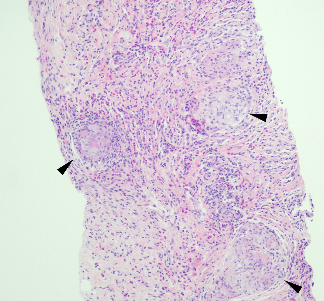
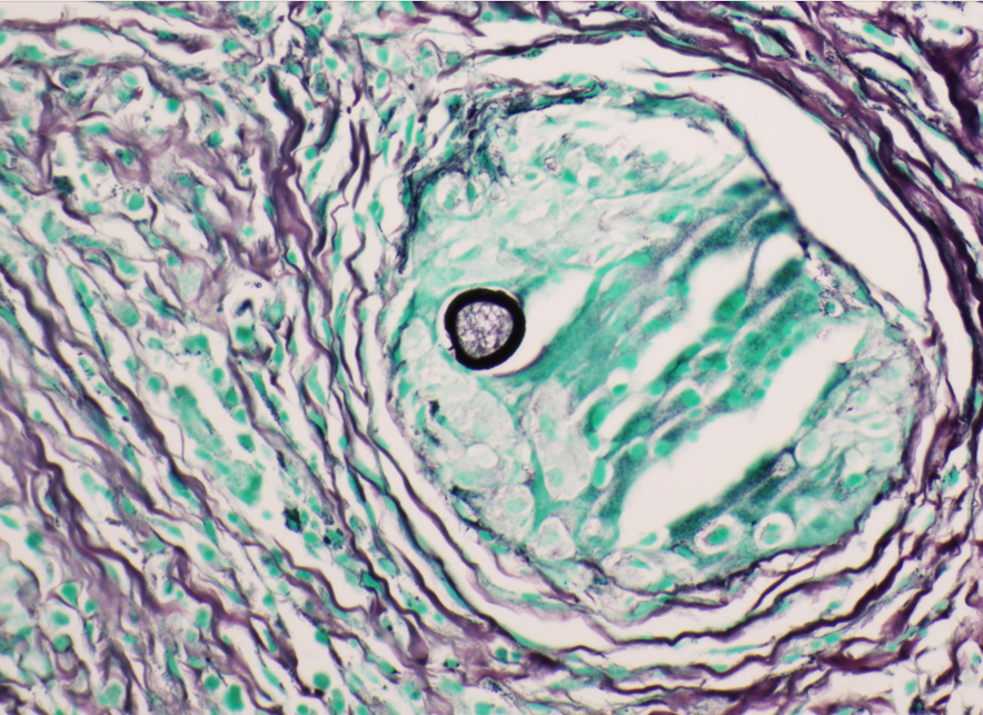
A 75 year old man came to the Emergency Room because of intractable hiccups.  He had a medical history of esophagitis, gastroesophageal reflux disease, gastric metaplasia diagnosed during a previous esophago-gastroduodenoscopy (EGD), and a significant episode of hiccups for several years. His esophagogastroduodenoscopy revealed diffuse edema and erythema on the duodenal mucosa. Histopathological examination of the duodenal biopsies (Figures 1-3) showed the presence of Strongyloides stercoralis within a few crypts of the duodenum, and adjacent eosinophil-rich inflammatory infiltrate within the mucosa. These findings provided an incidental finding of the parasite’s presence in the duodenal mucosa.  

Figure 1. H&E stain of the biopsy at 10X 
Figure 2. H&E stain of the biopsy at 400X  
Figure 3. H&E stain of the biopsy at 400X 

Discussion

Strongyloidiasis is a parasitic infection caused by the nematode Strongyloides – most commonly S. stercoralis. While it is commonly seen in tropical and subtropical regions, cases can also occur in temperate climates. Notably, our patient had a recent travel history to Jamaica, a known endemic region for Strongyloides infection. 

The life cycle of Strongyloides stercoralis involves both free-living and parasitic stages. The infectious filariform larvae penetrate the human skin typically after contact with contaminated soil or exposure to infected fecal matter. Subsequently, they migrate to the lungs through the bloodstream, and eventually reach the small intestine, where they mature into adult worms. The adult worms reside in the duodenal and proximal jejunal mucosa, reproducing asexually by parthenogenesis. Some of the eggs hatch within the intestine, releasing rhabditiform larvae into the feces. It causes autoinfection by penetrating the intestinal wall or the perianal skin area.  

The diagnosis of Strongyloides is typically accomplished by morphologic identification of larvae in the stool, duodenal aspirate, or sputum in disseminated cases. Strongyloides serologic testing is often performed in transplant patients who have a pertinent demographic and clinical history of potential exposure. The presence of eggs is rarely observed in the stool; therefore, microscopic examination of stool samples may have a lower sensitivity in uncomplicated infection with a low organism burden. In our case, stool samples were not collected for evaluation. Hyper-infection syndromes associated with disseminated Strongyloides could present as subclinical infection in patients under immunosuppression. As the larvae invade other organs, such as CNS, lungs, and blood stream, intestinal flora from the GI tract is carried along with the larvae, which causes super-infections, such as bacteremia and meningitis.  

No FDA-cleared molecular testing is available for Strongyloides while some reference laboratories may offer laboratory-developed-tests. Therefore, the laboratory diagnosis frequently relies on the morphologic identification of the filariform larvae or eggs from clinical samples.    In our case, the histopathological examination of the duodenal biopsies that were obtained to evaluate persistent hiccups revealed a significant eosinophil-rich inflammatory infiltrate within the mucosa, along with the presence of the larvae within the crypts. While hiccups can be due to various etiologies, including gastrointestinal disturbances and certain medications, and may not be directly related to parasitic infections, the diagnosis of Strongyloides in this case was purely incidental.  

References 

  1. Gulwani, Hanni. “Strongyloides Stercoralis.” Pathology Outlines – Strongyloides Stercoralis, Aug. 2012, http://www.pathologyoutlines.com/topic/smallbowelstrongyloides.html.  
  2. Carrada-Bravo, Teodoro. “Strongyloides Stercoralis: Vital Cycle, Clinical Manifestations, Epidemiology, Pathology and Treatment.” Revista Mexicana de Patolog, 1 Jan. 1970, http://www.medigraphic.com/cgi-bin/new/resumenI.cgi?IDARTICULO=16127.  
  3. “Strongyloides Stercoralis.” RCPA, 2023, http://www.rcpa.edu.au/Manuals/RCPA-Manual/Clinical-Problems/S/Strongyloides-stercoralis.  
  4. De la Cruz Mayhua, Juan Carlos, and Bisharah Rizvi. “Strongyloides Hyperinfection Causing Gastrointestinal Bleeding and Bacteremia in an Immunocompromised Patient.” Cureus, 24 June 2021, www.ncbi.nlm.nih.gov/pmc/articles/PMC8310433/.  

-Inas Mukhtar, MD, is from Sudan and graduated medical school from University of Khartoum and started a pathology residency in Sudan before applying here to the US. She is currently PGY-2 at Montefiore Medical Center. Her hobbies include watching documentaries and spending time with friends and family.

-Phyu Thwe, Ph.D, D(ABMM), MLS(ASCP)CM is Associate Director of Infectious Disease Testing Laboratory at Montefiore Medical Center, Bronx, NY. She completed her medical and public health microbiology fellowship in University of Texas Medical Branch (UTMB), Galveston, TX. Her interests includes appropriate test utilization, diagnostic stewardship, development of molecular infectious disease testing, and extrapulmonary tuberculosis.

Microbiology Case Study: Blastocystis in an Elderly Patient

A 68 year old female presented to our institution with complaints of lower right quadrant abdominal pain but an absence of nausea, vomiting, diarrhea and fever. The patient noted she thought she had a worm in her stool and submitted three stool samples for an ova and parasite exam. While the three stool samples were negative for worms and eggs, two of the three samples were positive for the organism seen below. The patient was treated with a regimen of metronidazole for ten days, and a repeat ova and parasite exam after treatment was negative for the parasite. The patient presented again three months later with similar complaints of abdominal pain, resulting in another ova and parasite exam which was again positive for the same organism.

Figure 1. Blastocystis spp. organism found in the most recent ova and parasite exam.
Figure 2. Another Blastocystis spp. organism found in the most recent ova and parasite exam (circled in red). The organism is in the vacuole form, with three nuclei visible on the periphery.

Originally discovered in 1911, Blastocystis species are anaerobic protozoan parasites commonly found in the human gastrointestinal tract.2 In addition to being found in humans, Blastocystis spp. has also been found in a variety of animals, with at least nine different genotypes described in literature.4 While historically these organism have been referred to as Blastocystis hominis when found in humans, genotyping and molecular studies have revealed immense genetic diversity in organisms isolated from humans, resulting in a more general designation of Blastocystis spp.1 These organisms have been observed worldwide, stemming from contaminated food, water and soil and transmitted in a fecal-oral route.2 Recently, there has been much debate as to whether the parasite is truly pathogenic or commensal due to the presentation of both symptomatic and asymptomatic patients found carrying this parasite.4

       Despite debate on the pathogenicity of Blastocystis spp., potential signs and symptoms from symptomatic patients have been documented. Such symptoms could include watery diarrhea, nausea, abdominal pain, bloating, excessive gas, loss of appetite, weight loss, and fatigue, among other symptoms.3 Diagnosis heavily relies on stool specimens using light microscopy to examine direct smears, with the vacuolar form of the parasite most commonly observed.1 In addition to the vacuolar form, granular, amoeboid and cyst forms have also been described in literature.4 The parasite itself lacks a cell wall, but contains mitochondria, Golgi apparatuses, and both smooth and rough ER.2 The vacuolar form, observed in Figure 1 and Figure 2, contains a large (5-40 mm) central vacuole-like body which compresses the nuclei to periphery of the cell.2

       Treatment is typically not needed if the patient remains asymptomatic.3 However, for individuals who present with symptomatic infection, a regimen of metronidazole or tinidazole is recommended.3 It is important to note that due to the controversial nature of Blastocystis spp. as a pathogenic parasite, additional pathogens or causes of disease should first be ruled out prior to treatment.2

References:

1 CDC. (2019). Blastocystis sp. [online]. Accessed on October 24th, 2023 from https://www.cdc.gov/dpdx/blastocystis/index.html#:~:text=Blastocystis%20is%20a%20genetically%20diverse,been%20classified%20as%20a%20stramenopile.

2 Leder, K. (2023). Blastocystis species [website]. Accessed online on October 26th, 2023 from https://www.uptodate.com/contents/blastocystis-species

3 Mayo Clinic. (n.d.) Blastocystis Hominis [website]. Accessed online on October 24th 2023 from https://www.mayoclinic.org/diseases-conditions/blastocystis-hominis-infection/symptoms-causes/syc-20351205

4 Wawrzyniak, I., Poirier, P., Viscogliosi, E., Dionigia, M., Texier, C., Delbac, F., & Alaoui, H. E. (2013). Blastocystis, an unrecognized parasite: an overview of pathogenesis and diagnosis. Therapeutic advances in infectious disease1(5), 167–178. https://doi.org/10.1177/2049936113504754

-Maggie King is a Masters Student in the Department of Pathology and Laboratory Medicine at The University of Vermont Larner College of Medicine.

-Christi Wojewoda, MD, is the Director of Clinical Microbiology at the University of Vermont Medical Center and an Associate Professor at the University of Vermont.

Microbiology Case Study: Middle Aged Woman with Abdominal Pain

Case Description

A 59 year old female presented with a one-month history of generalized abdominal pain, difficulty breathing, and early satiety. She was previously seen at an external facility and found to have liver hypointensity and abdominal distension which were concerning for liver cancer. Social history included travel to Sacramento and California deserts for hiking in the last few months. CT of the abdomen and pelvis demonstrated extensive peritoneal carcinomatosis with no primary etiology and a right hepatic lobe focus. MRI revealed diffuse omental caking and peritoneal thickening consistent with peritoneal carcinomatosis. Hepatic lesions also seen on MRI were consistent with hemangiomas. Laboratory evaluation was positive for Hepatitis B virus infection along with elevated liver enzymes (AST and ALT), CA-125, and normal levels of CEA. Paracentesis was performed removing 3 liters of yellow, cloudy fluid. Cytology demonstrated benign mesothelial cells, macrophages, and mixed inflammatory cells with a lymphocytic predominance in the peritoneal fluid. No malignant cells were identified. Initial aerobic, anaerobic, mycobacterial, and fungal cultures were negative. Serum Cryptococcus neoformans antigen testing was also negative. Ultrasound-guided core biopsy of the omental mass was performed; however, tissue was not sent for culture. Histopathology revealed granulomatous inflammation with eosinophilic infiltrate (Figure 1). A singular spherule was highlighted by GMS stain (Figure 2).  Fungal serology via complement fixation for Coccidioides was positive (titer: 1:8) while Blastomyces, Histoplasma, and Aspergillus serologies were negative. The patient was subsequently managed on fluconazole.

Figure 1. Core biopsy of omental tissue revealing granulomatous inflammation and diffuse eosinophilic infiltrate in a background of chronic inflammation, lymphocytes and fibrosis. Arrows indicate granuloma formation (H&E, 100x).
Figure 2. Core biopsy of omental tissue revealing a spherule with its thick wall containing endospores in a granuloma (GMS, 400x).

Discussion

Coccidioidomycosis is caused by the thermally dimorphic fungus Coccidioides that is endemic to the Southwestern United States, Mexico, and some regions in Central and South America. In endemic regions, farm workers, construction workers, military personnel, archaeologists, excavators, inmates, and correctional facility officers are most affected.1 Individuals with significant exposure to the outdoors in endemic regions may be disproportionately affected. Multiple species of Coccidioides can cause disease and geographic distribution is changing2; however, the clinical presentation and approach to treatment of coccidioidomycosis are the same.

The pathophysiology of Coccidioides infection requires inhalation of arthroconidia which may result in pulmonary disease. Once inhaled, the organism forms an endospore-filled spherule in host tissue. Mature spherules rupture and disseminate endospores into surrounding tissue, which can then undergo additional replication cycles within the host. Symptomatic pulmonary disease appears to be related to fungal burden and usually presents with fever, fatigue, cough, pleurisy, and dyspnea, or less commonly peripheral eosinophilia and high erythrocyte sedimentation rate.3  Extrapulmonary disease, also known as disseminated coccidioidomycosis, is observed in <1% of cases. Extrapulmonary spread may develop in the meninges, skin, bone, or joints via hematogenous or lymphatic spread. Rarer sites of disease include the peritoneum, glandular tissue, visceral organs, bone marrow, reproductive organs, the pericardium, kidney, and the bladder.3,4

The gold standard for diagnosis of coccidioidomycosis is culture; however, histopathology may yield definitive diagnosis, especially in cases where culture was not obtained. Following histopathological examination, polymerase chain reaction (PCR) from a variety of sources may also confirm the diagnosis, especially in cases without supporting clinical or laboratory data to suggest coccidioidomycosis.5 When culture is performed, Coccidioides is recovered in its environmental, or mold form. This phase is highly infectious and can pose a significant biosafety hazard; therefore, laboratory personnel should be appropriately notified when Coccidioides is suspected, especially since Coccidioides sp. can grow on solid media within the timeframe of conventional bacterial cultures.3 Serology can be used as an adjuvant test methodology in suspected cases. Serology may be performed on blood samples, CSF, joint, or pleural fluid and measured by immunodiffusion (ID), enzyme-linked immunoassay (EIA), or complement fixation (CF). ID uses gelatin as the medium in which antibodies are placed in one well and Coccidioides antigens are placed in another. Where the two meet through diffusion, a band will form from precipitated immune complexes. ID tests for both IgG and IgM antibodies.6 EIA, in contrast, uses enzyme-labeled antibodies to detect patientantibody complexed with bound antigen. CF incubates Coccidioides antigens with patient serum that has been heated (to inactivate endogenous complement). Following incubation, fresh, non-human serum containing complement is added. If immune complexes have formed between patient antibodies and the antigens, then the fresh serum complement will affix to those complexes and indicator red blood cells will remain intact when added. A positive result will have a suspension of intact red blood cells that can be titered. A negative test will result in lysed red blood cells. CF tests for IgG antibodies.6 Coccidioides antigen assays may be used to test the urine, blood, and CSF of patients with extrapulmonary disease. In immunocompromised hosts, serology may be negative, and coccidioidal antigen assay may be performed. Clinical management commonly involves long-term antifungal therapy with or without surgical intervention for high-risk individuals.4

References

  1. “Epidemiology.” UC Davis Center for Valley Fever, https://health.ucdavis.edu/valley-fever/about-valley-fever/epidemiology/index.html. Accessed 6 July 2023.
  2. Mazi PB, Sahrmann JM, Olsen MA, Coler-Reilly A, Rauseo AM, Pullen M, Zuniga-Moya JC, Powderly WG, Spec A. The Geographic Distribution of Dimorphic Mycoses in the United States for the Modern Era. Clin Infect Dis. 2023 Apr 3;76(7):1295-1301. doi: 10.1093/cid/ciac882. PMID: 36366776; PMCID: PMC10319749.
  3. Proia L. Chapter 28: The Dimorphic Mycoses. In: Spec A, Escota G, Chrisler C, Davies B., eds. Comprehensive Review of Infectious Diseases. > ed. Elsevier; 2020: 411-423.
  4. Johnson RH, Sharma R, Kuran R, Fong I, Heidari A. Coccidioidomycosis: a review. J Investig Med. 2021;69(2):316-323. doi:10.1136/jim-2020-001655.
  5. Binnicker MJ, Buckwalter SP, Eisberner JJ, Stewart RA, McCullough AE, Wohlfiel SL, Wengenack NL. Detection of Coccidioides species in clinical specimens by real-time PCR. J Clin Microbiol. 2007 Jan;45(1):173-8. doi: 10.1128/JCM.01776-06.
  6. “Explanation of Coccidioides Diagnostic Testing.” UC Davis Center for Valley Faver. https://health.ucdavis.edu/valley-fever/about-valley-fever/coccidioides-diagnostic-testing/index.html. Accessed October 26, 2023.

-Evelyn Ilori, MD, PhD is a third-year AP/CP resident at University of Texas Southwestern Medical Center in Dallas, Texas.

-Andrew Clark, PhD, D(ABMM) is an Assistant Professor at UT Southwestern Medical Center in the Department of Pathology, and Associate Director of the Clements University Hospital microbiology laboratory. He completed a CPEP-accredited postdoctoral fellowship in Medical and Public Health Microbiology at National Institutes of Health, and is interested in antimicrobial susceptibility and anaerobe pathophysiology.


-Clare McCormick-Baw, MD, PhD is an Assistant Professor of Clinical Microbiology at UT Southwestern in Dallas, Texas. She has a passion for teaching about laboratory medicine in general and the best uses of the microbiology lab in particular.

Three Safety Cultures Questions to ask Yourself, Your Staff and Your Leaders

Whether you are a newly graduated scientist or a seasoned individual starting at your fifth lab in your career, you might be surprised by the safety culture at the new facility. You could be so impressed by the safety culture at your new laboratory that you question how no one was seriously hurt at your former one. Or you could walk into the lab on your first day and immediately get a bad feeling in your gut. No matter how you feel on day one, two, or maybe day 32, just know that there are some things you can do to help understand your new perception of the culture. Any great piece of research starts with a question or two. Let’s examine some queries that can help you wrap your head around why some labs win, and others fall short when it comes to their safety culture.

First, let’s start with the why. When the safety culture does not look good, it is easy to assume that the deviant behaviors you witness are simply people taking advantage of the system. But not all bad behaviors are spawned from a desire to do harm. You need to find out what is influencing their unsafe behaviors. Most of the time, subpar safety behavior stems from a lack of understanding the consequences of unsafe actions. For example, some folks may not realize that handling their cell phone with gloved hands in the lab has the potential to transfer pathogens into the breakroom when they place that same phone on the table when they are eating their lunch. When the timing is right, you should have a conversation with the employee about what you saw and inquire if they are aware of the potential safety risks. You never know, you could discover that it was a topic skipped in safety training and you single-handedly just improved the quality of your safety training program!

The next question you should ask yourself is, are laboratory leadership aware of the safety issues present in their lab(s)? In most labs, the managers are often overburdened, spending most of their time chasing a schedule, trying to fill open positions, or putting out fires (figuratively we hope). Therefore, you should not assume that leadership is aware and allow unsafe practices to occur in the lab. Sometimes managers and supervisors are so hyper-focused on one thing, they might not be able to see a safety issue right in front of them. If you see unsafe habits, bring it up to lab leadership and share with them that your intentions are to avoid a potential harmful event from occurring. In some cases, managers are already aware of the situation and are trying to come up with solutions. Your conversation with them and perspectives about the safety concerns might be the missing piece that helps complete the puzzle they are trying to solve. So, you should feel comfortable bringing your concerns and be prepared to come with solutions to any problems you present to them.

Finally, ask yourself if the safety issues you see are isolated to a particular shift or certain individuals. Although it is the responsibility of laboratory leadership to champion the safety culture, it is up to the staff to feed and nurture its existence. When everyone works together, it is reflected positively in the safety culture, the audit results, and the injury and exposure reports. When gaps are present, there will be an increase in the negative indicators until the issues are identified and resolved. Instead of making assumptions about the safety culture of the entire lab, try to see where the gaps exist and then revisit the first question- why? It is a lot easier to coach a single individual that wears earbuds in the lab than the entire night shift crew that refuses to wear a lab coat until 5 minutes before the day shift supervisor appears. If that single person is the root of your safety concerns, don’t let their behavior go unchecked. As with negative attitudes in the department, poor safety habits can spread like wildfire. If a single individual’s behavior is not addressed, then others will soon follow suit. They will see that there are no repercussions to lax safety behaviors or worse, they will think nothing bad can come of cutting safety corners. Laboratory leaders and coworkers that normalize poor safety habits are only making the situation worse and damaging the safety culture of the lab while putting the entire staff at risk.

You should never assume a safety culture persists on its own. A good or bad safety culture is the sum of many different factors, and the reason behind the factors can be vast. So, before you are ready to write off a lab as unsafe, take the time to dig a bit deeper and find out what contributed to making the safety culture what it is today. If the lab has a great safety culture, find out why. The lab you are in today may not be the place at which you retire. Your path might lead to a different workplace that has an even worse safety culture than the one you left behind. By asking questions about what creates a great safety culture, you become equipped with the right tools and knowledge and will then be in a strong position to use what you know to improve the lives of others in your new lab.

-Jason P. Nagy, PhD, MLS(ASCP)CM is a Lab Safety Coordinator for Sentara Healthcare, a hospital system with laboratories throughout Virginia and North Carolina. He is an experienced Technical Specialist with a background in biotechnology, molecular biology, clinical labs, and most recently, a focus in laboratory safety.

It’s What’s on the Inside That Counts: Uses of Radiography at Autopsy

In several previous blogs, I’ve mentioned the topic of post-mortem radiography (or “x-rays”). While postmortem CT scanning is a hot topic in the field, plain films are a tool which has been in widespread use for decades. Autopsy standards of the National Association of Medical Examiners require, at a minimum, radiographs be performed on all infants, gunshot wound victims, explosion victims, and charred or decomposed remains. Let’s examine the reasons for these requirements and look at a few specific examples.

All infants must get full body x-rays to check for acute or healing rib and long bone (extremity) fractures, which could be indicative of physical abuse. The extremities are not usually dissected in the course of a typical post-mortem examination, but fractures that can’t be attributed to birth trauma (especially in a pre-mobile child) are a concerning finding that needs further investigation and dissection.

Radiographs taken of gunshot wound victims document the presence and location of retained projectiles, all of which need to be recovered as evidence.  

This x-ray of a gunshot wound victim shows two separate types of ammunition, seen as radio-opaque (white) in the image. The smaller, circular pieces are “birdshot” shotgun ammunition (blue arrows) while the larger pieces are traditional handgun ammunition (red arrow).

Radiographs are commonly performed in any type of penetrating trauma, including sharp force injury and explosive injuries, to identify retained foreign bodies (especially broken fragments of the blade, which may pose a risk to the pathologist). Similar to projectiles, these fragments need to be recovered as evidence. Radiographs can also document the presence of an air embolism, which can be missed at autopsy if special dissection techniques aren’t performed.  

This individual had sharp force injuries to their neck, which injured large veins. The x-ray in this case was performed to see if any fragments of the weapon were still in the body, but also showed a large air embolism in the right atrium and ventricle (blue arrows), seen as radiolucency (gray/black). Open injuries to veins in the head and neck can cause air emboli as breathing creates negative intra-thoracic pressure, drawing air inward though any open channels.

In pedestrians who have been struck by motor vehicles, radiography is the first step in examining trauma. As mentioned earlier, the extremities aren’t typically dissected during a traditional autopsy – but in pedestrians, lower extremity fractures can document the site of initial impact, and the distance of the fracture from the foot may indicate the bumper height of the car.

This x-ray of a pedestrian struck by a motor vehicle shows displaced fractures of the right femur, tibia, and fibula. Note the body bag zipper in the lower right corner.

Fire victims may have extensive thermal injuries and charring which can hide evidence of other injuries, and radiographs can help identify them. Radiographs are also one step toward identifying the victim. Decomposed bodies must be x-rayed for similar reasons – external and internal soft tissue alterations make assessment for trauma more difficult, and radiographs may be needed to confidently identify the body.

In this x-ray of a decomposed person, dental fillings are easily visible (red arrow), which are typically unique for an individual and can be used for identification. Note the irregular shapes of the fillings, which are just as important as their location. Also note the air-fluid level in the cranial cavity (blue arrow), indicating complete liquefication of the brain.

On occasion, radiographs can be performed on individual organs to help define anatomy or previous surgical alterations. Especially in the heart, radiographs can demonstrate coronary artery or valve calcifications, surgical clips from bypass grafts, or other radiopaque prostheses. Knowing the location of devices before dissection gives the pathologist a better chance at preserving and evaluating important structures.

This heart was x-rayed after removal, as the decedent had a history of heart disease with prior interventions. Coronary stents (red arrows) are easily visible; in practice, they can be difficult to find on gross dissection in patients with heavily calcified arteries.

Not all offices can afford the installation (or maintenance) of CT scanners, but access to x-ray machines is more widespread. As we’ve seen here, x-rays are a versatile tool which can document injuries, help identify decedents, and direct the pathologist to perform special autopsy procedures which aren’t part of the daily routine.

-Alison Krywanczyk, MD, FASCP, is currently a Deputy Medical Examiner at the Cuyahoga County Medical Examiner’s Office.