Microbiology Case: A 35 Year Old Male with Left Leg Cellulitis

Clinical History

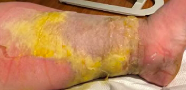
A 35 year old male with chronic bilateral lower extremity lymphedema due to obesity presented with a one-week history of subjective fevers and malaise with associated left lower extremity pain, swelling and erythema. The left leg was markedly edematous with erythema present above the knee down. The leg was tender to palpation, and multiple ruptured bullae and areas of severe desquamation with excessive serous drainage were observed. Importantly, no areas of purulence were noted (Image 2). A clinical diagnosis of severe non-purulent cellulitis was made, and the patient was admitted for parenteral antibiotic therapy of vancomycin and piperacillin-tazobactam. Necrotizing fasciitis was ruled out based on imaging, and significant clinical improvement was seen after 5 days of intravenous antibiotics. The patient was transitioned to oral therapy with amoxicillin-clavulanic acid and doxycycline for a total of 14 days of antibiotics.

Laboratory Workup

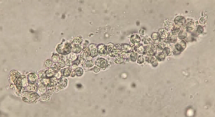
During the admission, urinalysis revealed turbid urine with elevated protein (30 mg/dL), and 2+ blood with 5 RBC/HPF on microscopic examination. Given the presence of protein with microscopic hematuria, causes of glomerulonephritis were investigated. Workup revealed a markedly elevated anti-streptolysin O (ASO) titer of 5310 (0-330) and a total complement (CH50) level of 14, which was low given his age. Urine sediment examination revealed red blood cell casts (Image 3). These clinical and laboratory findings were consistent with post-streptococcal glomerulonephritis (PSGN) due to Streptococcus pyogenes skin and soft tissue infection.

 
Image 1. Colony appearance and biochemical testing of S. pyogenes. A) Typical gram positive cocci in chains characteristic of streptococci. B) Growth on Sheep’s Blood Agar of small, translucent colonies with a wide zone of beta-hemolysis indicative of S. pyogenes. C) Catalase-negative S. pyogenes (left) compared to catalase-positive S. aureus (right). D) PYR-positive S. pyogenes (left) compared to PYR-negative S. aureus (right).
Image 2. Left lower extremity at presentation.
Image 3: Red blood cell cast seen in urine sediment.

Discussion

Streptococcus pyogenes are gram positive bacteria that appear in pairs and/or chains by microscopy (Image 1A). In culture, these organisms produce relatively small colonies which elaborate a large zone of beta hemolysis on blood agar plates; colonies are translucent with smooth edges (Image 1B). The beta-hemolytic activity of S. pyogenes is due to the activity of two hemolysins: Streptolysin-S (oxygen-stabile) and Streptolysin-O (oxygen-labile). S. pyogenes is the primary organism which expresses the Lancefield Group A carbohydrate antigen. Less frequently encountered strains of S. anginosus and S. dysgalactiae subsp. equisimilis may also express this antigen, so biochemical identification of S. pyogenes may be helpful for a definitive diagnosis. MALDI-TOF MS may also fail to discriminate between S. pyogenes and closely related β-hemolytic streptococci (including S. dysgalactiae and S. canis), necessitating adjunctive biochemical testing. Like other streptococci, S. pyogenes is catalase negative (Image 1C). Unlike other beta-hemolytic streptococci, S. pyogenes expresses pyrrolidonyl arylamidase (PYR) making this test a rapid and useful adjunctive diagnostic tool (Figure 1D). Bacitracin susceptibility was used historically but has been largely replaced by PYR testing due to concerns over specificity and prolonged turnaround time.

Globally, S. pyogenes is responsible for a large percentage of infection-related morbidity and mortality. The organism colonizes the skin and the nasopharynx of humans, but most colonized individuals do not develop active disease. Colonization however can lead to infection or dissemination to susceptible individuals. S. pyogenes infections exhibit a diverse range of clinical manifestations which can include pharyngitis, impetigo, erysipelas, cellulitis, necrotizing fasciitis, pyomyositis, streptococcal toxic shock syndrome, and bacteremia. S. pyogenes remains susceptible to penicillin, making β-lactams first-line drugs of choice for management. Conversely, rising levels of macrolide, lincomycin, tetracycline, and fluoroquinolone resistance has been observed. Susceptibility testing may be warranted if these agents are to be used, most often in the cases of severe penicillin allergy.

S. pyogenes infection can be complicated by multiple post-infectious immune-mediated sequelae including PSGN and rheumatic fever. Post-Streptococcus glomerulonephritis (PSGN) has a global incidence of > 470,000 individuals per year and occurs due to the deposition of immune complexes in the glomeruli resulting from previous S. pyogenes pharyngitis or soft tissue infection (as seen in this case). Typical clinical presentation of PSGN includes hematuria, proteinuria, edema, hypertension, elevated serum creatinine levels, hypocomplementemia, and general malaise. The elevated ASO titer (5310) was diagnostic of an S. pyogenes acute infection as the cause of this patient’s cellulitis. The development of proteinuria and hematuria following infection further supports a clinical diagnosis of PSGN. Treatment of PSGN is largely supportive with the focus on management of the underlying infection. Most individuals with kidney failure from PSGN recover to baseline renal function; however, there may be a link between PSGN and the later development of chronic kidney disease/end-stage renal disease.

References

  1. De la Maza LM, Pezzlo MT, Bittencourt CE, Peterson EM. 2020. Color Atlas of Medical Bacteriology, 3rd edition. ASM Press. Pg. 11-23
  2. Madaio MP, Harrington JT. 2001. The diagnosis of glomerular diseases: acute glomerulonephritis and the nephrotic syndrome. Arch Intern Med. 161(1):Pg. 25-34. doi: 10.1001/archinte.161.1.25.
  3. Stevens DL, Bisno AL, Chambers HF, Dellinger EP, Goldstein EJC, Gorbach SL, Hirschmann JV, Kaplan SL, Montoya JG, Wade JC. 2014. Practice Guidelines for the Diagnosis and Management of Skin and Soft Tissue Infections: 2014 Update by the Infectious Diseases Society of America. Clin Infect Dis. 59(2): Pg. e10-e52, https://doi.org/10.1093/cid/ciu296.
  4. Walker MJ, Barnett TC, McArthur JD, Cole JN, Gillen CM, Henningham A, Sriprakash KS, Sanderson-Smith ML, Nizet V. 2014. Disease manifestations and pathogenic mechanisms of Group A Streptococcus. Clin Microbiol Rev. (2): Pg. 264-301. doi: 10.1128/CMR.00101-13.
  5. Wong CH, Khin LW, Heng KS, Tan KC, Low CO. 2014. The LRINEC (Laboratory Risk Indicator for Necrotizing Fasciitis) score: a tool for distinguishing necrotizing fasciitis from other soft tissue infections. Crit Care Med. 32(7): Pg. 1535-41. doi: 10.1097/01.ccm.0000129486.35458.7d.

-John Markantonis, DO is the former Medical Microbiology fellow at UT Southwestern and has recently completed his clinical pathology residency. He is also interested in Transfusion Medicine and parasitic diseases.

-Andrew Clark, PhD, D(ABMM) is an Assistant Professor at UT Southwestern Medical Center in the Department of Pathology, and Associate Director of the Clements University Hospital microbiology laboratory. He completed a CPEP-accredited postdoctoral fellowship in Medical and Public Health Microbiology at National Institutes of Health, and is interested in antimicrobial susceptibility and anaerobe pathophysiology.

-Clare McCormick-Baw, MD, PhD is an Assistant Professor of Clinical Microbiology at UT Southwestern in Dallas, Texas. She has a passion for teaching about laboratory medicine in general and the best uses of the microbiology lab in particular.

Microbiology Case Study: Genotypic-to-phenotypic Discordant Results

Case History

Scenario 1: A 51 year old male with a history of diabetes, hypertension, coronary artery disease, gastric ulcer, chronic kidney disease and bilateral below knee amputation presented with epigastric pain, nausea, and vomiting. He was febrile and tachycardic. Computerized scan showed ascending/ transverse colitis and cholelithiasis. Blood cultures grew gram negative rods; the Biofire BCIDv2 panel reported Enterobacter cloacae with no genotypic, resistance markers detected. Phenotypic antimicrobial susceptibility testing (AST) from the Microscan Walkaway revealed resistance to ertapenem (>1mg/ml) but susceptibility to meropenem (£ 1mg/ml). Additionally, the isolate was resistant to 3rd-generation cephalosporins, fluoroquinolones, and intermediate-resistant to tetracyclines. Identification was confirmed by the MALDI-TOF MS upon growth on agar plates. The isolate was subbed with a meropenem disk to select for carbapenem resistance for further confirmatory testing. A Cepheid Carba-R test was ran on a sweep of the isolate growing near the carbapenem disk, which resulted in no carbapenemases detected. Results from E-tests with meropenem and ertapenem were consistent with original phenotypic result. Here, we reported the discrepant phenotypic result and genotypic results as is.

Image 1. Phenotypic testing results (E-test) for meropenem (MP,left) and ertapenem (ETP, right) of Enterobacter cloacae isolate described in scenario 1. E-test results were consistent with original phenotypic results which also identified the isolate as meropenem susceptible and ertapenem resistant. (Photo credit: Gizachew Demessie, Lead Tech, George Washington Hospital.)

Scenario 2: An 80 year old female underwent a Whipple procedure for a pancreatic mass. A wound culture was submitted from the operating room which grew both Streptococcus anginosus and Enterobacter cloacae complex. Phenotypic AST for the E. cloacae revealed susceptibility to ertapenem (≤0.5 mg/ml) but resistance to meropenem (4 mg/ml). The isolate was pan-susceptible to other drug classes (aside from intrinsic resistance). Similar to Case 1 above, identification was confirmed by the MALDI-TOF MS and the isolate was subcultured with selective pressure. A Cepheid Carba-R test did not detect any carbapenemases. However, upon repeating a phenotypic test, both ertapenem and meropenem were susceptible. Our investigation here led to the avoidance of reporting an incorrect phenotypic AST result.

Discussion

Genotype-to-phenotype discrepancies may occur in antimicrobial susceptibility testing. For example, an antimicrobial resistance (AMR) gene may be detected in a phenotypically susceptible isolate or an AMR gene may not be detected in a phenotypically resistant isolate. Such discordant results should be investigated so appropriate antimicrobial therapy is used on these patients. This leads us to an important question “What can laboratories do to solve these discrepancies?”

The first step in detection of discrepancies requires educating and teaching the lab staff to be vigilant in looking for odd susceptibility patterns (from results within a drug class and also the overall AST profile). Next, check if there was pure isolation of the organism on the purity plate; if not, each individual isolate should be subbed, identified and re-tested on both genotypic and phenotypic platforms. Of note, subbing the bacteria under selective antibiotic pressure (e.g. growing the isolate on agar plate with an antibiotic disk) can increase the potential of detecting resistance. Alternative methods (e.g. CarbaNP, mCIM, etc) could be considered if one is looking into specific resistant mechanisms. Due diligence in checking for clerical, transcription errors and contamination on equipment, especially when there is a consistent pattern of detection for a specific molecular target, is highly recommended. As such, a lab should maintain constant communication with the test manufacturer in case there are issues with batches or lots of reagents.1,2

While these rapid, genotypic panels tend to include the more common AMR mechanisms, there are still other mechanisms of resistance not on the panels. For gram negatives, AMR mechanisms such as AmpC beta-lactamases, porin mutations, efflux pumps and rare carbapenemases such as GES, IMI, and SME types are typically not included.3 Additionally, although the gene blaCTX-M is used as a marker for Extended Spectrum Beta-Lactamases (ESBL), different variants of ESBLs confer different cephalosporin (e.g. 3rd and 4th generation) phenotypes.4 A heteroresistant subpopulation, decreased or lack of expression of an AMR gene may also be potential explanations.

If a discrepancy is not resolved, it is suggested to report the isolate as resistant. If both the discrepant genotypic and phenotypic results are reported, one should consider recommending an infectious diseases consult or to contact the antimicrobial stewardship team.1 Additional information and suggested laboratory workflow can be found in Appendix H of the M100 guidelines from the Clinical Laboratory and Standards Institute.2 While molecular AMR approaches have many advantages such as a shorter turnaround time, phenotypic susceptibility testing can still offer valuable clinical information.5

  1. CLSI. Performance Standards for Antimicrobial Susceptibility Test. CLSI supplement M100. Wayne, PA: Clinical and Laboratory Standards Institute; 2022, Edition 32
  2. Yee R, Dien Bard J, Simner PJ. The Genotype-to-Phenotype Dilemma: How Should Laboratories Approach Discordant Susceptibility Results? J Clin Microbiol. 2021 May 19;59(6):e00138-20.
  3. Tamma PD, Sharara SL, Pana ZD, Amoah J, Fisher SL, Tekle T, Doi Y, Simner PJ. 2019. Molecular epidemiology of ceftriaxone non-susceptible Enterobacterales isolates in an academic medical center in the United States. Open Forum Infect Dis 6:ofz353.
  4. Paterson DL, Bonomo RA. 2005. Extended-spectrum beta-lactamases: a clinical update. Clin Microbiol Rev 18:657–686.
  5. Dien Bard J, Lee F. 2018. Why can’t we just use PCR? The role of genotypic versus phenotypic testing for antimicrobial resistance testing. Clin Microbiol Newsl 40:87–95. 10.1016/j.clinmicnews.2018.05.003. 

Rami Abdulbaki, MD is a Pathology Resident (PGY-3) at The George Washington University Hospital. His academic interest includes hematopathology and molecular pathology.

-Rebecca Yee, PhD, D(ABMM), M(ASCP)CM is the Chief of Microbiology, Director of Clinical Microbiology and Molecular Microbiology Laboratory at the George Washington University Hospital. Her interests include bacteriology, antimicrobial resistance, and development of infectious disease diagnostics.

What’s NOT New in Cancer Care?

In June of 2017 just at the start of the annual American Society of Clinical Oncology (ASCO) meeting in Chicago, Illinois, there were at least 7 new FDA approvals for immuno-oncology agents targeting PD-L1 in cancer. At that time (2017), there were 2030 potential agents targeting 265 different targets across cancer including the modalities of t-cell targeted and other immunomodulators, cell therapy, cancer vaccines, oncolytic viruses, and CD3-targeted bispecific antibodies. Just three years later (2020), prior to the COVID-19 pandemic, this landscape had increased to 4720 potential agents targeting 504 targets across the same spectrum. That represents a 233% growth in these agents. Although only a fraction of these is “approved” (i.e., FDA approved and in use in patients clinically), many these agents are in clinical trials that require patient recruitment using pathology and other testing data. What does this mean for pathologists and laboratory professionals? Depending on the tumor being targeted and the target, there may or may not be a specific laboratory test that needs to be performed which may be routine, like histology parameters or immunohistochemistry, or may require advanced methods, like unique antibodies/clones, specific quantification methods, or molecular testing. The range of testing is not even unique to a specific therapy—for example, pembrolizumab uses staining for PD-L1, MSI, or no testing at all depending on tumor type. For the sub-specialized pathologist that focuses on one or two organs only, mastering the rapid pace and required diagnostic-therapeutic pairings is still a challenge. Imagine what it is like to be a general surgical pathologist in a community setting serving a community cancer center. Moreover, the diagnosis of a specific tumor is often completely disconnected for any biomarkers that may be indicated at the time of collection or several months later depending on therapeutic outcomes. This poses a range of problems in logistics and processing that are still being worked out at the individual system level. Still, the plethora of new treatments for cancer patients is very exciting.

In 2017, the largest group of targets (which was heterogenous) were tumor associated antigens (TAA) which are molecules that are not normally found in the human body produced by tumor cells as the result of changes to cellular processes. Whether it is hybrid proteins, glycosylation, or phosphorylation products, etc., these unique antigens held amazing promise as something we could target and destroy without fear of hurting normal human cells. However, the bulk of these approaches were for tumor vaccines (>90%) in 2017, dropping to 58% in 2020 (and from a total of 265 to only 198). To date, however, only a handful of cancer vaccines have been fully approved including sipuluecel-T for metastatic prostate and T-VEC for advance melanoma. This example category creates a complex set of challenges for pathologists and laboratory professionals. What data is needed about a patient or their tumor before a vaccine can be used? Does it require special studies that are not easily available or are costly? After vaccination, what follow-up tissue or blood studies are needed to follow the patient? Who dictates which tests are required before treatment: industry or medicine? But the more important challenge is: When do we, as the laboratory, pull the trigger to develop and disseminate such information and on-board new tests? Certainly, we are not going to look at Phase I trials and start taking about needs for future diagnostics. But by Phase III (where there is still a high dropout rate before full FDA approval) the number of potential agents and tests may still be daunting. If we wait until approval, now we are behind because our clinical colleagues will start immediately wanting to use the therapy. Tumor vaccines are an interesting category because we assume, for the most part, that there is likely only a diagnostic role needed. But then consider targets like CD-19, PD-L1, PD-1, CD3, Her2, CTLA-4, CD20, MUC1, CD22 and so on which are very familiar to our laboratory family because we often have already a test for these markers.

But is it the correct clone?

Do we have to score or interpret it differently?

When the agent is for cell therapy (the largest growth area of therapy development with 294% growth alone), what role does the transfusion medicine team play in administering or monitoring the patient?

As with the prior example, at what point do we, as a specialty of diagnosticians, dig into the forthcoming clinical trial results to plan? If our colleagues are in academic centers and are part of the clinical trials, they often are aware of and are administering the very tests that determine trial entrance. But if one reads just a few clinical trials of these agents, you may find that the inclusion criteria require a large battery of tests; however, on the other end when it is clinical ready for prime time, only one biomarker may be needed. Such a clustered landscape of information poses frustrating challenges for the clinical team and laboratory team in trying to find the way forward to get patients the life-saving therapies that are quickly arriving.

There is no question that the collision of targeted therapeutics and evolving diagnostics (i.e., precision cancer medicine) has demonstrated phenomenal growth with ever increasing benefits for patients. Affordability and access to these therapeutics aside*, studies continue to be completed and published including combinations therapies and hybrid therapies which show incredible promise. At ASCO 2022, the results of the DESTINY-Breast04 Phase III trial showed that trastuzumab deruxtecan (HER2-directed antibody and topoisomerase inhibitor conjugate) show a 49% reduction in the risk of disease progression or death versus physician’s choice of chemotherapy for patients with HER2-low metastatic breast cancer. That finding should be read a few times to make sure that the impact of this statement is very clear for pathologists and the laboratory. Previously, how we report HER2 (0, 1+, 2+, 3+) was complicated and often required FISH for questionable cases to look directly for HER2 amplification. This new category of patients requires reporting accurately 1+ or 2+ (FISH negative) disease, as it has incredible implications for patients. This news follows the recent new indications for CDK inhibitors in breast cancer related to Ki-67 mitotic score. Just when we thought breast cancer was straightforward, there is more to know and, more importantly, more time and tedium and standardization needed to report it for each patient. And, of course, early triple-negative breast cancer can also be treated with checkpoint inhibitors after PD-L1 testing is performed…but that’s literally old news as the data was release in 2020 at the start of the pandemic.

Outside of therapeutics, diagnostics are evolving quite rapidly with the COVID-19-induced ability to use digital pathology more readily creating a super-highway for artificial intelligence products to be validated for clinical use. PaigeAI has two such products (one for prostate and the second for breast lymph node evaluation released March of 2022) and many others are sure to follow. In parallel, screening, imaging, and surgery have also had advancements that continue to improve patient care and outcomes. So, it seems that everything feels new in cancer but is that the case?

The bulk of tumors diagnosed in the US (and elsewhere) are done with simply H&E staining (up to 75%) with another 20% being further confirmed by a few IHC tests (bringing the total up to 95%). This is not new and, most importantly, is the standard of care for the time being that we use to classify tumors. That classification has dictated, to some degree, the correct NCCN or other cancer protocol that oncologists used to treat patients. At some point, however, sufficient data on the bulk of all tumor types will likely point precision medicine treatments at all cancers. At that point, will a tissue biopsy be necessary with full histology or will a fine needle aspiration with molecular testing dictate the care? The credible assumption is that standard histology and IHC will remain in practice for the foreseeable future because so much billing, accreditation, and compliance is tied closely to them. But we CAN envision a “histology-free” oncopathology approach that matches patients to treatments with a panel of biomarkers. Sounds amazing but also stressful from the point of view of your typical anatomic pathologist.

*But the final thought on this, and perhaps the most important, is cost. Much like the domestic energy market is facing a dwindling pool of customers who agree to pay more and more for “traditional power” while their neighbors pump excessive kilowatts into the grid with their solar panels and windmills enjoying essentially “free power”, progress in cancer screening, detection, and treatment should be dwindling the pool of potential patients and increasing the costs to deliver care to the remainder. However, data and trends suggest that cancer is increasing globally. Why, if we are spending so much money and development on cancer care? Poverty and access. Cancer care is both expensive (in the US) and relatively expensive (in LMICs) with a focus on a small group of patients (0.55% of a population per year develop cancer). Projections of populations who need certain therapeutics are calculated using payer pools and markets that are existing and reliable. That does not include the bulk of LMICs. So, when we consider the cost of the PD-L1 checkpoint inhibitor class per year per patient is upwards of $125,000 USD, how can we even consider that an option for impoverished patients living off $1 USD per day? But if we don’t sort that out and treat these patients, we are assuming that persons who are impoverished are less valuable than persons who can afford expensive care. That evil logic, however, doesn’t hold true because even individuals in the US often become destitute or lose the bulk of their fiscal well-being when they must pay for cancer care—a situation that simply does not occur in countries with socialized medicine and/or universal healthcare.

Cancer care is rapidly evolving and the new tools and therapies available are incredible and miraculous for many patient types who would have faced a death sentence even 10 years ago. But with this amazing progress, we cannot ethically let people with limited resources succumb to these diseases over something so trivial as money. To do so poses harm and sets us up for failure as a species. It is for these reasons that ASCP engages in global health outreach. We are excited to have recently launched the Access To Oncology Medicines (ATOM) program with UICC and more than 2 dozen partners which will rapidly bring high-quality generic cancer therapeutics to low- and middle-income countries. In parallel with the St. Jude/WHO efforts on pediatric cancer globally, we will deliver quality cancer diagnosis and treatment to all patients everywhere.

If you want to learn more about PD-L1 testing and/or overcoming barriers to I-O in persons of color, new education from ASCP is available at no cost at https://www.ascp.org/content/learning/immuno-oncology/.

You can also check out our free educational resources on HER2-low breast cancer and Ki-67 testing in breast cancer at https://www.ascp.org/content/learning/breast-cancer.

Special thanks this month the Kellie Beumer (instructional design) and Melissa Kelly (monitoring and evaluation) from the ASCP medical education grants team for their thoughtful inputs into this piece.

References

  1. https://www.cancerresearch.org/en-us/scientists/immuno-oncology-landscape
  2. https://www.mskcc.org/cancer-care/diagnosis-treatment/cancer-treatments/immunotherapy/cancer-vaccines
  3. https://www.astrazeneca.com/media-centre/press-releases/2022/enhertu-efficacy-results-in-her2-low-breast-cancer.html
  4. https://www.urmc.rochester.edu/news/story/what-is-ki-67-in-breast-cancer
  5. https://www.nejm.org/doi/full/10.1056/NEJMoa1910549
milner-small


-Dan Milner, MD, MSc, spent 10 years at Harvard where he taught pathology, microbiology, and infectious disease. He began working in Africa in 1997 as a medical student and has built an international reputation as an expert in cerebral malaria. In his current role as Chief Medical officer of ASCP, he leads all PEPFAR activities as well as the Partners for Cancer Diagnosis and Treatment in Africa Initiative.

A Quick Primer on BK Virus

While SARS-CoV-2 testing may be dominating discussions, I wanted to highlight other important, but lesser known molecular microbiology tests, starting with BK virus.

About BKV

BK virus (BKV), a member of the Polyomaviridae family, has a tropism for uroepithelial cells and causes disease in immunosuppressed patients, particularly those who have undergone renal transplants.1,2,3 The vast majority of immunocompetent adults are infected with BKV, with estimates up to 90%, and the bulk of cases are entirely asymptomatic.1,3 The exact method of transmission is unknown,3,4 but respiratory transmission is hypothesized. BKV can remain latent after initial infection and can reactivate when immunosuppressed.4 Intermittent asymptomatic viral shedding in urine is particularly common in pregnant individuals or elderly individuals.2

In renal transplant patients, BKV can lead to significant damage to the transplanted kidney and graft failure.1 Polyomavirus-associated nephropathy (PVAN) can occur.2 In bone marrow transplant recipients, hemorrhagic cystitis can occur as a result of this virus.2 Other organ systems can be impacted although much more infrequently.4

The Lab’s Role in Diagnosis and Monitoring BKV

Given the profound impact on renal transplant patients in particular, these patients are routinely screened for BKV both in the blood and the urine. Importantly, BKV can be shed asymptomatically in the urine and thus correlation with BKV detection in the blood is essential. Molecular testing is the method of surveillance. There are currently no FDA approved assays for BKV so labs that perform testing use laboratory-developed tests with analyte specific reagents or research use-only kits.1

Quantification is necessary for monitoring. As with any quantitative assay, there must be at least one negative control, one high positive control, and one low positive control included per run. All controls should fall within the linear range of the assay. To monitor for amplification inhibition, an internal control should be included for each sample.1

Image 1. Example of low (left) and high (right) positive controls.

We perform a BKV LDT assay here using Diasorin reagents and instrumentation. The green line is BKV target while the purple line is the internal control (IC). We run a low positive, high positive, and negative control with every run. Director review for all control and patient results is required. We use commercial BKV positive controls, which have established acceptable range that the quantification of controls must fall within for the run to be considered valid.

The Results and How They Impact Patient Care

Currently, there are no targeted treatments for BKV. In renal transplant individuals, modulation of immunosuppression is the main approach for managing BKV.3,4 A delicate balance must be achieved as reducing immunosuppression can lead to organ rejection while high levels of BKV can cause organ failure.

References

  1. 2016. 12.3 Molecular Methods for Identification of Cultured Microorganisms, Leber AL Clinical Microbiology Procedures Handbook, 4th Edition. ASM Press, Washington, DC. doi: 10.1128/9781683670438.CMPH.ch12.3
  2. Gregory A. Storch and Richard S. Buller, 2019. Human Polyomaviruses, In: Carroll KC, Pfaller MA Manual of Clinical Microbiology, 12th Edition. ASM Press, Washington, DC. doi: 10.1128/9781683670438.MCM.ch108
  3. Furmaga J, Kowalczyk M, Zapolski T, et al. BK Polyomavirus-Biology, Genomic Variation and Diagnosis. Viruses. 2021;13(8):1502. Published 2021 Jul 30. doi:10.3390/v13081502
  4. Mark D. Reploeg, Gregory A. Storch, David B. Clifford, BK Virus: A Clinical Review, Clinical Infectious Diseases, Volume 33, Issue 2, 15 July 2001, Pages 191–202, https://doi.org/10.1086/321813

-Paige M.K. Larkin, PhD, D(ABMM), M(ASCP)CM is the Director of Molecular Microbiology and Associate Director of Clinical Microbiology at NorthShore University HealthSystem in Evanston, IL. Her interests include mycology, mycobacteriology, point-of-care testing, and molecular diagnostics, especially next generation sequencing.

Laboratory Ergonomics: Safe Today, Healthy Tomorrow

Ergonomics is a safety topic that gets little respect in the laboratory, but it can become very important over time. The effects of poor ergonomics are cumulative, and they can appear suddenly. When they arise, the pain and treatment are often difficult, and as people age, healing is slower as well. Because the consequences of repetitive motion injuries are slow to appear, it can be a challenge to raise concerns and create solutions regarding ergonomics. Education and action today can prevent a great deal of future injuries and staff shortages.

There are several areas in the lab where a focus on ergonomics can create benefits, and creating healthy movement and comfort does not need to be expensive or difficult. Laboratory workstations have a primary and secondary work zone.  Keep the most frequently used objects in the primary zone (within 18 inches of reach) and less frequently used in the secondary zone (within three feet).  Every employee is a different size. Teach staff to take a minute before beginning work to adjust the chair and other work items to make the workstation more comfortable.  Eliminate clutter beneath the workstation to allows room to stand or sit allowing for foot and leg comfort.

Chairs should have 4-way and preferably 6-way adjustability and come in a variety of sizes to fit the employees who work in the lab.  Chairs should have five legs with casters that are appropriate for the surface being used (e.g.: hard casters on carpet and soft casters on tile).  The backrest should flex between 90 and 113 degrees with arm rests removed on chairs in the technical area to allow the chair to get closer to the benchtop. 

The tops of computer monitors should be at eye level.  Since many employees may use the same monitor, having it on a movable arm will help each user move the monitor to an acceptable level.  Any glare on the monitor screen can be reduced with a glare screen or by reducing the light in the department.  Keyboards should lay flat to allow the hands and wrist to work in a neutral position and the arms to work at a 90 degree level for comfort.

When using a centrifuge, stand directly in front and work over the top when loading and unloading, and use two hands to close the lid.  Centrifuges should be placed low enough so that employees can see into the body of the machine easily. Place antifatigue mats in front of laboratory equipment that requires standing for long periods of time. These mats relieve lower back and leg discomfort.  When bending and lifting, employees should lift using their thighs and not the back. Teach staff to hold objects close to the body when lifting.  Never lift more than 50 pounds without assistance from other employees or an assistive device such as a hand truck.

Capping and uncapping tubes for an extended period, phlebotomy, and transcription are laboratory tasks that require the use of the same muscle groups in the hands.  When working in these areas, it is important to vary the tasks every 2-3 hours per day and take mini-breaks to stretch fingers and arms in order to prevent carpal tunnel issues.

Breaks are an important part of overall ergonomic health.  It is better to take a five minute break every hour than to take a 15 minute break every four hours.  It is especially important if you are using a microscope or a computer for an extended period of time.  Remember the 20-20-20 rule: Every 20 minutes look up to focus on something 20 feet away and blink your eyes 20 times.  This will allow you to moisturize your eyes and give them a short rest. This can help to prevent ergonomics issues such as Computer Vision Syndrome which can result in neck pain, vision problems, and headaches.

Ergonomics safety is important on all areas of the laboratory, and the best way to ensure good work practices is to perform an ergonomics assessment. An ergonomic assessment should include identifying physical work activities or conditions of the job that are associated with work-related musculoskeletal disorders (MSDs) and how to eliminate these hazards.  For additional information, review the Occupational Safety and Health (OSHA) laboratory ergonomics fact sheet (https://www.osha.gov/sites/default/files/publications/OSHAfactsheet-laboratory-safety-ergonomics.pdf).

Over one third of all U.S. worker injuries are related to MSDs caused by poor ergonomics. Laboratory employees are valuable resources, now more than ever, and preventing time away from work, surgeries and medical bills for laboratorians should be a priority. The results of poor ergonomic practices in the lab do not show up today, but they will have effects tomorrow if we don’t pay attention to them. Those effects can be career-altering, career-ending, and they can interfere with the happy and healthy retirement that we all want to enjoy. Take steps today to prevent that future- provide training, raise awareness, and perform ergonomics assessments to make sure staff remains comfortable and healthy for all of their tomorrows.

Dan Scungio, MT(ASCP), SLS, CQA (ASQ) has over 25 years experience as a certified medical technologist. Today he is the Laboratory Safety Officer for Sentara Healthcare, a system of seven hospitals and over 20 laboratories and draw sites in the Tidewater area of Virginia. He is also known as Dan the Lab Safety Man, a lab safety consultant, educator, and trainer.