By the Book

One of my favorite parts of being a cytotechnologist is the delight of having cytology students rotate through our institution as a practicum site. The pandemic caused a clinical rotation hiatus for the safety of both our staff and students, but thanks to widespread healthcare vaccination, we were able to bring in some fresh minds to experience the variety of interesting cases we enjoy every day. I think what I love most about having students here is reminiscing of when I was in their shoes seven years ago. I remember going into my rotations using nothing but morphologic criteria I memorized from lecture and labs. My clinicals served as a rude awakening that we rarely see any textbook perfect cases. Cancer is like a shape-shifter – one melanoma looks entirely different than another. Two lung squamous cell carcinomas from the right upper lobes from two different patients could look entirely different. The unique variation within and between cancer types is what makes this field so beautifully fascinating. The first time a cytotechnology student shows me a case, tells me their thoughts, works through the criteria, and lists the differentials, I look up and say, “nothing is quite by the book.” How often we fall into a routine of relying on criteria, closing our minds to certain diagnoses because it doesn’t quite look like the clinical impression. When the pathologic and clinical impressions divide, more diagnostic tests are performed, CPT codes fill our billing tab, and we start to panic. “It’s supposed to be adenocarcinoma, so why doesn’t it look like adenocarcinoma?!?

A few weeks ago, the lab received a left pleural fluid from a patient who presented with a history of small cell cervical cancer. I remember learning about this in my first semester of grad school – how rare a finding of small cell carcinoma is, accounting for less than 5% of cervical cancers. It essentially mimics small cell carcinoma of the lung and other neuroendocrine carcinomas, where you should be able to identify the telltale salt-and-pepper chromatin, nuclear molding, scant cytoplasm, loosely cohesive or isolated, necrosis, usually an absence of nucleoli, a high proliferation index with mitotic figures, etc. It’s an aggressive disease to say the least, just like its lung counterpart. When this cancer metastasizes, it takes its same characteristics with it, spreading rapidly without care.


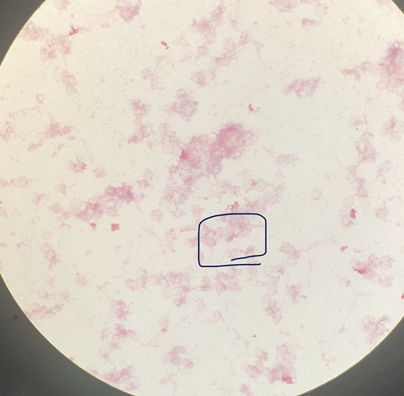
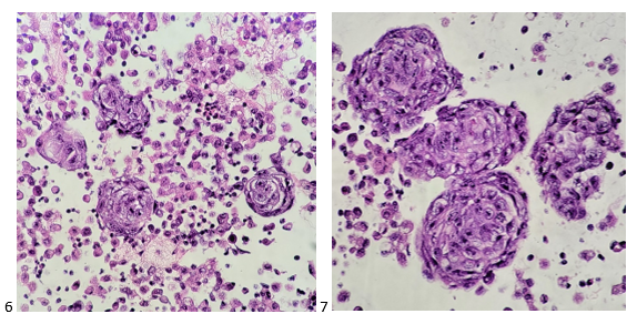
The first step in processing a fluid is to prepare a fresh, air-dried, Diff-Quik-stained cytospin to triage the specimen and decide whether the specimen should be processed routinely or hand-prepped and stained with overtly positive fluids to prevent cross-contamination. There was one cluster identified on the Diff-Quik preparation, but compared to the background of mesothelial and inflammatory cells, the tumor content was insufficient to push it up to hand-processing. The bluish cytoplasm caught my attention as a feature of neuroendocrine tumors AND lymphomas, but the nuclear molding had me favoring neuroendocrine.

Image 1. Pleural fluid, left. DQ-stained cytospin.

That afternoon, I examined the pap-stained smears and SurePath liquid-based preparation, identifying similar cells of interest. However, despite the presence of nuclear molding and scant cytoplasm, the nuclei presented with prominent nucleoli. An interesting feature, to say the least.

Images 2-5. Pleural fluid, left. 2-3, Pap-stained smears (2, lightened to highlight nucleoli); 4-5, Pap-stained SurePath liquid-based preparation.

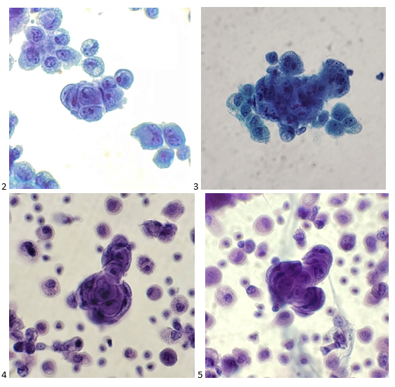
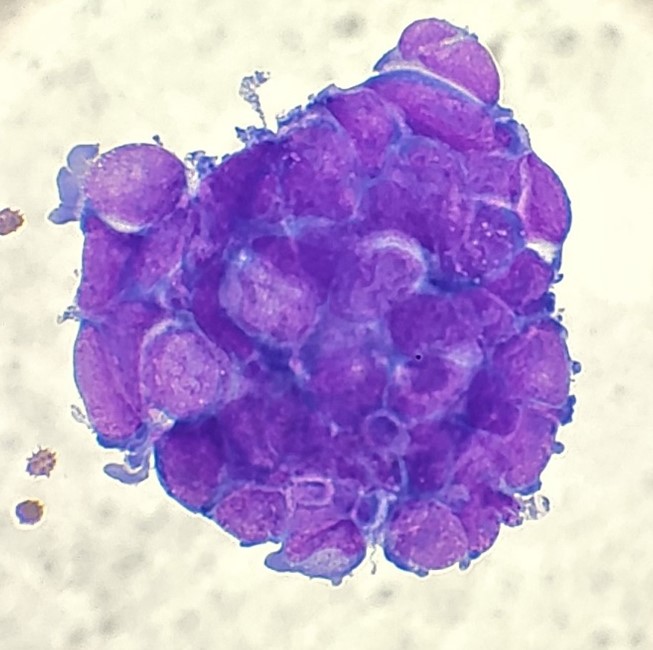
The following morning, I screened the cell block slides and came across molded groups of cells (appearing as a garden aerial view). Still the prominent nucleoli baffled me, and I thought, “Why doesn’t this look like a classic small cell carcinoma? They clinical history even included known lung mets from the patient’s small cell cervical cancer!”

Images 6 and 7: pleural fluid, left. 6, H&E cell block section 100X; 7, H&E cell block section 400X.

When I sent the case for review by the pathologist, I wrote up a diagnosis of Positive for Malignant Cells; Carcinoma, small cell? Recommend correlation with IHC.” My attending was just as intrigued. She ordered a thorough panel of immunohistochemistry stains based on the morphologic findings.

Images 8-11. Pleural fluid, left. 8, synaptophysin+; 9, CD56+; 10, TTF-1+; 11, BerEP4+.

The tumor cells are positive for synaptophysin, CD56, TTF-1, and BerEP4, focally positive for CK7 and chromogranin (not shown), and negative for calretinin, PAX-8, and p40 (also not shown). The findings support the diagnosis of metastatic high grade carcinoma with neuroendocrine differentiation.

While the stains support a diagnosis of small cell carcinoma, the morphologic diagnosis was mildly questionable. I went back to the patient’s record to see what we may have missed in the clinical history. It turns out the patient initially presented with Stage IB2 HPV+, moderately-differentiated cervical adenocarcinoma in 2020. After completing brachytherapy and one cycle of chemotherapy, but could not tolerate additional treatments due to leukopenia and elevated LFTs. Shortly thereafter the patient complained of abdominal pain and a liver mass and bulky lymphadenopathy were identified on imaging. An FNA of a supraclavicular lymph node confirmed not only metastasis of the patient’s cervical cancer, but discovered a small cell/neuroendocrine transformation. And this is why proper documentation of clinical history is so important to pathologists and laboratory professionals. In one of my earlier posts, I preached that cancer doesn’t discriminate; so why should we? Keeping an open mind is paramount to both succeeding in and enjoying the field of cytopathology. If it looks like a duck, and it walks like a duck, it might actually have transformed into a goose.

-Taryn Waraksa, MS, SCT(ASCP)CM, CT(IAC), has worked as a cytotechnologist at Fox Chase Cancer Center, in Philadelphia, Pennsylvania, since earning her master’s degree from Thomas Jefferson University in 2014. She is an ASCP board-certified Specialist in Cytotechnology with an additional certification by the International Academy of Cytology (IAC). She is also a 2020 ASCP 40 Under Forty Honoree.

Hematology Case Study: Presenting a Double Feature Starring Chronic Myelogenous Leukemia

One of the reasons I love working in Hematology is because when we have unexpected results they are often accompanied by visuals… and a picture is worth a thousand words! Unusual or critical results in Chemistry can be interesting, sometimes there are dilutions to perform, results to compare or puzzles to solve, I have always loved working up a good antibody or complicated multiple antibodies in Blood Bank or calculating how many units I may need to screen to find compatible ones, gram stains of unusual organisms in Microbiology can be exciting, but nothing beats some of the cells we see in Hematology! It’s always fascinating when we find unusual cells and follow up with smear reviews with our pathologists. And, being able to save these visuals in CellaVision or saving the slides for teaching, is a plus. These cases are a gift that keeps on giving! Lately I’ve had my share of “exciting” specimens, usually on a Saturday or Sunday afternoon! It never fails to get the adrenaline going when you are the first one to see a CBC with a WBC of 400,000, a differential that is over 90% blasts, rare lymphoma cells, malarial parasites, or a body fluid smear full of malignant cells. The following 2 cases are a very remarkable looking smear and a not so remarkable one, from 2 different patients with the same diagnosis.

The first patient is a 71 year old male who had a routine CBC done by his primary care physician. The blood was collected as an outpatient on a Saturday morning, and brought to our lab by a routine courier that afternoon (of course, right before change of shift!). We had one previous CBC result on this gentleman, from several years earlier, which was essentially normal. CBC result shown below:

Table 1. Case 1, CBC results. [Editor’s note: a previous version of this table noted a Hct of 231.8. The correct result is 31.8.]
Table 2. Case 1, Manual Differential results.
Image 1. Peripheral smear, Case 1, WBC 363.14.

As soon as I saw the results, I called the provider with the WBC and alerted them that I would be contacting the pathologist on call and calling back with the differential. Our pathologist confirmed blasts on the peripheral smear and requested that the sample be sent out for flow cytometry. The pathology report stated “Marked leukocytosis with left shift and <5% blasts. The presentation is suspicious for a myeloproliferative neoplasm (e.g. chronic myelogenous leukemia (CML)). Immunophenotypic studies have been ordered and will be reported separately. Clinical correlation and Hematology consult recommended.” Flow cytometry results showed left shifted maturation and FISH studies demonstrated t(9;22) BCR-ABL with 98% of positive nuclei in bone marrow. No other mutations were detected. Diagnosis: chronic myelogenous leukemia. Five days later, we had a bone marrow scheduled on a 50 year old male. A CBC done 2 weeks earlier showed a mild leukocytosis and thrombocythemia. (WBC 12.4, Hgb 17.8, Hct 52%, PLT 539). Diagnoses under consideration were possible CML, polycythemia or a myeloproliferative neoplasm (MPN). The patient’s CBC the day of the procedure is shown below.

Table 3. Case 2, CBC results.
Table 4. Case 2, Manual Differential results.

Cytogenetic analysis showed an abnormal clone characterized by the Philadelphia chromosome translocation, t(9;22). The BCR/ABL gene rearrangement was detected by FISH, with 78% of positive nuclei in bone marrow. The bone marrow was negative for other mutations. Flow cytometry analysis reported no evidence of abnormal myeloid maturation or increased blast production. There was no evidence for a lymphoproliferative disorder. Diagnosis: chronic myelogenous leukemia.

In 1959, at a time when techniques for preparing chromosomes for visualizing under the microscope were still very unsophisticated, 2 researchers in Philadelphia detected a tiny abnormality in the chromosomes of patients with CML. They noticed that part of chromosome 22 appeared to be missing. It was not until 1970, when techniques for chromosome banding became available, that this discovery was shown to be a translocation between chromosomes 22 and 9. The shortened chromosome 22 was named the Philadelphia (Ph) chromosome after the city where it was discovered.

Image 2. The Philadelphia chromosome. A piece of chromosome 9 and a piece of chromosome 22 break off and trade places (cancer.gov).

At diagnosis, over 90% of CML cases have the t(9;22) translocation which has become a hallmark for a diagnosis of CML. However, the Ph chromosome is also detected in about 30% of adult acute B cell lymphoblastic leukemia (B-ALL), and a very small number of acute myeloid leukemias (AML) and childhood B-ALL so testing must be done for differentiation. t(9;22) is a translocation of the proto-oncogene tyrosine-protein kinase ABL1 gene on chromosome 9 and the breakpoint cluster region BCR gene on chromosome 22. The newly formed chromosome 22 with the attached piece of chromosome 9 is called the Philadelphia chromosome. The BCR-ABL oncogene is formed on the Philadelphia chromosome and the product of the Ph translocation is an abnormal fusion protein, p210, which has increased tyrosine kinase activity. This, in turn, is responsible for the unregulated proliferation of cells seen in CML. Tyrosine kinase inhibitors (TKIs) have been developed as targeted therapy for Ph+ CML.1

So, how can these 2 patients with very different CBC results both be diagnosed with CML? CML can be classified into phases of CML-chronic phase (CML-CP), accelerated phase (CML-AP), and blast crisis (CML-BP). The WHO Classification of 2017 proposed a system of cutoffs to define each phase. The phases are based mainly on the number of blasts in the blood or bone marrow. Progression from CML-CP to CML-AP is also generally recognized to correlate with an increase in BCR-ABL1 levels. Several studies have been done that discuss another phase, pre-leukemic (pre-clinical) CML. These pre-leukemic patients have the Philadelphia chromosome, the genetic hallmark of CML, without other abnormalities. They have a normal to mildly elevated WBC and are asymptomatic. In these cases, progression to CML-CP can be several months to several years. One interesting factor common in this phase, which can help in diagnosis, is the presence of an absolute basophilia (ABC) >200/mm3. This basophilia is also seen in CML-CP and often progresses with the disease.2

Results from both patients are compared below. While we may more readily recognize a new CML that presents with very high WBC, left shift, and blasts, FISH, flow and cytogenetics of both these patients indicated a diagnosis of CML. This second patient may be one that could be classified as a pre-CML. The patient is certainly fortunate to have physicians who suggested further workup so he can benefit from his early diagnosis.

Table 5. Comparison of results from 2 cases.

References

  1. Huma Amin*, Suhaib Ahmed. Characteristics of BCR–ABL gene variants in patients of chronic myeloid leukemia. Open Medicine, 2021.16:904-912.
  2. Aye, Le Le; Loghavi, Sanam; Young, Ken H et al. Preleukemic phase of chronic myelogenous leukemia: 2. morphologic and immunohistochemical characterization of 7 cases Annals of Diagnostic Pathology. April 2016 21:53-58 Language: English. DOI: 10.1016/j.anndiagpath.2015.12.004.
  3. Kuan JW, Su AT, Leong CF, Osato M, Sashida G. Systematic review of pre-clinical chronic myeloid leukaemia. Int J Hematol. 2018 Nov;108(5):465-484. doi: 10.1007/s12185-018-2528-x. Epub 2018 Sep 14. Erratum in: Int J Hematol. 2018 Nov 7;: PMID: 30218276.
  4. https://www.cancer.gov/publications/dictionaries/cancer-terms/def/philadelphia-chromosome
Socha-small

-Becky Socha, MS, MLS(ASCP)CMBBCM graduated from Merrimack College in N. Andover, Massachusetts with a BS in Medical Technology and completed her MS in Clinical Laboratory Sciences at the University of Massachusetts, Lowell. She has worked as a Medical Technologist for over 40 years and has taught as an adjunct faculty member at Merrimack College, UMass Lowell and Stevenson University for over 20 years.  She has worked in all areas of the clinical laboratory, but has a special interest in Hematology and Blood Banking. She currently works at Mercy Medical Center in Baltimore, Md. When she’s not busy being a mad scientist, she can be found outside riding her bicycle.

Just A Little Too Much – Unrelated Cluster of Brucella Positive Cases in a Community Hospital

A 65 year old, patient 1, who did not have a travel history outside the United States, presented at the ED for hematuria, left upper quadrant pain, generalized weakness, and a fever of 100°F. He was admitted for suspected viral illness causing idiopathic thrombocytopenia and acute cholecystitis. However, CT scan and RUQ ultrasound upon admission was negative for cholecysitits. His AST and ALT were elevated. Blood parasite smears were ordered to rule out Babesia, which were negative. HIV and Ehrlichia antibodies were also negative. Blood cultures (BC) were also drawn on hospital day (HD) 1.

His BC became positive 3 days after incubation, with the initial Gram stain showing tiny gram negative coccobacilli (Figure 1). FilmArray Blood culture Identification panel-2 (BCID-2) returned negative. BC broth was sub-cultured to Blood, chocolate, and MacConkey agar plates and a haze of growth was observed on the BAP (Figure 2) and Choc plates after 48 hours of incubation. No growth on MAC. Our laboratory could not rule out Brucella or Burkholderia after a battery of biochemical tests (oxidase: strong positive, catalase positive, no satellite growth, nitrite positive) performed in BSL3. The isolate was then referred to the state department of health (DOH), which provided a final identification of Brucella suis. The history revealed that he ate raw beef and sushi a week before his symptoms appeared.

One week after patient 1 admission, a 50 year old male (patient 2) presented to ED with 2 weeks of unexplained fever, chills, headache and occasional vomiting. Blood cultures were drawn prior to admission. Malaria screening was also performed due to his recent travel history of South Eastern Africa. Patient 3, a 36 year old female also presented with similar symptoms 2 days after patient 2 was admitted. However, Patient 3 had a recent travel to Eastern Africa. Her blood cultures were also drawn. In both patients, HIV Ag/Ab screening, malaria, Babesia, Erhlichia PCR, and Quantiferon were negative. Liver panels were also performed on both patient 2 and 3. Both ALT and AST were elevated, which is a common theme in all three patients. Both patients 2 and 3 drank unpasteurized camel milk during their trips. Similar to patient 1, the blood cultures of patient 2 and 3 became positive three days after incubation. Since the growth pattern and Gram stain characteristics of blood bottle subcultures was similar to that of patient 1, the isolates were sent to the DOH as soon as a haze of growth appeared on the media. Both isolates were identified as Brucella melitensis.

Discussion

Brucella spp. are facultative, small gram negative coccobacilli and tend to appear as clusters on the Gram stain. Brucella melitensis is the most common cause of human infection followed by B. suis and B. abortus although the latter two can be more prevalent in certain geographical regions. B. canis infection in human is rare. Brucellosis is a zoonotic disease. Most industrialized countries, with effective public health measures, have managed to control the infection. According to 2019 report by CDC, 165 cases of Brucellosis were reported in the United States. Brucella infections in certain parts of the world are particularly associated with drinking raw unpasteurized goat or camel milk.

Brucellosis symptoms appear between 1-4 weeks after exposure. Clinical presentations of Brucellosis are often nonspecific and can mimic malaria or typhoid fever in those returning from the endemic areas; therefore, Brucellosis is considered “the disease of mistakes.” One of the organ systems commonly affected in Brucellosis is liver, with the manifestation of acute liver failure and unexplained thrombocytopenia. Approximately 25 to 35 percent of Brucellosis patients have high AST and ALT associated with low platelet levels. A similar profile was observed in our patients presented here.

While person-to-person transmission is rare, Brucella is the most common laboratory acquired infection (LAI). LAI occurs via aerosolization of Brucellae upon manipulation of isolates on open benches. Manipulation includes picking colonies for rapid biochemical tests, MALDI-ToF, or susceptibility testing on an open bench. Notable LAI’s due to Brucella are reported by New York State Department of Health in 2015-2017, when more than 200 cases of laboratory workers were exposed. Because the infectious dose for Brucella is extremely low (5CFU/mL), it is considered a highly pathogenic category B Bioterrorism agent. LAI can be prevented if clinicians notify the clinical laboratory of suspicious Brucella cases when samples are sent for cultures.

Most laboratories today utilize rapid molecular blood culture panels for initial identification of positive blood cultures. Microbiology laboratories should implement a bio-alert “rule out or refer” protocol to minimize exposure when 1) rapid molecular blood culture identification multiplex panels fail to detect any organisms; 2) prolonged incubation time (blood culture bottles and subculture media) in most cases; 3) atypical Gram stain characteristics (small coccobacilli that typically tend to cluster in positive blood cultures and sometimes appear as gram variable); and 4) growth only on blood and chocolate, but not on MacConkey agar.

Identification of Brucella by MALDI-ToF or any commercial methods in sentinel laboratories is highly prohibited. Biochemical tests must be performed in a biosafety cabinet to rule out potential biothreat agents. In most cases, MALDI-ToF systems can misidentify select bioagents. One salient example was a case report by the Yale University Clinical Microbiology Laboratory, where Brucella was misidentified as Ochrobactrum anthropi by Vitek MS.

Because of a diverse range of clinical symptoms, definitive diagnosis of Brucellosis is mainly achieved by laboratory findings by means of serology or isolation of organisms in culture. Serologic diagnosis requires two serum samples – the first sample taken during the acute phase of illness and the latter should be taken 2-4 weeks. A rise in antibodies of four fold or higher is considered positive Brucellosis. The Laboratory Reference Network and Center for Disease Control (CDC) can perform Brucella microagglutination test (BMAT) for B. melitensis, B. suis, and B. abortus. American Society of Microbiology (ASM), Association of Public Health Laboratories (APHL), and Laboratory Response Network (LRN) have diagnostic protocols and guidelines for ruling out or refer potential Brucella from culture isolation.

Figure 1. Small clusters of gram negative coccobacilli from patient 1’s positive blood culture.
Figure 2. 48 hour old slow growing Brucella on Blood Agar Plate (Image courtesy: BioThreat agent bench cards for Sentinel Laboratory by Texas Department of State Health Services).

References

  1. Kazak E, Akalın H, Yılmaz E, Heper Y, Mıstık R, Sınırtaş M, Özakın C, Göral G, Helvacı S. Brucellosis: a retrospective evaluation of 164 cases. Singapore Med J. 2016 Nov;57(11):624-629. doi: 10.11622/smedj.2015163. Epub 2015 Nov 13. PMID: 26768063; PMCID: PMC5331138.
  2. Ackelsberg J, Liddicoat A, Burke T, Szymczak WA, Levi MH, Ostrowsky B, Hamula C, Patel G, Kopetz V, Saverimuttu J, Sordillo EM, D’Souza D, Mitchell EA, Lowe W, Khare R, Tang YW, Bianchi AL, Egan C, Perry MJ, Hughes S, Rakeman JL, Adams E, Kharod GA, Tiller R, Saile E, Lee S, Gonzalez E, Hoppe B, Leviton IM, Hacker S, Ni KF, Orsini RL, Jhaveri S, Mazariegos I, Dingle T, Koll B, Stoddard RA, Galloway R, Hoffmaster A, Fine A, Lee E, Dentinger C, Harrison E, Layton M. BrucellaExposure Risk Events in 10 Clinical Laboratories, New York City, USA, 2015 to 2017. J Clin Microbiol. 2020 Jan 28;58(2):e01096-19. doi: 10.1128/JCM.01096-19. PMID: 31694974; PMCID: PMC6989065.
  3. Manual of Clinical Microbiology. 11th edition. 2018.
  4. Poonawala H, Marrs Conner T, Peaper DR. The Brief Case: Misidentification of Brucella melitensis as Ochrobactrum anthropi by Matrix-Assisted Laser Desorption Ionization-Time of Flight Mass Spectrometry (MALDI-TOF MS). J Clin Microbiol. 2018 May 25;56(6):e00914-17. doi: 10.1128/JCM.00914-17. PMID: 29802238; PMCID: PMC5971538.
  5. https://www.cdc.gov/brucellosis/exposure/areas.html

-Phyu M. Thwe, Ph.D., D(ABMM), MLS(ASCP)CM is Microbiology Technical Director at Allina Health Laboratory in Minneapolis, MN. She completed her CPEP microbiology fellowship at the University of Texas Medical Branch in Galveston, TX. Her interest includes appropriate test utilization and extra-pulmonary tuberculosis.

Lab Safety Whiplash

The world seemed like a brighter place just a few short weeks ago. The pandemic seemed to be nearing an end, and life was returning to normal. In laboratories, the COVID-19 testing volumes decreased, wearing surgical masks all day long at work was no longer the norm, and the workday had that old feeling of familiarity again. Then, suddenly, it all came roaring back. The COVID-19 Delta Variant, loading its victims with over 1000 times more viral particles than the original could, came to visit. Now masking and social distancing are back with a vengeance, and everyone holds their collective breath as we wait to see what other cancellations and restrictions will come our way. It is almost worse this time because we know what the future will bring, and it isn’t pretty.

So how do we deal with it in the laboratory? How do we manage our lab safety program as our staff deals with this physical and mental whiplash? Many labs already saw the fatigue workers exhibited in the past 18 months. People stopped distancing from each other, they became less diligent about hand hygiene in the department, and PPE use became a bigger compliance issue than it had been when the pandemic began.

Fortunately, this is not a new challenge for lab safety professionals. Even without a pandemic, maintaining an awareness for the importance of lab safety has been a consistent need. Those who have been in the field for years and have never had a chemical exposure or a needle stick become complacent about the hazards where they work. Formaldehyde is treated like it was water, and contaminated blood tubes are handled with no gloves. This “disease” spreads also, when new employees observe these poor safety behaviors and emulate them. A poor safety culture does not have to become a pandemic, however, there is a cure, even in times such as these.

First, determine where your lab safety culture lies on the spectrum- is it very broken, or does it just need a little boost? Make an assessment of the overall culture using surveys or by talking to lab staff and leadership directly. Review your findings with the staff so that they are clear about why you are tackling the issues. That act alone raises awareness in the department. If possible, obtain a commitment from staff to improve the overall safety culture. Find safety champions who will work with you on the on-going project. Be sure safety is being discussed daily and is placed in front of the staff. Use huddles, e-mails and safety boards to promote a positive culture.

Unsafe behaviors in the laboratory can easily have consequences that may affect others in the department. Spills and exposures are just some incidents that may occur. Messy lab areas can create trips or falls, and improper storage of chemicals or hazardous wastes can be dangerous as well. Perhaps laboratory staff don’t think enough about the dangerous consequences because there isn’t enough training about them. Perhaps they don’t think about the potential consequences to others because they haven’t been told about the possible physical, environmental, or financial consequences. Maintaining awareness of these issues is always key.

The COVID-19 pandemic and its apparent rebound has made for some very long months for employees in healthcare, and the struggles do not appear to be ending anytime soon. As safety leaders, it is important for us to do what we can to help staff build resilience against the whiplash and to reinvigorate them to continue with good safety practices. We must remind them that despite all of the changes in safety guidelines in the recent past that the basics – PPE use, using engineering controls and work practice controls- are there to help us get safely through the day so that we can still go home healthy and to be able to enjoy our lives so that we can see the end of these unusual times.

Dan Scungio, MT(ASCP), SLS, CQA (ASQ) has over 25 years experience as a certified medical technologist. Today he is the Laboratory Safety Officer for Sentara Healthcare, a system of seven hospitals and over 20 laboratories and draw sites in the Tidewater area of Virginia. He is also known as Dan the Lab Safety Man, a lab safety consultant, educator, and trainer.

Microbiology Case Study: An Adult Male with HIV Presents with Shortness of Breath

Case History

An adult male presented to the Emergency Department with hemoptysis and shortness of breath. The patient was previously diagnosed with HIV-1, but was non-compliant on antiretroviral medications. At the time of admission, HIV-1 viral load was greater than 1,000,000 copies/mL and his CD4 T+ cell count was 3 cells/µL. The following microbiology tests were ordered and were negative: acid fast bacilli culture, fungal culture, and Mycobacterium tuberculosis complex PCR on sputum as well as a Legionella urinary antigen. A bronchoalveolar lavage (BAL) was obtained and sent for a laboratory-developed Pneumocystis jirovecii PCR, which was positive.

Pneumocystis jirovecii

Previously named Pneumocystis carinii, Pneumocystis jirovecii was originally thought to be a parasite. Although this organism cannot be grown in routine fungal culture, molecular analysis has revealed that Pneumocystis is a fungus. Asymptomatic colonization is common, which may play a role in transmission or in disease development when the immune system is suppressed. Colonization is more common in children than adults.

Pneumocystis jirovecii is the causative agent of Pneumocystis jirovecii pneumonia (PJP or PCP). Patients with PJP often present with cough, fevers, and dyspnea. Diffuse bilateral infiltrates are commonly observed on chest X-rays. Patients are typically treated with trimethoprim-sulfamethoxazole, which is also used for PJP prophylaxis in high risk populations. Pneumocystis can very rarely infect extrapulmonary sites, including lymph nodes, spleen, bone marrow, and liver.

Any immunosuppressed person is at risk for PJP. In particular, patients with HIV and AIDS are at a high risk for PJP, especially those with a CD4+ T cell count less than 200 cells/µL. Prior to effective antiretroviral medications and routine PJP prophylaxis in AIDS patients, PJP was one of the top causes of infections and death in those with HIV and AIDS. In the highlighted case, our patient had a CD4 T+ cell count of 3 cells/µL, which put him in the high risk category.

When PJP is suspected, a respiratory sample, either BAL or induced sputum, should be collected. The gold standard is to perform microscopy on respiratory samples using histopathology stains (Grocott-Gomori methenamine silver (GMS), hematoxylin and eosin (H&E), Papanicolaou-stained, or immunohistochemistry) and microbiology stains (calcofluor white stain). On GMS stains, Pneumocystis appears as thin-walled spheres measuring 2 – 5 microns with intracystic bodies while foamy eosinophilic exudates can be observed on the H&E stain. In the microbiology lab, fluorescein-conjugated monoclonal antibody kits are often used, which can stain the cyst and/or trophic form of Pneumocystis, depending on the kit. However, the immunofluorescent stain lacks sensitivity, especially in the non-HIV population. Molecular assays have been developed, but are not widely available or standardized. In comparison to fluorescent stains, molecular assays are highly sensitive and specific for Pneumocystis DNA, but important caveats do exist. There are no FDA-cleared Pneumocystis PCR assays, meaning that methodology, and subsequent sensitivity and specificity, varies lab to lab. While not available yet, the development and use of quantitative PJP assays have been proposed, which could offer fungal burden information and help distinguish between colonization and infection. Serology options are available, but are not specific to PJP. One serological test, (1,3)-β-D-glucan (BDG), can be used as an aid for diagnosis of PJP. BDG is estimated to be 94-96% sensitive in PJP patients. While BDG testing is non-invasive, it is positive for a variety of fungal infections including Candida spp. and Aspergillus spp. Thus, additional PJP studies are needed to support a PJP diagnosis.

Sources:

  1. Morris A, Norris KA. Colonization by Pneumocystis jirovecii and its role in disease. Clin Microbiol Rev. 2012;25(2):297-317. doi:10.1128/CMR.00013-12
  2. Bateman M, Oladele R, Kolls JK. Diagnosing Pneumocystis jirovecii pneumonia: A review of current methods and novel approaches. Med Mycol. 2020;58(8):1015-1028. doi:10.1093/mmy/myaa024
  3. Miller JM, Binnicker MJ, Campbell S, et al. A Guide to Utilization of the Microbiology Laboratory for Diagnosis of Infectious Diseases: 2018 Update by the Infectious Diseases Society of America and the American Society for Microbiology. Clin Infect Dis. 2018;67(6):e1-e94. doi:10.1093/cid/ciy381
  4. Zhang SX, Babady NE, Hanson KE, et al. Recognition of Diagnostic Gaps for Laboratory Diagnosis of Fungal Diseases: Expert Opinion from the Fungal Diagnostics Laboratories Consortium (FDLC). J Clin Microbiol. 2021;59(7):e0178420. doi:10.1128/JCM.01784-20
  5. Onishi A, Sugiyama D, Kogata Y, et al. Diagnostic accuracy of serum 1,3-β-D-glucan for pneumocystis jiroveci pneumonia, invasive candidiasis, and invasive aspergillosis: systematic review and meta-analysis. J Clin Microbiol. 2012;50(1):7-15. doi:10.1128/JCM.05267-11
  6. Theel ES, Doern CD. β-D-glucan testing is important for diagnosis of invasive fungal infections. J Clin Microbiol. 2013;51(11):3478-3483. doi:10.1128/JCM.01737-13
  7. Guarner J, Brandt ME. Histopathologic diagnosis of fungal infections in the 21st century. Clin Microbiol Rev. 2011;24(2):247-280. doi:10.1128/CMR.00053-10

-Paige M.K. Larkin, PhD, D(ABMM), M(ASCP)CM is the Director of Molecular Microbiology and Associate Director of Clinical Microbiology at NorthShore University HealthSystem in Evanston, IL. Her interests include mycology, mycobacteriology, point-of-care testing, and molecular diagnostics, especially next generation sequencing.