A COVID Reflection

Usually, I talk about some of the more administrative happenings in the laboratory world (accreditation, competency, etc.). Today, however, as there is seemingly a glimmer of light at the end of the nation’s pandemic tunnel, I thought I would reflect on what we have collectively experienced.

 Like much of the nation, it has been a difficult journey for laboratorians. It has been particularly trying for those who were asked, who were required, to rise up and meet the unprecedented challenges of the times while suffering from the same burdens of fear, uncertainty, and physical ailments as those they were serving.

Dying Alone

One year ago, my uncle died from COVID-19. He died alone and afraid in the nursing home where he never wanted to be. We visited him after being given special permission from the president of the company operating the nursing home. After being told about how unusual it was to be allowed to see him, we dressed in full PPEs and went into his room. We found him curled in a fetal position, dead and cold to the touch. It was so unfair.

I think about all of the laboratorians who had to endure similar or worst experiences: those who lost close family members and even those who themselves suffered through the disease.

Unseen Warriors

Laboratorians have always been the silent warriors in the life-long battle to defeat pain and disease. More often, nurses and doctors received public gifts of admiration and praise for their service to patients. With quiet satisfaction, laboratory technologists, technicians, and support personnel are dedicated 24 hours a day, seven days a week, to providing the information on which 70% of medical decisions are based. Information that no other group of professionals can provide.

I think about all of the effort and skill required in the mad rush to set up tents and collection sites needed across the nation. And then, too, there were the laboratories needing to scale up testing or create entirely new testing areas with new instruments and new tests kits. The chaos was magnified by constantly changing guidelines, reagent shortages, and a lack of trained personnel.

Amid all the confusion, misinformation, and anger, laboratorians were themselves experiencing disease, death, and social isolation. Yet still, they delivered the results the nation needed to understand the pandemic’s depth and breadth.

Needless Death

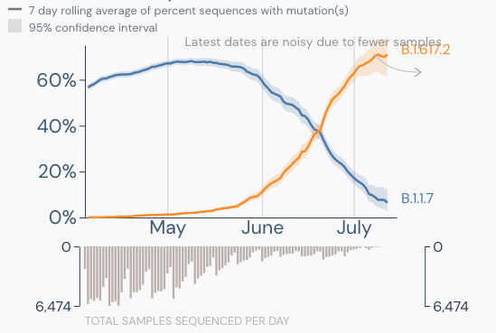
Now the Delta variant has taken hold just when the nation thought the disease, if not bested, had at least been brought under some semblance of control. Unfortunately, the refusal of many to get vaccinated contributes to the virus’s persistence. More will suffer, and more will die.

How many needless deaths will the nation have to experience? Will there ever be a point when everyone who can be vaccinated will be? Or, two years later, will we be mourning preventable COVID-19 deaths. Will we still have to watch our loved ones perish with a tube down their throat, or worst, alone in a room far away surrounded by cold walls and quiet indifference?

 Sigh.

Regardless of where this pandemic leads or how the nation reacts, laboratorians will continue to remain steadfast in their dedication to their profession and their patients. We have often considered ourselves the stepchild of the healthcare industry because, despite the criticality of what we do, we go unnoticed and unremarked on as long as we deliver the results our patients need. We are okay with that.

We are also tired and worn.

Conclusion

Thanks, fellow laboratorian, for reading this minor soliloquy of frustration and sadness. I will probably be back next quarter discussing inspections, competency, or some other administrative aspect of laboratory operations. I hope, also, to discuss how the nation has reached or is close to reaching the theoretical goal of herd immunity because of high vaccination levels. However, if I were honest, I know the likelihood of this happening is disappointingly low.

If you can get vaccinated, please do.

-Darryl Elzie, PsyD, MHA, MT(ASCP), CQA(ASQ), has been an ASCP Medical Technologist for over 30 years and has been performing CAP inspections for 15+ years. Dr. Elzie provides laboratory quality oversight for four hospitals, one ambulatory care center, and supports laboratory quality initiatives throughout the Sentara Healthcare system.

Pitfalls of Artificial Intelligence for COVID-19 Variant Classification

While you have surely heard about all of the SARS-CoV-2 variants and how concerning they are, I would bet that you may not know how they are classified. Sure, from my last post, the technical aspects of whole genome sequencing and targeted approaches have been described, but bioinformatic (big data) analyses are essential to assign lineages. Furthermore, the advances of machine learning have been integrated into this system for SARS-CoV-2 lineage assignment.

How VOC lineages are given

First, phylogenetic trees (circular example below) are formed to demonstrate relatedness of strains based on how many mutations they share. The more similar they are, the closer they are together. These trees are not new nor do they rely on artificial intelligence, but they can give visual clues as to whether a lineage is new.  For instance, when the first variant of concern B.1.1.7 (now called Alpha) was discovered, it branched away from other limbs of the phylogenetic tree.

Within these new viral variants, there are a set of mutations that are present in most of the viral variants. For instance, there are 17 protein coding changes in Alpha variant. However, these exact 17 mutations may not be in every Alpha variant. Individually, mutations may be present in 98% of isolates or lower; the spike gene deletion of amino acids 242-244 of the Beta variant (B.1.351, South Africa origin) is only present in 88% of specimens sequenced. This could be due to issues in sequencing, data processing, or just the prevalance/biology of the virus.

As there are many mutations that fit into certain variants, it would be difficult for a human to process all of this information in a probabilistic manner to assign lineages. Thus, machine learning tools (most common SARS-CoV-2 program is Pangolin) have been added onto the end of bioinformatic analyses to assign the lineage to a sample.

How machine learning works

The subject of machine learning has been discussed in a previous post about Protein folding prediction. Briefly, it is helpful to remember that machine learning is a process to create algorithms that give an outcome based on training data. The more diverse, large, and well curated the data, the better the accuracy of the program. One pitfall is they are based on previous data, which works well for many situations: using AI to find a lung cancer on chest x-ray would work well, because lung cancers have consistent characteristics.

However, with COVID-19, new variants keep arising and current variants are evolving (think Delta and Delta “plus”). Furthermore, if the classifier Pangolin is trained on high quality data, then trying to interpret lower quality data (missing genome regions, few sequencing reads) may confuse Pangolin and lead to inaccurate results. What follows is an example of how this occurred at our institution.

Case study

We have been sequencing COVID-19 positive specimens at UT Southwestern for the last several months. Many of the cases have been the Alpha variant (B.1.1.7, origin U.K.). However, it was around this time that Delta (B.1.617.2, origin India) cases started to arise. In one week, we found two specimens that were classified as B.1.95. This was an unusual variant I had not heard of before. There are several “wild type” strains that are B.1.1/ B.1.2 and other derivations, but I had not seen anything like this before.

Clinical history

Two specimens sequenced belonging to Hispanic, adolescent brothers whose mother had recently been hospitalized with COVID-19. There was information on mother’s travel history.

Therefore, I performed manual review of the specific variants. Many of the diverse mutations occur in the spike protein, so this was analyzed first. Immediately, I noticed two classic mutations of the Delta variant: a 2 amino acid deletion in the spike gene (S:Del157_158) and a receptor binding site mutation (S:L452R) also seen in the variant from California (B.1.429). Other mutations could be evaluated, but the combination of these two mutations is unique to Delta variant.

One suspected cause was that the Pangolin lineage classifier had an issue. Specifically, it had not been updated since February 2021- when Delta did not exist. Thus, there was no data for the program to classify the variant properly. Upgrading to the latest version of Pangolin provided the correct lineage classification.

A Few weeks later…

Once again, I was checking the lineages reported by the classifier and there were several B.1.617.2 and B.1.617.1. Both of these are variants from India (before the helpful WHO Delta designation), but they are distinct sub-variants. It was odd to see B.1.617.1, because this was found to be less infectious compared to the dominant B.1.617.2 variant (later named Delta) and B.1.617.1 was not spreading across the globe.

Intervention:

Therefore, I once again went to the sequence data for the spike protein to compare some mutations. Although these are sub-variants from the same original variant, they have several mutually exclusive mutations in the spike protein. The figure below compares the prevalence of specific mutations in the spike protein of B.1.617.1 and B.1.617.2 (dark purple = common in a variant, white = rare).

Upon manual review, all of the spike gene mutations were specific to B.1.617.2. So why was there an issue in classification? Again, there were few sequences for either of these sub-variants at that time, so the classifier wasn’t as well trained. Updating the Pangolin version brought the benefit of new data and more accurate classifications.

Take away messages

  1. Updating Lineage classification software (Pangolin) on a regular basis is needed for accurate results.
  2. Manual review is essential for any abnormal findings- a typical process for pathologists, but also plays an important role in COVID-19 variant monitoring.
  3. Know what you’re looking for and know which mutations differentiate the variants.
  4. Delta is now the dominant strain in the U.S. (graphic below).

References

  1. Outbreak.info
  2. https://pangolin.cog-uk.io/

Jeff SoRelle, MD is Assistant Instructor of Pathology at the University of Texas Southwestern Medical Center in Dallas, TX working in the Next Generation Sequencing lab. His clinical research interests include understanding how lab medicine impacts transgender healthcare and improving genetic variant interpretation. Follow him on Twitter @Jeff_SoRelle.

Microbiology Case Study: 70 Year Old Man with a History of Papillary Urothelial Carcinoma

Case Description

A 70 year old male with a past medical history of hypertension and non-invasive multifocal high grade papillary urothelial carcinoma was being followed closely for recurrence and underwent magnetic resonance imaging (MRI) of the abdomen and pelvis. The report described a 2.6 x 3.9 x 5.2 cm lobulated cystic mass involving the left psoas muscle. Furthermore, there was encasement of the left common iliac artery and less involvement of the left common iliac vein (Image 1). Further evaluation of the lesion was pursued to determine the etiology. An important aspect of this case to consider is the patient’s prior cancer treatment regimen, which included intravesical Bacille Calmette-Guerin (BCG) for 5/6 cycles. The final BCG treatment was held because the patient developed “BCG-osis” comprised of chest pain, rigors, chills and hypotension. Given the only pathology to date on the patient was non-invasive papillary carcinoma (even though it is high grade), the oncology group did not think the psoas muscle lesion was a metastasis. Fine needle aspiration (FNA) was pursued and the CT-guided aspiration demonstrated “abundant histiocytes and acute inflammation with necrotic debris…Acid fast organisms identified on AFB (acid fast bacilli) stain…Negative for malignant cells” (Image 2). Microbiology cultures were obtained at the time of FNA. The AFB smear showed 1+ AFB. AFB grew in culture and reacted with the Mycobacterium tuberculosis complex probe. The patient’s interferon gamma response assay (IGRA) was negative the year prior. Antimicrobial susceptibilities (AST) using the Mycobacterial Growth Indicator Tube system with single drug concentrations revealed susceptibility to isoniazid, rifampin, and ethambutol with resistance to pyrazinamide. Per protocol, the isolate was sent to a reference laboratory for identification which returned as BCG. During the interim, before AST was available, the patient was referred to our Infectious Diseases outpatient clinic where he was started on R(ifampin)-I(soniazid)-P(yrazinamide)-E(thambutol) therapy and then followed up his care with the county health department.  

Image 1. Magnetic resonance imaging of the pelvis demonstrating an enhancing multilobulated cystic mass overlying the left psoas muscle Top: transverse plane (orange arrow). Bottom: sagittal plane (orange arrow).
Image 2. Photomicrographs of the drained fluid from the psoas muscle abscess.
Top) hematoxylin and eosin photomicrograph demonstrating abundant neutrophils and necrotic debris (10x objective).
Bottom) AFB stain demonstrating multiple AFB (see arrows) within the necrotic cellular debris (50x oil immersion objective).

Discussion

BCG is an attenuated strain of Mycobacterium bovis that has, historically, been used as a vaccine to Mycobacterium tuberculosis (MTB) most often used in areas of endemicity. M. bovis is a member of the MTB complex so hybridization probes used in clinical laboratories can distinguish MTB complex from Mycobacterium kansasii or Mycobacterium avium complex but cannot distinguish the MTB complex members from one another. Reference laboratories have molecular techniques including PCR or sequencing that can separate the differing members of the MTB complex. Another traditional distinguishing characteristic between M. bovis (including BCG) and MTB is susceptibility to pyrazinamide. MTB carries a pyrazinamidase which is required to activate the antibiotic; M. bovis does not, so it is intrinsically resistant to pyrazinamide. A laboratorian or clinician should be cognizant of this when a culture result returns as MTB complex that is susceptible to rifampin, ethambutol and isoniazid but is resistant to pyrazinamide alone. Furthermore, IGRAs were designed, in part, to distinguish those who have been vaccinated with BCG versus those exposed to MTB or wild type M. bovis who are latently infected. IGRAs will be negative in those exposed to BCG but would be reactive with either MTB exposure or M. bovis (non-BCG strains).

This case describes an uncommon complication of BCG immunomodulator therapy as a treatment of superficial papillary urothelial carcinoma. BCG’s use as a therapeutic intervention for malignancy is unique. It is postulated that the instillation of the organism stimulates the host’s immune response which can cause inflammation and sloughing of the bladder lining (urothelial cells), which effectively removes foci of superficial pre-cancerous in situ lesions or other intact foci of superficial urothelial carcinoma. The typical course of treatment is 6 cycles of BCG. Local inflammation resulting in cystitis is the most common complication experienced in 27-95% of patients.1 However, approximately 19% of patients undergoing BCG therapy experience severe enough complications to prematurely terminate BCG therapy, according to one study by the European Organization for Research and Treatment of Cancer.2 The patient described developed systemic symptoms during the course of his BCG therapy which prompted his physicians to terminate it. Although far less common than local genitourinary symptoms, mycotic aneurysms can occur in an estimated 0.7-1.4% of cases.1 Psoas abscesses are thought to arise from mycotic aneurysm leak.3

References

  1. Liu Y, Lu J, Huang Y, Ma L. Clinical spectrum of complications induced by intravesical immunotherapy of bacillus Calmette-Guerin for bladder cancer. Journal of Oncology. 2019. DOI: 10.1155/2019/6230409
  2. Sylvester, R, Brausi M, Kirkels W, et al. Long-term efficacy results of EORTC genito-urinary group randomized phase 3 study 30911 comparing intravesical instillations of epirubicin, bacillus Calmette-Guerin, and bacillus Calmette-Guerin plus isoniazid in patients with intermediate- and high-risk stage Ta T1 urothelial carcinoma of the bladder. European Urology. 2010. 57:5; 766-773.
  3. Leo E, Molinari A, Rossi G, et al. Mycotic abdominal aneurysms due to Mycobacterium bovis after intravesical bacillus Calmette-Guerin therapy. Annals of Vascular Surgery. 2015. 29:6;1381.e1-1318.e6.

-Dominick Cavuoti is a Professor at UT Southwestern Medical Center who specializes in Cytopathology, Infectious Disease pathology and is a medical director of the Microbiology laboratory at Parkland Health and Hospital System.

-Kelley Carrick is a Professor at UT Southwestern Medical Center who specializes in Cytopathology and Gynecologic pathology. She is the Chief of Cytopathology at Parkland Health and Hospital System.

-Clare McCormick-Baw, MD, PhD is an Assistant Professor of Clinical Microbiology at UT Southwestern in Dallas, Texas. She has a passion for teaching about laboratory medicine in general and the best uses of the microbiology lab in particular.

Microbiology Case Study: Salads, Stool, and Special Staining Studies

Case History

A woman in her 40s presented to her primary care physician in summer 2020 with mild abdominal pain, diarrhea, nausea, and headache. She experienced loose bowel movements 3 – 4 times per day for the past 18 days. She denied bloody stools, travel, consumption of raw or undercooked meats or unpasteurized dairy, contact with animals, or recent antibiotic use. SARS-CoV-2 PCR was negative. A stool sample was collected and sent for an enteric panel PCR (Salmonella, Shigella, Campylobacter, and Shiga toxin), a bacterial stool culture (Aeromonas, Plesiomonas, and Vibrio), and an ova and parasite (O&P) exam with a request to perform a modified acid-fast stain. While the enteric panel and bacterial stool culture were negative, the following organism was observed on the modified acid-fast stain (Image 1). This organism measured approximately 9 µm, was variably modified acid-fast, and had a wrinkled-cellophane appearance. The organism was identified as a Cyclospora cayetanensis oocyst. The patient later shared that she had consumed a bagged salad mix that was implicated in the ongoing Cyclospora outbreak.

Image 1. Cyclospora cayetanensis.

Cyclospora cayetanensis

Cyclospora cayetanensis, a coccidian protozoan, is transmitted through ingestion of food or water contaminated with infectious oocysts. While infected humans shed oocysts in their stool, these oocysts are unsporulated and non-infectious at the time of excretion. In order to sporulate and become infectious, these excreted oocysts must incubate in the environment for 7 – 15 days post-excretion. Due to the required incubation post-excretion, direct fecal-oral transmission cannot occur.

Endemic areas include Central and South America, Middle East, South East Asia, and the Indian subcontinent. In non-endemic areas, travelers make up a large proportion of cases. Local outbreaks in non-endemic areas are often due to contaminated food sources. Most commonly the source of these outbreaks arise from consumption of raw fruits and vegetables that are difficult to thoroughly clean. These include leafy green vegetables (salad mixes, lettuce), herbs (basil, cilantro), and raspberries. Moreover, Cyclospora is resistant to many disinfectants used in the food industry. As exposure to this parasite is through contaminated food and water, infected patients are also at risk for other food and waterborne parasites including Cryptosporidium.

Once infectious oocysts are ingested, symptoms are typically observed after a one week incubation. Clinical presentation of Cyclospora infection includes diarrhea, nausea, fatigue, low grade fever, and weight loss. Although Cyclospora causes infections in both immunocompromised and immunocompetent individuals, symptoms may be severe and prolonged in immunocompromised patients, particularly in HIV and AIDS patients. Children and elderly individuals are also at higher risk for severe disease. Trimethoprim-sulfamethoxazole is the standard treatment. If untreated, symptoms can last for 10 – 12 weeks and may exhibit a relapsing pattern.

Stool samples should be submitted to the clinical microbiology laboratory for microscopic and/or molecular studies. To increase recovery of the organism during intermittent or low burden shedding, multiple stool specimens should be submitted over 2 -3 days. When viewed under a UV fluorescent microscope, Cyclospora oocysts autofluoresce and appear blue or green. While safranin-based stains or UV fluorescent microscopic examination can be used, modified acid-fast staining is commonly performed for the microscopic identification of Cyclospora. Cyclospora oocysts are modified acid-fast variable and measure 8 – 10 µm in diameter, unlike Cryptosporidium oocysts which are modified acid-fast positive and measure 4 – 6 µm in diameter. It is important to alert the clinical microbiology lab if suspecting Cyclospora as stains used in routine O&P exam, including trichrome stains, are not effective in highlighting Cyclospora. Although lab developed tests and FDA cleared multiplex gastrointestinal pathogen panels including Cyclospora are available, molecular assays are not yet routinely used for identification of Cyclospora due limited widespread availability.

References

  1. Almeria S, Cinar HN, Dubey JP. Cyclospora cayetanensis and Cyclosporiasis: An Update. Microorganisms. 2019;7(9):317. Published 2019 Sep 4. doi:10.3390/microorganisms7090317
  2. Hadjilouka A, Tsaltas D. Cyclospora Cayetanensis-Major Outbreaks from Ready to Eat Fresh Fruits and Vegetables. Foods. 2020;9(11):1703. Published 2020 Nov 20. doi:10.3390/foods9111703
  3. Ortega YR, Sanchez R. Update on Cyclospora cayetanensis, a food-borne and waterborne parasite. Clin Microbiol Rev. 2010;23(1):218-234. doi:10.1128/CMR.00026-09
  4. Garcia LS. Diagnostic Medical Parasitology. 6th Edition. 2016.
  5. Casillas SM, Bennett C, Straily A. Notes from the Field: Multiple Cyclosporiasis Outbreaks — United States, 2018. MMWR Morb Mortal Wkly Rep 2018;67:1101–1102. DOI: http://dx.doi.org/10.15585/mmwr.mm6739a6

Paige M.K. Larkin, PhD, D(ABMM), M(ASCP)CM is the Director of Molecular Microbiology and Associate Director of Clinical Microbiology at NorthShore University HealthSystem in Evanston, IL. Her interests include mycology, mycobacteriology, point-of-care testing, and molecular diagnostics, especially next generation sequencing.

The Red Queen’s Gambit: Helping the Lab Avoid Burnout

In Lewis Carroll’s book Through the Looking Glass, Alice is being given a tour of Looking-Glass Land by the Red Queen when this happens:

Alice never could quite make out, in thinking it over afterwards, how it was that they began: all she remembers is, that they were running hand in hand, and the Queen went so fast that it was all she could do to keep up with her: and still the Queen kept crying “Faster! Faster!” but Alice felt she could not go faster, though she had not breath left to say so.

However, after running until Alice feels absolutely exhausted she looks around in surprise to find that they are exactly in the same place where they had begun.

Carroll, Lewis (1991) [1871]. “2: The Garden of Live Flowers”. Through the Looking-Glass (The Millennium Fulcrum Edition 1.7 ed.). Project Gutenberg.

“Well, in our country,” said Alice, still panting a little, “you’d generally get to somewhere else—if you ran very fast for a long time, as we’ve been doing.”

“A slow sort of country!” said the Queen. “Now, here, you see, it takes all the running you can do, to keep in the same place. If you want to get somewhere else, you must run at least twice as fast as that!”

Laboratory medicine is one of many areas of healthcare where more is constantly expected to be done with less, where the inhabitants of our looking-glass land have to run as fast as we can just to maintain the status quo. Also like many areas of medicine, our already strained workforce suddenly became victims of an unprecedented global COVID-19 pandemic stressing and stretching our capabilities. The gamble then, is expecting members of our incredible laboratory medicine community to run so fast that they ultimately burn themselves out.

From the December 2020 New York Times article on laboratory workers in the time of COVID-19 titled, “‘Nobody Sees Us’: Testing-Lab Workers Strain Under Demand“:

Morale in the labs has flagged as the country continues to shatter records for caseloads, hospitalizations and deaths. The nation’s testing experts know these statistics better than anyone: They count the numbers themselves, sample by sample. But they are also easy targets of criticism and complaint.

“There is always this undercurrent of, it’s never good enough,” said Dr. Abbott, of Deaconess Hospital in Indiana. “It’s devastating. We’re working as hard as we can.”

In April 2020, just a few weeks after COVID-19 was officially declared a global pandemic, the April issue American Journal of Clinical Pathology opened with two timely editorials, one from Dr. Jeanette Guarner discussing the three emergent coronavirus diseases of the past two decades (SARS, MERS and COVID-19) and the next by Dr. Steven H Kroft titled “Well-Being, Burnout, and the Clinical Laboratory.”

In this issue were three different articles, the results of extensive surveys conducted by the ASCP to determine the job satisfaction, well-being and burnout prevalent among 1) pathologists, 2) pathology residents and fellows, and 3) laboratory professionals. Knowing now what clinical laboratories, leaders and trainees were about to go through thanks to COVID-19, made these publications about the stress and satisfaction felt by those in lab medicine was timely (if not grimly ironic).

What is shown in those excellent publications, and what we can only assume has become more true, is “burnout,” (the “combination of emotional exhaustion, depersonalization, and loss of sense of personal accomplishment”) prevalent in laboratory medicine, with the majority of pathologists, residents and fellows, and professionals reporting having experienced it if not experiencing it as an ongoing problem.

There is no single solution to burnout in the laboratory. As Dr. Kroft outlines in his editorial, these surveys can be seen as initial steps to understanding the problem and plotting potential courses forward (“a roadmap for what workplace landmines to try to avoid.”). But several meaningful pieces of data emerged from these surveys as well: Overwhelmingly, pathologists and lab professionals enjoy their work (91% and 86%) and feel valued by their colleagues (79% and 71%). Also telling is the fact that while well over 90% of laboratory professionals reported “a little bit of stress” to “a lot of stress,” 2/3rds of them reported feeling either “somewhat satisfied” or “very satisfied” with their jobs. Clearly, no one knows the value of laboratory medicine better than those of us doing it. But recognition and support coming from within the laboratory space should be seen as a good first step to acknowledging these contributions.

Recognition is needed from outside lab leadership as well, and especially should be accompanied by both stress-reducing measures (filling labor gaps, adequate compensation and benefits etc.) and opportunities to feel ownership and personal investment in the contributions we make to healthcare. Healthcare leaders, professional organizations, and all of those who were vocal supporters of labs’ contributions during the worst of the pandemic, should continue to advocate on behalf of laboratory staff’s well-being.

Even as vaccines and other mitigation efforts are providing more widespread pandemic relief in the United States, it’s clear that we are now through a COVID-19 looking glass. The lab was already running as fast as it could, but to get us to where we are now, many of us started running twice as fast. Hopefully we will both continue to run and also be supported in that ongoing race to stay where we are.

-Dr. Richard Davis, PhD, D(ABMM), MLS(ASCP)CM is a clinical microbiologist and regional director of microbiology for Providence Health Care in Eastern Washington. A certified medical laboratory scientist, he received his PhD studying the tropical parasite Leishmania. He transitioned back to laboratory medicine (though he still loves parasites!), and completed a clinical microbiology fellowship at the University of Utah/ARUP Laboratories in Utah before accepting his current position. He is a 2020 ASCP 40 Under Forty Honoree.