Trust Your Gut

A 20 year old female patient referred herself to a surgical oncologist specializing in sarcomas after she presented to an outside hospital for a sudden onset of epigastric pain. The patient also reported a one-year history of decreased appetite without nausea, vomiting, or weight loss. The outside institution performed an abdominal ultrasound and identified a large nonvascular heterogenous masslike lesion in the left upper quadrant not definitively associated with the spleen or kidney. The mass measured 12.1 x 9.9 x 10.7 cm. The radiologist’s overall impression was a hematoma; however, a CT scan with contrast was recommended to further classify the lesion. Instead, an MRI was performed, and the same radiologist described the lesion as having a thick irregular enhancing rind with enhancing septations and central necrosis. With the lesion appearing distinct from adjacent organs, a retroperitoneal sarcoma was posited on imaging. Reviewing the outside imaging and clinical history, the surgical oncologist referred the patient to interventional radiology for an ultrasound-guided biopsy of the left-sided retroperitoneal mass.

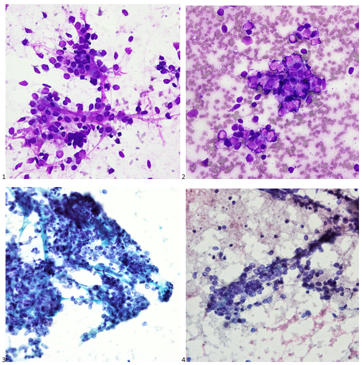
When the cytologist arrived in the procedure room for the time-out, the radiologist informed her of the surgical oncologist’s and outside radiologist’s opinions of a retroperitoneal sarcoma. A 17-gauge coaxial needle was advanced into the peripheral and non-necrotic aspect of the retroperitoneal mass, and multiple 22-gauge fine needle aspirations were obtained and handed to the cytologist. She prepared two air-dried smears and two alcohol-fixed slides. The air-dried smears were stained in our Diff-Quik (DQ) set-up and deemed adequate. The pathologist’s immediate cytologic evaluation was “tumor cells present.”

Images 1-3: Retroperitoneum, Left-side, Ultrasound-guided FNA: DQ-stained smear.

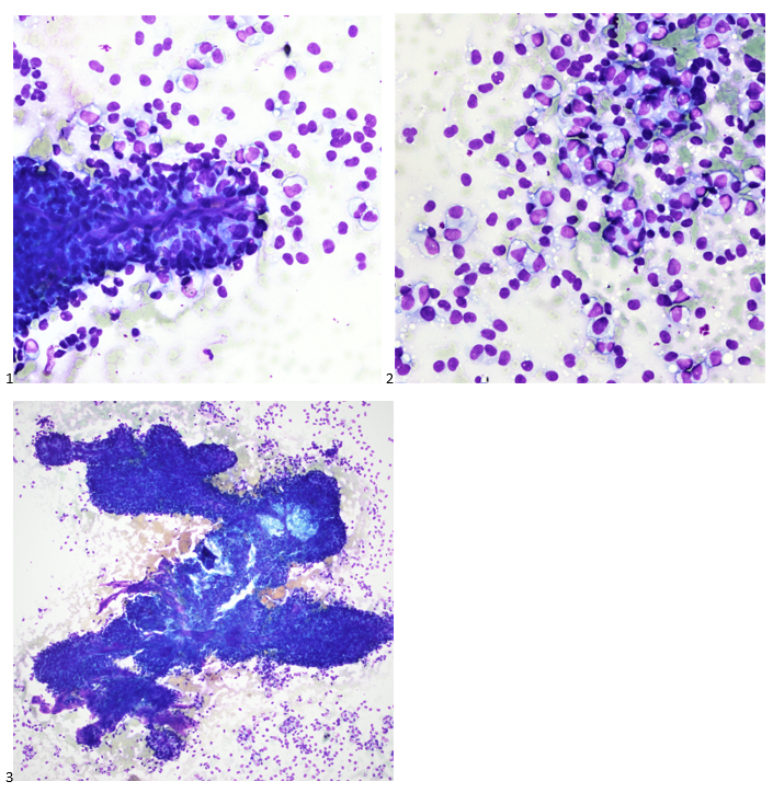
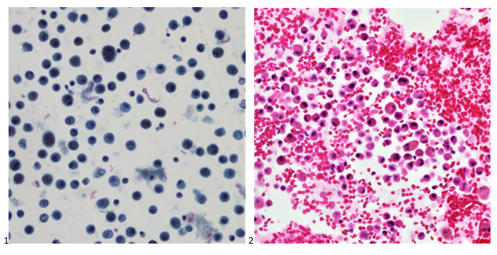
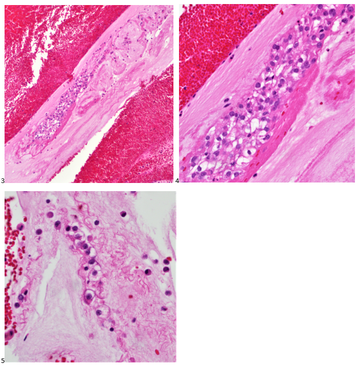
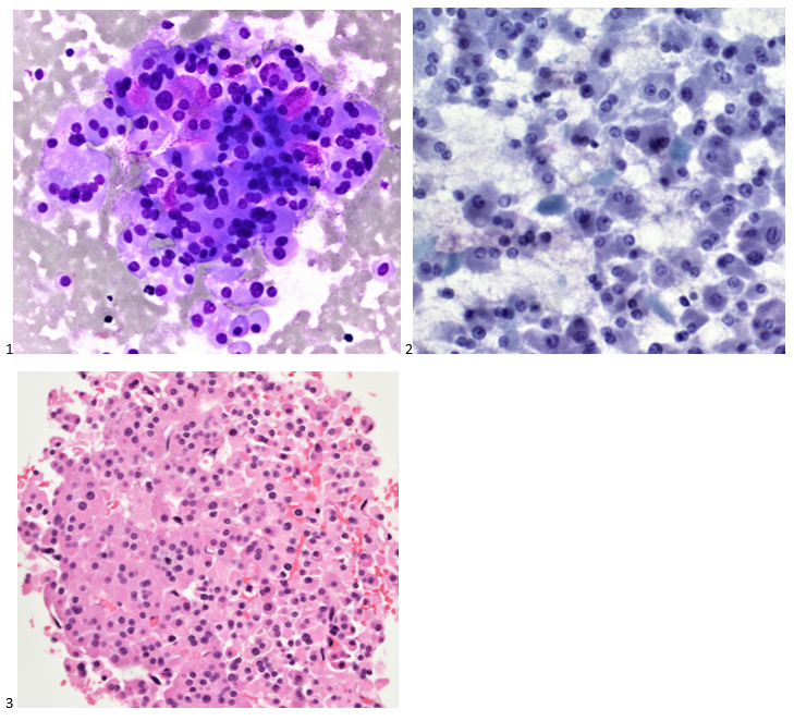
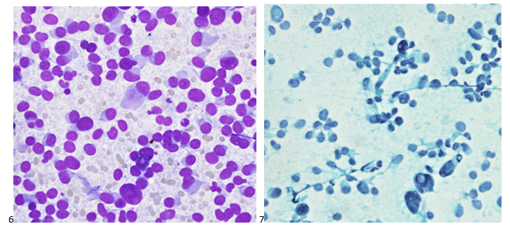
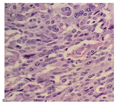
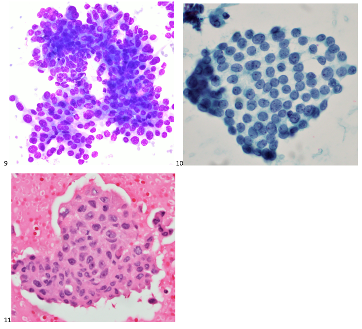
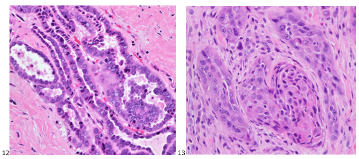
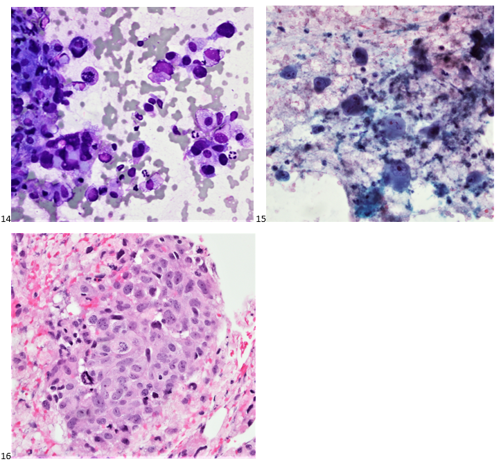
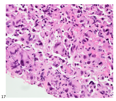
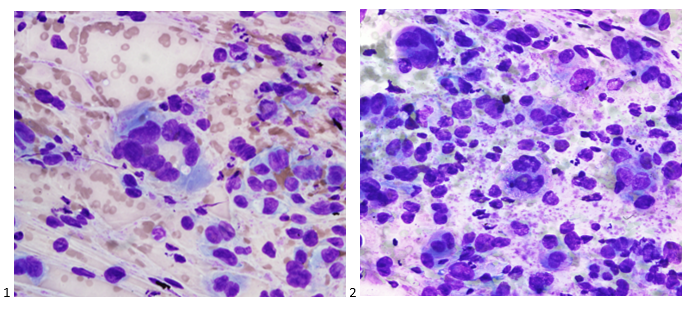
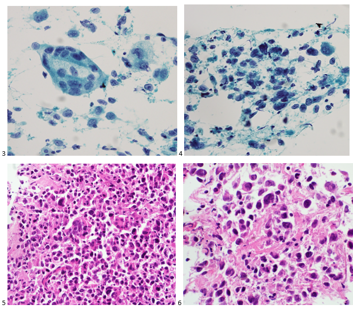
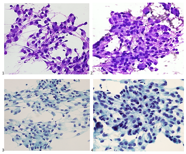
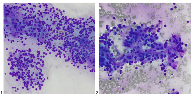
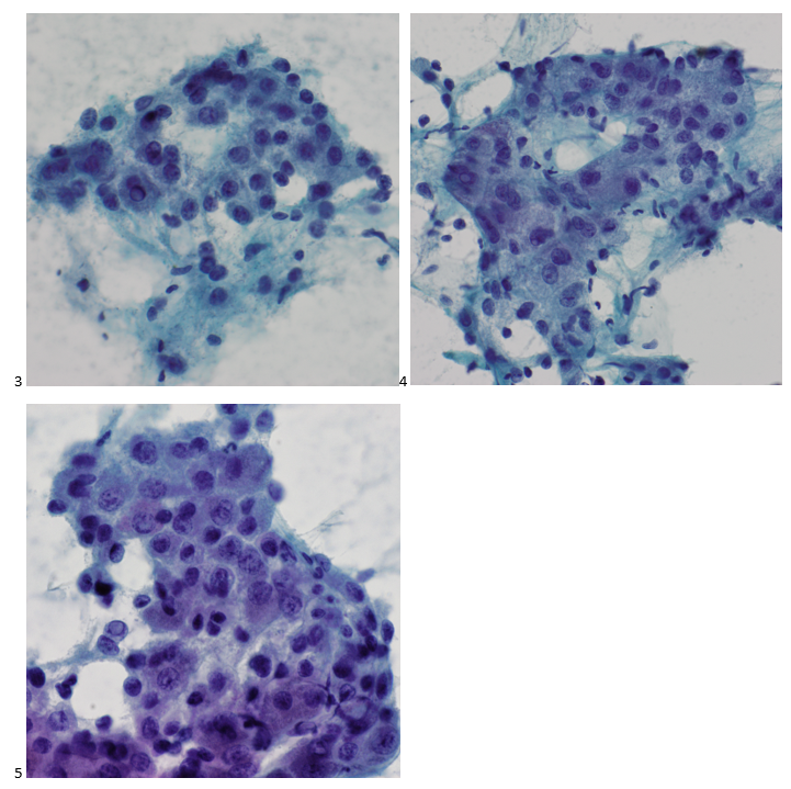
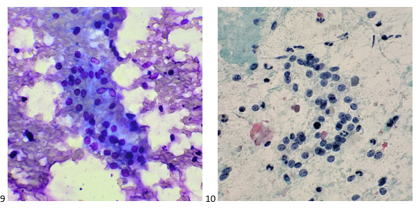
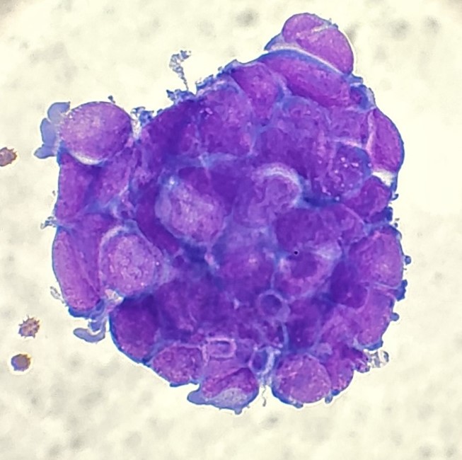
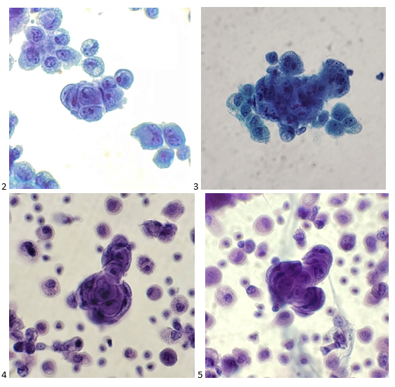
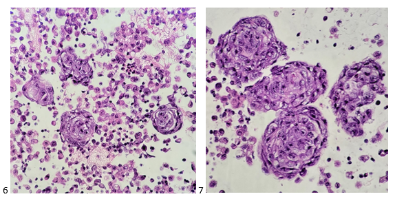
The following morning, the cytologist primary screened the Papanicolaou-stained slides and H&E-stained cell block sections in addition to the DQ smears, with the former preparations presented below.

Images 4-7: Retroperitoneum, Left-side, Ultrasound-guided FNA. 4-5: Pap-stained smear; 6-7: H&E Cell Block section (400X).

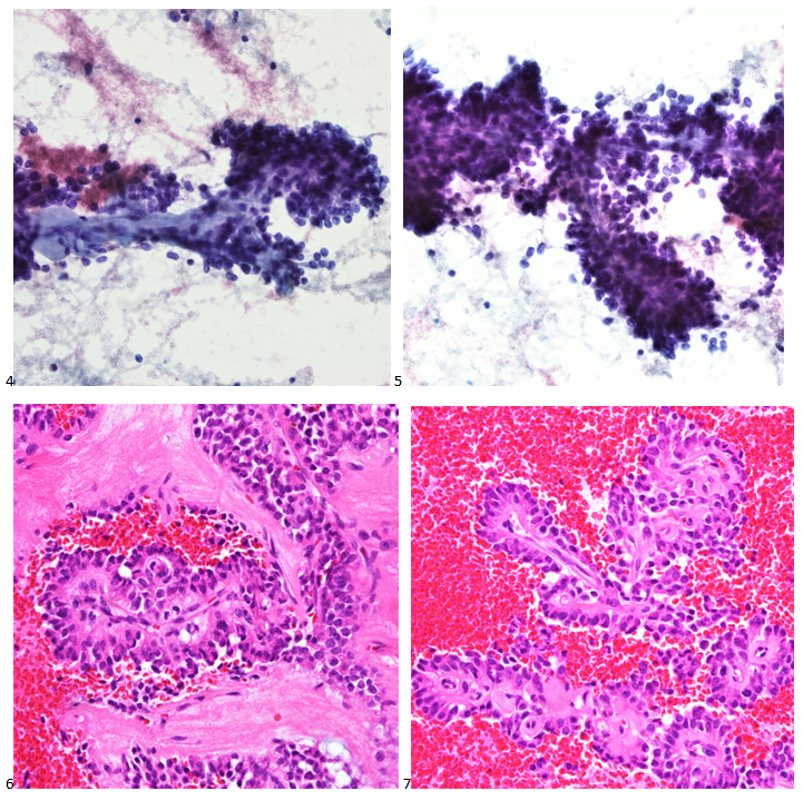
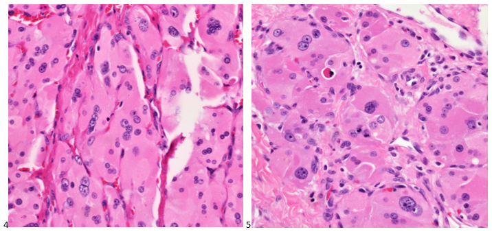
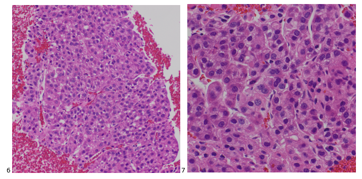
The cytologist entered her results as positive for malignant cells with a note of “atypical cells in papillary fragments” and gave the case to the pathologist for the final interpretation. The pathologist reviewed the slides prior to ordering immunostains. He paused and thought, “there’s something about the morphology and her age… it just doesn’t make sense for this to be a retroperitoneal sarcoma. It doesn’t look like a sarcoma. The cells are just too round or ovoid, bland, and poorly cohesive, and the fibrovascular cores – I just don’t think this is a sarcoma. Maybe a melanoma? Or some type of renal tumor? The cytoplasmic vacuolization could suggest this, but the mass is distinct from the kidney, so it can’t be. The nuclear grooves are intriguing, almost like a papillary thyroid carcinoma. A neuroendocrine tumor is also possible, the delicate papillary fronds though… Hmm. But where would it be originating from? How could this be distinct from other organs in the abdominal cavity?” He hemmed and hawed, glancing over our list of in-house immunostains. With only nine pre-cut unstained sections associated with the three H&E cell block levels, the pathologist ordered additional unstained recuts. He knew this was going to be a challenge due to the discrepancy between the clinical history and the morphology. 

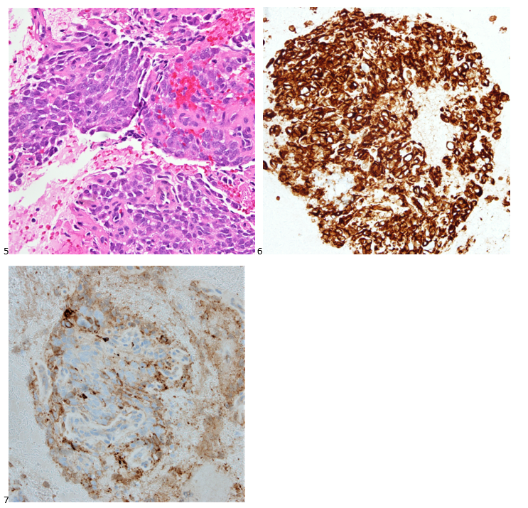
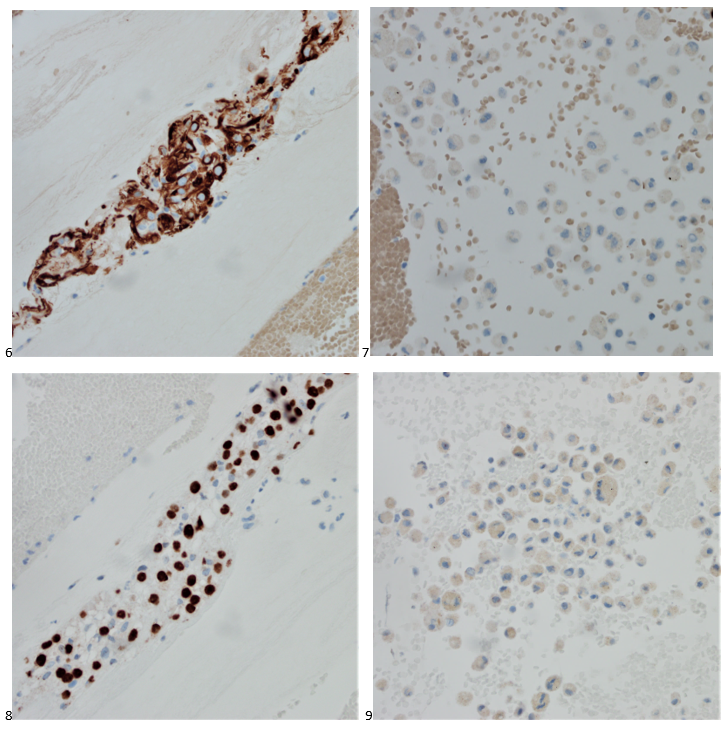
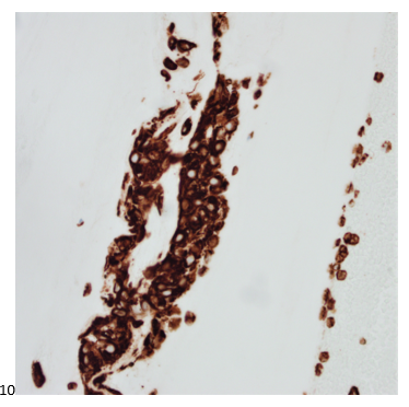
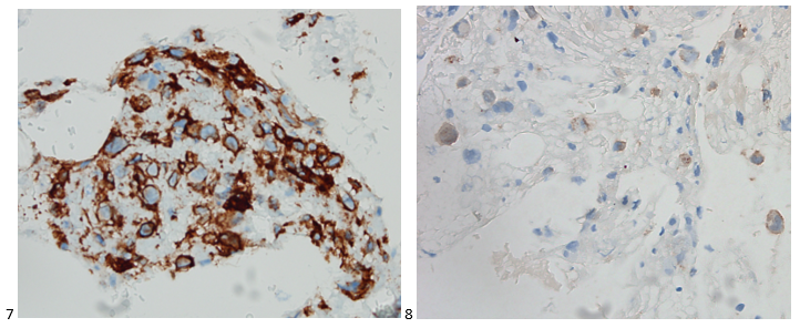
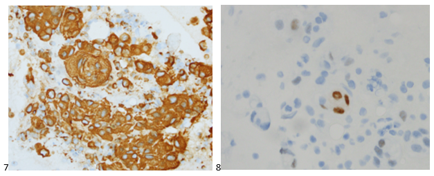
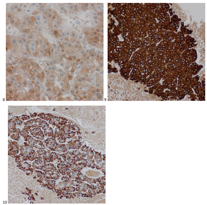
With proper positive and negative controls, the tumor cells show positive staining for AE1/AE3, Cam 5.2, vimentin, CD99 (dot-like), CD56, beta catenin (nuclear), PR, AMACR, and SOX11, while negative staining for CK7, CK20, PAX-8, RCC, chromogranin, synaptophysin, GATA-3, EMA, GFAP, S100, calretinin, WT-1, E-cadherin, and p53 (wild type pattern). The proliferative index by Ki-67 is low at <1%.

Images 8-10: Retroperitoneum, Left-side, Ultrasound-guided FNA. 8: beta catenin (nuclear)-positive; 9: AMACR-positive; 10: SOX11-positive.

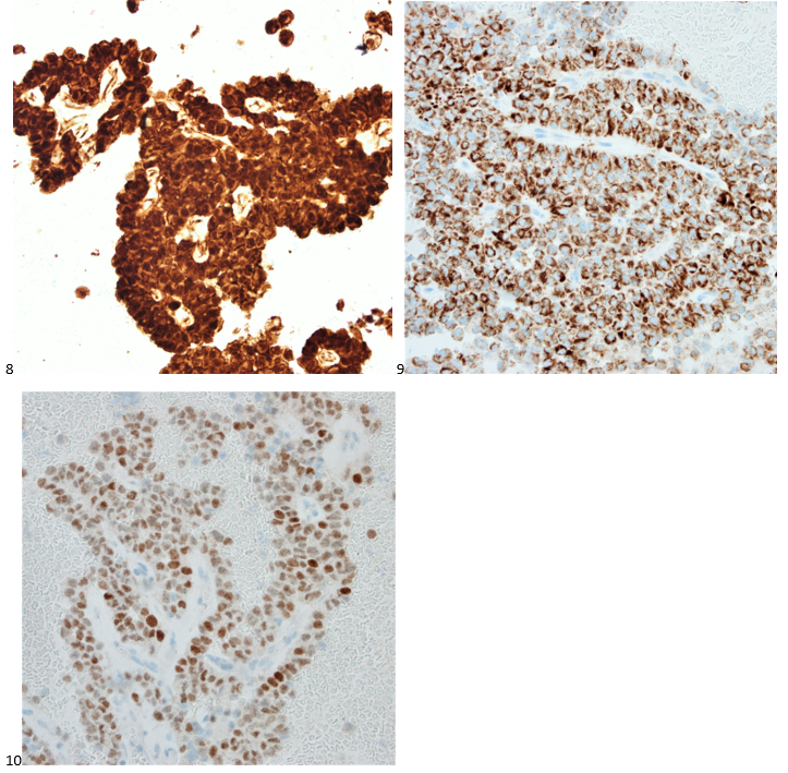
The combination of morphology with the extensive immunoprofile of the tumor is consistent with solid pseudopapillary neoplasm (SPN) of the pancreas.

Had there been any mention of the tumor involving or replacing the pancreas, this diagnosis and workup would have been much more straightforward. SPNs, albeit rare, account for 30% of tumors in women within their third or fourth decade of life.1 This patient presented with the most common SPN symptoms of abdominal pain and early satiety, but the mass appearing extrapancreatic on imaging posed a diagnostic challenge, as extrapancreatic SPNs are rare.2-3 Fortunately, SPNs are low-grade malignant neoplasms that respond well to surgical resection, and this patient is doing just fine after her distal pancreatectomy. In this case, both the patient and our pathologist listened to their guts with the patient pursuing advanced medical care for something much more complicated than a hematoma and the pathologist relying on his morphology expertise despite an odd clinical presentation.

References

  1. La Rosa S, Bongiovanni M. Pancreatic solid pseudopapillary neoplasm: key pathologic and genetic features. Archives of Pathology & Laboratory Medicine. 2020;144(7):829-837. doi:10.5858/arpa.2019-0473-ra
  2. Dinarvand P, Lai J. Solid pseudopapillary neoplasm of the pancreas: a rare entity with unique features. Archives of Pathology & Laboratory Medicine. 2017;141(7):990-995. doi:10.5858/arpa.2016-0322-rs
  3. Cheuk W, Beavon I, Chui D, Chan JKC. Extrapancreatic solid pseudopapillary neoplasm. International Journal of Gynecological Pathology. 2011;30(6):539-543. doi:10.1097/pgp.0b013e31821724fb

-Taryn Waraksa-Deutsch, MS, SCT(ASCP)CM, CT(IAC), has worked as a cytotechnologist at Fox Chase Cancer Center, in Philadelphia, Pennsylvania, since earning her master’s degree from Thomas Jefferson University in 2014. She is an ASCP board-certified Specialist in Cytotechnology with an additional certification by the International Academy of Cytology (IAC). She is also a 2020 ASCP 40 Under Forty Honoree.

Weirdoma

A 36 year old male was referred to gastroenterology after presenting to the emergency room for hematemesis and severe fatigue. He was pale and tachycardic, and the CBC showed a hemoglobin of 4.5. An esophagastroduodenoscopy (EGD) at the time demonstrated erosive esophagitis with a visible vessel and was treated with a PPI. A repeat scope the following month no longer demonstrated a vessel but identified a 10 cm ulcerative gastric cardia mass at the GE junction. Forcep biopsies showed a gastric ulcer with granulation tissue, and the stains performed yielded results that were not consistent with carcinoma or lymphoma; however, the biopsy material was limited, and the patient was referred to our GI clinic for further workup.

The interventional gastroenterologist requested cytology be present for the patient’s endoscopic ultrasound to ensure an adequate specimen was obtained for a definitive diagnosis. During the rapid onsite evaluation (ROSE), we determined the Diff-Quik smear was adequate, and the pathologist could confidently suggest that tumor cells were present. We collected additional FNA passes in our cell block tube to run ancillary studies.

The following morning, all we could make of the case was that it was a poorly-differentiated malignant neoplasm with spindle and epithelioid features. The cytoplasm was minimal and fairly wispy while the nuclei were hypochromatic and fragile with nuclear grooves and nucleoli.  On the Diff-Quik smears, the cytoplasm looked blue, which pointed us in the direction of possibly lymphoma or neuroendocrine, but the clustering made me favor neuroendocrine. With the pap-stained smears, were torn between carcinoma and a neuroendocrine tumor, maybe even an epithelioid GIST, albeit an odd location. And of course, there’s always the differential of melanoma, the great mimicker. Off to IHC we go!

Images 1-4: Stomach, GE Junction, EUS-FNA. 1-2: DQ-stained smear; 3-4: Pap-stained smear.

When our immunostains were delivered later that afternoon, our pathologist came up to me and said, “I got it! I know what it is!” Ecstatic, I replied, “What is it? Lymphoma? Carcinoma? GIST? MELANOMA?” “No, it’s a WEIRDOMA! Nothing is staining positive. No epithelial markers, no definitive lymphoid markers… nothing. It’s a weird case. I have to run additional stains.”

Images 5-7: Stomach, GE Junction, EUS-FNA. 5: H&E Cell Block section (600X); 6: Vimentin-positive; 7: CD56-positive

Back to the drawing board and 20 additional recut sections later, more immunostains were ordered and a mixed profile led us down a more confusing path. The tumor cells show positive staining for vimentin, CD56, and CD10 (focal), and negative staining for AE1/AE3, Cam5.2, CK7, desmin, SMA, HHF35, CD34, CD117, DOG-1, S100, SOX-10, synaptophysin, SALL4, CD45, CD68, and CD21. Proliferative index by Ki-67 was approximately 35%. The morphology and immunoprofile of the tumor were highly unusual, suggesting a mesenchymal neoplasm, possibly a sarcoma.

The concurrent forcep biopsies demonstrated rare atypical cells that were difficult to classify due to the limited number of cells, the non-specific morphology, and the following non-specific immunophenotype: positive staining for CD99, partial positive staining for D2-40 and NSE, and focal or weak positive staining for Cam5.2, while negative for AE1/AE3, CK7, EMA, S100, CD31, desmin, PAX8, BCL2, and myogen. The biopsy tissue consisted of predominantly ulcerative tissue and a fragment of squamous mucosa with a lamina propria infiltrate of atypical cells with spindle and epithelioid morphology.

Due to the FNA cell block consisting of 80% tumor compared to the limited forcep biopsy tissue, we sent FFPE cell block sections for RNA fusion studies to help us further classify the tumor. An EWSR1::ERG (in-frame) rsa(22;21)(q12.2;q22.2) gene fusion was detected in the tissue sample, which has been reported in extraskeletal Ewing sarcoma.

Stomach, GE Junction, EUS-FNA Final Diagnosis: Ewing Sarcoma

Fortunately, the patient’s PET scan did not demonstrate any evidence of metastatic disease, and the patient along with his care team decided to pursue systemic therapy as these tumors tend to be chemosensitive. The need for radiation therapy will be reviewed depending on the tumor’s response to systemic therapy. A strange presentation, this visceral Ewing sarcoma, and a reason why immunostains and molecular profiling are so important to rendering a definitive diagnosis. In our study set files, however, it will forever be dubbed my favorite weirdoma.

-Taryn Waraksa-Deutsch, MS, SCT(ASCP)CM, CT(IAC), has worked as a cytotechnologist at Fox Chase Cancer Center, in Philadelphia, Pennsylvania, since earning her master’s degree from Thomas Jefferson University in 2014. She is an ASCP board-certified Specialist in Cytotechnology with an additional certification by the International Academy of Cytology (IAC). She is also a 2020 ASCP 40 Under Forty Honoree.

A Diamond in the Mucin

A 35 year old male and current every day smoker transferred his care to our genitourinary (GU) clinic after undergoing a partial nephrectomy for clear cell renal cell carcinoma. A PET scan was performed, and a 5 millimeter FDG-avid right upper lobe (RUL) lung nodule was identified. While the mediastinal and hilar lymph nodes were mildly enlarged on the chest CT, they did not demonstrate FDG-avidity on the PET scan. The patient was referred to pulmonary for a workup of his subcentimeter lung nodule. Given the patient’s age and clinical history, the care team and the patient decided to pursue a lung biopsy via robotic-assisted bronchoscopy and subsequent lymph node sampling via endobronchial ultrasound (EBUS).

After the timeout, the patient was intubated, and the pulmonologist performed an airway inspection. Upon entering the right upper lobe, the pulmonologist noticed blood streaking along the bronchial wall. A right upper lobe bronchoalveolar lavage (BAL) was collected and sent for culture and cytology. Following the trachea and lung survey, the respiratory team began the robotic-assisted bronchoscopy. After confirming the correct location with both radial EBUS and fluoroscopy, multiple fine needle aspirates (FNAs) were collected from the right upper lobe lung nodule. Only benign bronchial cells were identified on rapid onsite evaluation (ROSE). A right upper lobe bronchial brushing was attempted and sent directly to cytology. Forceps were used to obtain transbronchial biopsies from the radiologically-suspicious area. The team then switched over to linear EBUS for the lymph node sampling portion of the procedure. Lymph nodes were sampled at the following stations: 11L, Level 7, and 4R. On ROSE, only lymphocytes and anthracotic pigment were identified for all three lymph nodes.

Upon e, the cytologist brought the specimens back to the laboratory for accessioning and processing. The following morning, the same cytologist screened all cytology specimens and the respective cell blocks associated with the case. The lymph nodes were signed out as negative for malignancy, with lymphocytes and anthracotic pigment present. The RUL FNA was inadequate for diagnosis as the material consisted mainly of bronchial cells and alveolar macrophages. The FNA findings were correlated with the transbronchial biopsy, which was signed out as benign pulmonary parenchyma with hemorrhage, fibrin clot, and respiratory bronchiolitis. Similarly, the bronchial brushing was negative as only benign bronchial cells were identified. In screening the BAL, there was an overabundance of hemosiderin-laden alveolar macrophages on the smear, cytospin, and SurePath liquid-based preparations (Image 1). When screening the cell block slides, abundant macrophages were also identified (Image 2).

Images 1-2. Lung, Right Upper Lobe, Bronchoalveolar Lavage 1: Pap-stained SurePath Liquid-based Prep; 2: H&E Cell Block Section (400X).

However, in the cell block sections, the cytologist also identified cells stranded within areas of mucin (Image 3). It may have been easy to dismiss these as macrophages, but contrary to the dusty hemosiderin-laden macrophages in this specimen, these cells had abundant vacuoles, and irregular nuclei with more prominent nucleoli (Images 4-5). The pleomorphic nuclei and larger vacuoles distinguished these cells from lipid-laden macrophages most often seen in aspiration pneumonia.

Images 3-5. Lung, Right Upper Lobe, Bronchoalveolar Lavage 3: H&E Cell Block section (100X); 4: H&E Cell Block section (400X); 5: H&E Cell Block section (600X).

The cytologist, marking the areas of interest, swiftly rendered a diagnosis of positive for malignant cells, favoring metastatic clear cell renal cell carcinoma. She enthusiastically described the case to the attending cytopathologist, who immediately ordered confirmatory immunostains on the unstained paraffin sections of the cell block.

With adequate controls, CD68 highlights the alveolar macrophages (not shown). The tumor cells show positive staining for AE1/AE3 and PAX-8 (Images 6-7), while macrophages show negative staining for AE1/AE3 and PAX-8 (Images 8-9). The tumor cells also show positive staining for vimentin (Image 10) and RCC (focal), and negative staining for TTF-1.

Images 6-10. Lung, Right Upper Lobe, Bronchoalveolar Lavage 6: AE1/AE3-positive tumor cells; 7: AE1/AE3-negative macrophages; 8: PAX-8-positive tumor cells; 9: PAX-8-negative macrophages; 10: Vimentin-positive tumor cells.

RUL BAL Final Diagnosis: Positive for malignant cells. Metastatic clear cell renal cell carcinoma.

When we attend a biopsy of an area clinically suspicious for metastatic renal cell carcinoma, we prepare ourselves for an overtly bloody sample. Even if the smears consist mainly of blood during ROSE, we rest assured that we will usually find tumor cells in the cell block sections. By obtaining a BAL of the blood-streaked RUL, the pulmonologist was able to pinpoint an area of lymphangitic spread. We often give little credit to BALs in cytology, especially when using concurrent techniques such as FNAs to sample a subcentimeter lung nodule without bulky disease. However, as seen in this case, sometimes it’s the only specimen that can help us render a diagnosis of metastatic cancer.

-Taryn Waraksa-Deutsch, MS, SCT(ASCP)CM, CT(IAC), has worked as a cytotechnologist at Fox Chase Cancer Center, in Philadelphia, Pennsylvania, since earning her master’s degree from Thomas Jefferson University in 2014. She is an ASCP board-certified Specialist in Cytotechnology with an additional certification by the International Academy of Cytology (IAC). She is also a 2020 ASCP 40 Under Forty Honoree.

Thyroid Tales (First Edition)

I’ve found that our cytologists have a love-hate relationship with thyroids. Pathologists do too. Or it could be that we see so many goiters (50%) and follicular lesions or atypia of undetermined significance (35%) that the rare papillary thyroid carcinoma is a gem in our eyes. Minimally-invasive thyroid FNAs are instrumental in the management of thyroid nodules. It’s important to note that due to lack of architecture and assessment of capsular invasion, cytologic diagnoses may be limited, and prior to referring the patient for a potentially unnecessary surgery, various molecular tests can be utilized. The ongoing evolution of molecular testing on thyroid FNAs help classify indeterminate and suspicious cytology diagnoses (Bethesda Categories III and IV), examining the risk of malignancy or detecting the presence of genetic alterations, which help guide surgical intervention versus surveillance. This post (the first edition) features a series of our classic Bethesda Category VI specimens, which bypassed the need for risk classification and defaulted in surgical intervention based on guidelines at the time of diagnosis. It is worth mentioning that many of these cases occurred prior to the implementation of the Thyroid Imaging Reporting and Data System (TI-RADS), so to preserve the accuracy of patient history, a TI-RADS score will not be assumed.

Case 1

Okay, I know I said Bethesda VI, but let’s kick this series off with a Bethesda Category IV case. Thankfully, the patient decided to undergo a partial thyroidectomy, yielding a beautiful tissue follow-up. A 59-year-old male with newly diagnosed melanoma of the neck underwent imaging for staging purposes. A left thyroid nodule was identified measuring 3.0 centimeters. The patient presented for an ultrasound-guided fine needle aspiration.

Images 1-3: Thyroid, Left Lobe, FNA 1: DQ-stained smear; 2: Pap-stained smear; 3. H&E Cell Block section (400X).

Abundant oncocytic Hürthle cells, some with mild atypia, were identified, suggestive of Hürthle cell neoplasm (Images 1-3). With a lack of lymphocytes, we did not feel comfortable suggesting Hashimoto’s (lymphocytic) thyroiditis. Immunostains performed on cell block sections show the tumor cells are positive for TTF-1, focally positive for thyroglobulin and AE1/AE3 (rare), and negative for calcitonin. The morphology and immunohistochemical profile support the above diagnosis.

The patient underwent a left lobectomy and isthmusectomy. Pathology showed a 3.2 cm Hürthle cell carcinoma (Images 4-5) in the left lobe of the thyroid (encapsulated with a foci of capsular invasion without vascular invasion) as well as a 0.3 cm micropapillary carcinoma. Since Hürthle cell carcinoma does not typically concentrate radioiodine, the patient would not be responsive to treatment with radioactive iodine. Therefore, there would be less benefit derived from treating the smaller right lobed nodules (which don’t meet biopsy criteria) from remnant ablation. The patient had a clinically limited stage thyroid cancer. The patient is monitored with neck ultrasounds rather than serum thyroglobulin testing (due to the remaining right lobe).

Images 4-5: Thyroid, Left Lobe with Isthmus, Excision: H&E section (600X).

Cytology diagnosis: Hürthle cell neoplasm.

Pathology diagnosis: Hürthle cell carcinoma.


Case 2

A 53-year-old female with no prior history presented with fatigue and a self-palpated right thyroid nodule and normal thyroid function tests. She reported an extensive family history of hypothyroidism. On thyroid ultrasound, the right upper pole thyroid nodule measured 2.0 x 2.0 x 1.8 cm and was mostly solid and hypoechoic with microcalcifications. Pre-intervention serum calcitonin measured 1634 pg/mL. The patient underwent an FNA of the thyroid nodule and the smears are depicted below.

Images 6-7: Thyroid, Right Lobe, Upper Pole, FNA: 6: DQ-stained smear; 7: Pap-stained smear.

Cells appear plasmacytoid and appear both isolated and in clusters (Image 6). The nuclei are eccentrically placed, and the chromatin has a salt and pepper appearance akin to a neuroendocrine tumor (Image 7-8). Also identified were pink granules and intranuclear pseudoinclusions (Images 6-7). We performed immunohistochemical stains on paraffin sections of the cell block. Tumor cells show positive staining for calcitonin, chromogranin, mCEA, and TTF-1, while negative staining for thyroglobulin and CD45.

Following the diagnosis, the patient had a CT scan for staging purposes. Multiple lymph nodes in the right cervical chain were identified. the patient at a clinical stage IVA diagnosis. In the interim, the patient had a total thyroidectomy which revealed medullary thyroid carcinoma of the right lobe measuring 2.2 cm, a micropapillary carcinoma of the left lobe measuring 0.1 cm (Image 8). Lymphovascular invasion was not identified, the inked surgical resection margins are free of carcinoma, and metastatic medullary carcinoma was identified in 6 of the 77 lymph nodes removed during the central compartment lymph node dissection, and bilateral cervical lymphadenectomies. The calcitonin level dropped to 23 pg/mL postoperatively.  Genetic testing was performed to assess for Multiple Endocrine Neoplasia Type 2 (MEN2), and although her result was indeterminate, a RET mutation was not identified.

Image 8: Thyroid, Excision: H&E section (600X).

Case 3

A 67-year-old male with no pertinent medical history presented to the endocrinology clinic after his primary care physician identified a large lump in the patient’s neck. A 7.0 cm hypoechoic right thyroid mass with macrocalcifications was noted on ultrasound imaging. The patient was referred to diagnostic imaging for a thyroid FNA. The smears and cell block section are depicted below. While the papillary formation of Image 9 is not evident on the pap-stained slide (Image 10), the nuclear grooves and pseudoinclusions along with irregular nuclear membranes and powdery chromatin are highlighted. A separate needle pass was collected for molecular testing, which revealed a BRAF V600E mutation in the tumor cells.

Images 9-11: Thyroid, Right Lobe, FNA 9: DQ-stained smear; 10: Pap-stained smear; 11. H&E Cell Block section (600X).

Two weeks after the FNA diagnosis, the patient was scheduled for a partial thyroidectomy of the right lobe. While 60% of the mass demonstrated well-differentiated papillary thyroid carcinoma (Image 12), 40% of the tumor contained poorly differentiated thyroid carcinoma with squamous features (Image 13). No sarcomatous components or giant tumor cells were identified. Carcinoma with squamous features invaded into the surrounding tissue, strap muscle, thymus, and right paratracheal lymph node. Interestingly, the right-sided levels 3 and 4 lymph nodes contained predominantly well-differentiated papillary thyroid carcinoma with rare foci of poorly differentiated thyroid carcinoma with squamous differentiation.

Images 12-13: Thyroid, Right Lobe, Excision: H&E section (600X).

Five months after the excision, the patient developed a left-sided pleural effusion. A diagnostic thoracentesis was performed and metastatic thyroid carcinoma was identified. Immunostains performed on the cell block slides with adequate controls show that the tumor cells are positive for PAX8, and negative for TTF-1, and thyroglobulin. The findings support the diagnosis. While patients with papillary thyroid carcinoma tend to have better disease-free survival rates, the poorly differentiated tumor was difficult to control and eventually resulted in widespread metastasis.

Cytology diagnosis: Papillary thyroid carcinoma.

Pathology diagnosis: Poorly differentiated thyroid carcinoma with squamous features in a background of well-differentiated papillary thyroid carcinoma.


Case 4

A 73-year-old female presented with a rapidly growing and painful thyroid mass that measured 8 cm on imaging. Originating from the right lobe, multiple needle passes targeted various areas of the mass via ultrasound-guidance. The smears and cell block section are presented below. Smears (Images 14-15) feature pleomorphic nuclei in a background of inflammation and necrosis. The cell block section (Image 16) demonstrates increased mitotic figures and neutrophils.

Images 14-16: Thyroid, Right Lobe, FNA: 14: DQ-stained smear; 15: Pap-stained smear; 16. H&E Cell Block section (600X).

We performed immunocytochemical stains on paraffin sections of the cell block. Tumor cells how positive staining for p53, focal staining for cyclin D1, and negative staining for AE1/AE3, thyroglobulin, and BCL-2. Rare tumor cells show staining for TTF-1. The proliferation index by Ki-67 immunostaining is approximately 70%.

While not a standard procedure for thyroid specimens, core biopsies (Image 17) were also obtained from this mass.

Molecular testing on the core biopsy sample identified a high mutation burden, with the tissue harboring both TP53-inactivating and TERT promoter mutations. Imaging demonstrated widespread metastasis, and this patient did not survive the extensiveness of her disease.

Cytology Diagnosis: Undifferentiated (anaplastic) thyroid carcinoma.

Pathology Diagnosis: High-grade carcinoma consistent with anaplastic carcinoma (interchangeable diagnoses).


That’s enough for our classic thyroid cases. Stay tuned for the second edition featuring thyroid FNAs with unsuspecting findings!

-Taryn Waraksa-Deutsch, MS, SCT(ASCP)CM, CT(IAC), has worked as a cytotechnologist at Fox Chase Cancer Center, in Philadelphia, Pennsylvania, since earning her master’s degree from Thomas Jefferson University in 2014. She is an ASCP board-certified Specialist in Cytotechnology with an additional certification by the International Academy of Cytology (IAC). She is also a 2020 ASCP 40 Under Forty Honoree.

Histio Makes History

An 81 year old female presented to the head and neck clinic after being diagnosed with cutaneous T cell lymphoma of the posterior mid-parietal scalp at an outside institution. She was initially treated with Brentuximab every three weeks but developed significant toxicities. The patient’s previous “T cell lymphoma” material was reviewed at our institution and the immunophenotypic report described the neoplastic cells as being positive for CD45, CD2, CD4, BCL6+, CD3 (subset), and CD123 (scattered), while negative for CD7, CD8, CD20, CD30, CD56, EBER ISH, PAX5, and lysozyme. Immunohistochemical slides were not provided for review. Flow cytometric analysis determined that there was no immunophenotypic evidence of a clonal T cell population in the patient’s peripheral blood.

A second scalp biopsy was performed at another outside institution, and the findings were similar to the parietal scalp; however, there were atypical pleomorphic cells which displayed irregular contours, hyperchromasia, and multiple nucleoli. The atypical cells were predominantly positive for CD4 and diffuse positivity for CD1a. These same pleomorphic cells were negative for CD3, CD8, CD20, CD30, ALK1, BCL6, CD56, EBER, AE1/AE3, SOX10, Desmin, PAX5, MUM1, CD5, and Cam 5.2.

The smears contained large, highly pleomorphic cells with irregular, elongated, and multilobated nuclei, frequent nuclear grooves and folds, fine chromatin, prominent nucleoli, and variable amounts of pale, eosinophilic cytoplasm, alt.

The outside tissue block on the original scalp biopsy was requested, and our pathology department performed additional immunostains. The neoplastic cells of interest were positive for CD1a, S100, CD68 (a small subset), and negative for lysozyme, CD21, CD30, and CD3. Ki67 proliferation index was interpreted at approximately 60%. An unstained FFPE tissue section was sent to a reference laboratory, and the neoplastic cells were strongly positive for Langerin.

While the Brentuximab treatment initially appeared to have a positive impact on the overall disease burden, the PET CT following 3 cycles showed a mixed response, including resolution of cervical lymphadenopathy and identification of multiple new lung nodules and bulky mediastinal lymphadenopathy. Between that and numerous reported toxicities, the treatment protocol was discontinued. The patient was then referred to radiology for a CT-scan guided right lower lobe lung biopsy measuring 2.2 x 1.3 centimeters with an SUV or 29.6.

In the CT Scan suite, we received multiple FNA passes from the interventional radiologist and made air-dried and alcohol-fixed smears, rinsing the residual needle material into a tube of balanced salt solution for a cell block preparation. We determined our specimen was adequate for scant tumor cells, as depicted on the Diff-Quik smears below.

Images 1-2. Lung, right lower lob, CT-guided FNA. Diff-Quik stained smears.

In comparison to the material from the second scalp biopsy, the cells from the lung biopsy appeared identical. Our Pap-stained smears and H&E cell block sections also demonstrated the highly pleomorphic cells described above.

Images 3-6. Lung, Right Lower Lobe, CT-guided FNA. 3-4: Pap-stained smears, 5-6: H&E sections (5: 100x, 6: 400x).

Immunostains performed on the cell block slides with adequate controls show that the tumor cells are positive for CD1a, CD4, partially positive for CD45 and S100, negative for AE1/3, TTF-1, and p40.

Images 7-8. Lung, Right Lower Lobe, CT-guided FNA. Cell block section immunohistochemistry. 7: CD1a-positive; 8: partially S-100-positive.

Our pathologists felt the cells from the second scalp biopsy and the lung biopsy were representative of a Langerhans cell sarcoma, a form of malignant histiocytosis, rather than a T-cell lymphoma. It is possible that the first scalp biopsy’s diagnosis of T-cell lymphoma was due to sampling error and the pleomorphic cells of interest were missed. The Ki-67 proliferative index of 60% helped to distinguish between Langerhans cell histiocytosis and Langerhans cell sarcoma.

Molecular testing performed on the core biopsy was negative for a BRAF mutation and positive for an NF1 inactivating mutation. The tumor may then be sensitive to mTOR inhibitors and MAPK pathway inhibitors, such as MEK inhibitors. Appeals for a MEK inhibitor were denied by insurance, but fortunately, the tumor also demonstrated high PD-L1 expression at 90%, making this specific patient a candidate for pembrolizumab, which was fully covered by insurance.

____________________________________________________________________________________________

I can’t help but think about the disparities associated with cancer and the inaccessibility of potentially lifesaving or life-prolonging treatments. Sure, there may be viable alternatives, such as this case, but what if we had equal access to cutting edge, personalized therapies? What if the only therapy available was too costly to bear? Just because a cancer might be rare, such as Langerhans cell sarcoma, it doesn’t mean access to a proven effective therapy should also be rare. Even with drug assistance programs, so many patients face the harsh reality of tapping into their life savings to just to save their own life. When we became medical laboratory professionals, we promised to provide timely and accurate for all of our patients. Now, it’s time that pharmaceutical companies and our healthcare system as a whole work together to provide high quality, low-cost, readily accessible and personalized treatment options to every patient. They deserve that chance to overcome or at least manage their cancer.

-Taryn Waraksa-Deutsch, MS, SCT(ASCP)CM, CT(IAC), has worked as a cytotechnologist at Fox Chase Cancer Center, in Philadelphia, Pennsylvania, since earning her master’s degree from Thomas Jefferson University in 2014. She is an ASCP board-certified Specialist in Cytotechnology with an additional certification by the International Academy of Cytology (IAC). She is also a 2020 ASCP 40 Under Forty Honoree.

Tumor on the Brain

Back in my Master’s program at Jefferson, I fondly remember the week we covered central nervous system (CNS) tumors. I was fascinated by the mnemonic tools we would use to identify different CNS tumors, such as “fried eggs” for oligodendrogliomas, perivascular pseudorosettes in ependymomas, and the whorling associated with meningiomas. Fortunately, for our patients, and unfortunately, for our diagnostic curiosity, we rarely see CNS tumors at my institution. Brain lesions resulting from metastatic carcinomas are typically well-identified via imaging and treated appropriately by the surgical, medical, and radiation oncology teams, but cytologists are available to screen cerebrospinal fluids (CSFs) for CNS involvement. For primary CNS tumors, however, we’re left recollecting the core memory of the second semester of our didactic phase. When a metastatic CNS tumor made its way into our lab, our cytology team swooned with excitement. (Yes, I know, but please introduce me to a lab professional who doesn’t embrace their quirks.) A 27-year-old male patient presented to radiation oncology three years after surgical debulking of a brain tumor at an outside institution. The patient, who was referred to radiation oncology at to treat the residual tumor at the original institution, did not follow up and developed an 8 centimeter recurrence a year after the initial resection. At this point, the patient experienced complete vision loss and underwent a biparietal-occipital craniectomy. A repeat brain MRI was performed a year later, and once again, a large enhancing extra-axial mass was identified along with multiple smaller masses also increasing in size. The patient received radiation after worsening difficulty with ambulation. After almost completing the planned fractions of radiation, the patient elected to stop their radiation therapy due to worsening seizures. A left neck mass was identified six months prior, and while the mass had not grown or caused pain, the patient was referred to head and neck surgical oncology for evaluation. Surveillance imaging demonstrated an enlarged left level 5A lymph node, suggestive of metastatic disease. Multiple ultrasound-guided fine needle aspiration biopsies were obtained from the lymph node, and ROSE was performed. The Diff-Quik-stained and concurrent Pap-stained smears demonstrated lesional tissue, although everything from epithelioid histiocytes to spindle cell melanoma to a renal primary were considered as a differential. Based on the location, a salivary gland primary was also a possibility for this case. The streaked cytoplasm and pseudoinclusions in both smears were concerning for a metastasis of the patient’s primary CNS tumor, but we were still hesitating to make the call.

Images 1-4. Lymph Node, Neck, Left, Level 5A, US-guided FNA. 1-2: Diff-Quik-stained smears, 3-4: Pap-stained smears.

The following morning, the H&E-stained FFPE cell block sections demonstrated the characteristic whorls expected for the patient’s primary, although the idea of metastasis was uncanny.

Images 5-6. Lymph Node, Neck, Left, Level 5A, US-guided FNA. H&E sections (6: 100x, 7: 400x).

We then used immunohistochemical studies to confirm our morphologic diagnosis. Immunostains performed on the cell block slides with adequate controls show that the tumor cells are positive for vimentin and PR (focal), while negative for AE1/AE3, EMA, CK7, CK20, TTF-1, Napsin A, p40, Pax8, synaptophysin, and S-100. The Ki-67 proliferation index fell at 18%, which is consistent with intermediate aggressive disease in a WHO Grade 2 atypical meningioma.

Images 7-8. Lymph Node, Neck, Left, Level 5A, US-guided FNA. Cell block section immunohistochemistry. 7: Vimentin-positive; 8: focally PR-positive.

The patient had next gen sequencing performed on his tissue, which demonstrated an NF-2 mutation, indicating he may benefit from MTOR inhibitors, but he elected not to pursue systemic therapy.

Where meningiomas account for 36% of primary brain tumors, atypical meningiomas comprise only 5-15% of all meningiomas (Cai et al., 202. Extracranial metastasis of atypical meningioma is a rare event, with only a few cases documented in the literature. While meningioma metastases are uncommon, a thorough collaboration between clinical impression and pathologic interpretation is necessary to ensure the possibility is not entirely excluded.

References

Cai C., Kresak J.L., Yachnis A.T. (2021) Atypical meningioma. Pathology Outlines. Retrieved October 11th, 2022, from https://www.pathologyoutlines.com/topic/cnstumoratypicalmeningioma.html.

P.S. I’d like to take this opportunity for a shameless plug. My Doctor of Health Science (DHSc) research survey is live now through November 23rd, 2022. If you’re a medical laboratory professional or pathologist, please consider contributing to our field of laboratory medicine! Click the following link to read the consent form and take the one-time anonymous survey. Thank you for your time!

https://www.surveymonkey.com/r/leadinglabs

-Taryn Waraksa-Deutsch, MS, SCT(ASCP)CM, CT(IAC), has worked as a cytotechnologist at Fox Chase Cancer Center, in Philadelphia, Pennsylvania, since earning her master’s degree from Thomas Jefferson University in 2014. She is an ASCP board-certified Specialist in Cytotechnology with an additional certification by the International Academy of Cytology (IAC). She is also a 2020 ASCP 40 Under Forty Honoree.

Triaging Times

As a clinical instructor and lead cytologist at my institution, I like to remind our newer cytologists and cytology students of the importance of being prepared for FNA biopsies so they develop good habits or best practices as they become more experienced. This level of preparation helps to create a culture of ongoing learning and improvement, which is necessary for the laboratory. In my experience, I’ve met some cytologists who prefer to go into a case blind, with the mindset that knowing the patient’s clinical history in advance muddies their knowledge, skills, and abilities, limiting their mindset by excluding the possibility of other diagnoses. While diving into the unknown might seem exciting, it is also a hindrance and could result in errors, especially when the clinical history helps us triage the patient’s sample. For example, knowing that the patient has a history of lymphoma or that the presentation state includes bulky lymphadenopathy prompts us to collect additional needle passes to send for flow cytometry analysis. Another concern is not knowing whether the patient has a history of breast, gastric, or esophageal cancer, and consequently processing the specimen routinely, which may result in an extended cold ischemic time. This delay in fixation along with insufficient formalin fixation can yield false negatives on ER/PR IHC in breast cancers and HER2 FISH in breast, gastric, and esophageal cancers, which could restrict the use of hormone therapies, such as tamoxifen and aromatase inhibitors for hormone receptor-positive (HR+) cancers, or trastuzumab for HER2+ cancers. I cannot overemphasize the importance of familiarizing yourself with clinical history and communicating case specifics while you act as a mediator between clinician and pathologist.

Whether the clinical history impacts the pre-analytical phase, such as specimen collection (limiting cold ischemic time or collecting additional needle passes for ancillary studies) or the analytical phase, as such processing (formalin fixation) and diagnosis (selecting an appropriate immunoprofile), we must remain vigilant and proactive in laboratory medicine. In this case, knowing the patient’s clinical history was of the utmost significance as it helped to reduce the number of immunostains and ancillary studies necessary to make the diagnosis. Using morphologic criteria in tandem with the patient’s clinical history narrowed the differential diagnoses to just two possible types of cancer, presented below.

A 59 year old male patient presented to the emergency room after an automobile accident. On imaging, the X-ray and CT scan identified a left humerus mass and fracture, and bloodwork was performed. His medical record was sparse and uneventful with no recent visits or encounters. To build a more comprehensive wellness profile and prepare for surgery, he was also offered a one-time screening for Hepatitis C, as an adult who was born between 1945 and 1965.

The left humerus mass was biopsied via CT-scan guidance and two passes were obtained. The Diff-Quik stained smears demonstrate large polygonal cells, some with abundant, granular cytoplasm and some isolated cells with naked nuclei. Vessels also appear to traverse some of the cell groups.

Images 1-2: Bone, Humerus, Left, CT-guided FNA. Diff-Quik-stained smears.

The Pap-stained smears also demonstrate polygonal cells with granular cytoplasm, nuclei with coarse chromatin, and prominent nucleoli. An interesting feature frequently identified in this case is the intranuclear inclusions, and in hindsight, a focus on these may have further reduced the number of immunostains performed.

Images 3-5: Bone, Humerus, Left, CT-guided FNA. Pap-stained smears.

The H&E-stained cell block sections show trabeculae with endothelial wrapping around the cell cords. While renal cell carcinoma was listed as a differential diagnosis due to its telltale oncocytic cytoplasm and vascularity, hepatocellular carcinoma was favored.

Images 6-7: Bone, Humerus, Left, CT-guided FNA. H&E sections (6: 100x, 7: 400x).

Immunostains were performed using proper positive and negative controls on the cell block sections, and the tumor cells show positive staining for Arginase, cam5.2, and Hepar1, while negative staining for CK7 and PAX8 (not shown).

Images 8-10: Bone, Humerus, Left, CT-guided FNA. Cell block section immunohistochemistry. 8: Arginase-positive; 9: cam5.2-positive; 10: Hepar1-positive.

Fortunately, before ordering immunostains, both our cytologist and pathologist working on the case peered into the patient’s medical record and noticed that he had recent bloodwork which demonstrated a positive Hepatitis C screening. This diagnosis was as recent as the identification of his humerus mass. Had it not been for his car accident, I can’t imagine how long he would have gone undiagnosed for both hepatitis and metastatic hepatocellular carcinoma. Incidental findings save lives, folks.

Granted, in settings of unknown primaries with widespread metastatic disease, such as carcinomatosis, an extensive workup is almost always inevitable. Narrowing down possible etiology based on information such as gender, age, and environmental or occupational exposure can help, but that doesn’t always yield a definitive answer as time- or cost-effectively as possible. In this case, that one clue of untreated Hepatitis C was all the cytopathology team needed. A rarity, sure, but as we are asked to do more personalized tests with less material, think of the patient’s specimen as a puzzle and keep your eye out for a clue both under the microscope and behind the computer. You never know what you might find that reduces errors and unnecessary testing while efficiently leading to a definitive diagnosis.

-Taryn Waraksa-Deutsch, MS, SCT(ASCP)CM, CT(IAC), has worked as a cytotechnologist at Fox Chase Cancer Center, in Philadelphia, Pennsylvania, since earning her master’s degree from Thomas Jefferson University in 2014. She is an ASCP board-certified Specialist in Cytotechnology with an additional certification by the International Academy of Cytology (IAC). She is also a 2020 ASCP 40 Under Forty Honoree.

Peritoneal Problems

A 74 year old male patient with an extensive cardiac history initially presented to the ER with black stool, warranting a CT scan, upper endoscopy, and colonoscopy, identifying a large, obstructive mass in the colon, smaller, yet unresectable polyps, and subcentimeter liver lesions and lung nodules. The colonic mass was biopsied, consistent with adenocarcinoma; however, the liver lesions were too small to characterize. One month after the onset of symptoms, a right hemicolectomy was performed, and the pathology was signed out as moderately differentiated adenocarcinoma, microsatellite stable, with evidence of lymphovascular and perineural invasion, placing the patient’s stage at IIA (pT3, pN0, cM0). Through shared decision-making, the medical oncologist and patient elected for surveillance due to multiple comorbidities. Forgoing adjuvant therapy, the patient was discharged to physical therapy/rehabilitation. The patient returned for imaging 4 months after his hemicolectomy, demonstrating an enlargement in one of the liver lesions, but then, the patient was lost to follow-up for 20 months.


The patient reestablished care and surveillance imaging, which demonstrated a hypodense liver lesion (in a background of poorly visualized subcentimeter liver lesions), a nonocclusive thrombus in the right portal vein, a heterogenous enhancement of the left portal vein (suggestive of an underlying tumor thrombus), and an 8 cm heterogenous right adrenal mass. Based on the most recent CT scan, the differential diagnoses of the adrenal mass include metastatic disease or a primary adrenal lesion including adrenal cortical carcinoma or pheochromocytoma (for which biochemical analysis should be performed before attempting a biopsy). Extensive peritoneal lymphadenopathy was visualized as well. The area of the right hemicolectomy, however, did not show evidence of recurrence. After biochemical evaluation for metanephrines ruled out a pheochromocytoma, the patient underwent a CT scan-guided adrenal FNA and core biopsy.

The Diff-Quik smear assessed at the time of biopsy revealed a highly cellular specimen, some cells with bare nuclei, enlarged nuclei, and some pseudoglandular structures.

Images 1-2: Adrenal Gland, Right, Fine Needle Aspiration. 1-2: DQ-stained smears

Telepathology confirmed an adequate sample of tumor cells present, and core biopsies were obtained.

The following morning, the pap-stained smears and H&E cell block sections were screened. The cells appeared polygonal with a high N/C ratio and prominent macronucleoli. Cell arrangements formed thickened trabeculae. However, the cytoplasm is more granular than the lipid-rich cytoplasm seen in an adrenal cortical carcinoma. The H&E cell block sections depicted a beautiful trabecular pattern with endothelial cells wrapping the periphery.

Images 3-6: Adrenal Gland, Right, Fine Needle Aspiration. 3-4: Pap-stained smear; 5-6: H&E Cell Block sections.

The preliminary morphology was interpreted as carcinoma, and both cytotechnologist (or cytologist, as we now prefer to be called) and pathologist suggesting features of adrenal cortical carcinoma; however, the IHC markers proved otherwise!

Images 7-9: Adrenal Gland, Right, Fine Needle Aspiration, IHC Cell Block Sections. 7:HepPar1+; 8: Arginase+; 9: pCEA (canalicular pattern)+.

Other differential diagnoses considered renal cell carcinoma and pheochromocytoma (to be safe). The IHC profile ruled out adrenal cortical carcinoma as the cells of interest were negative for inhibin, calretinin, and Melan A. Negative PAX-8, EMA, AE1/AE3, and vimentin staining ruled out renal cell carcinoma, and negative chromogranin, synaptophysin, GATA-3, vimentin, and S100 staining enabled us to safely say that a pheochromocytoma was out of the equation as well. Positive staining for HepPar1, arginase, pCEA (canalicular pattern), and CAM5.2 supported the unlikely diagnosis of metastatic hepatocellular carcinoma (HCC).

This diagnosis placed the patient at Stage IV HCC. It came to light that the patient has a remote history of hepatitis and a high-risk history of drinking, contributing to a poor prognosis. Due to the patient’s condition, they held off on HCV antiviral therapy and decided to observing viral load through regular blood work. The patient and clinician discussed the risks and benefits along with alternatives of systemic therapy, as his multiple comorbidities still pose a significant risk. Immunotherapy was determined to be the best option to delay the progression of his cancer and maintain quality of life.

-Taryn Waraksa, MS, SCT(ASCP)CM, CT(IAC), has worked as a cytotechnologist at Fox Chase Cancer Center, in Philadelphia, Pennsylvania, since earning her master’s degree from Thomas Jefferson University in 2014. She is an ASCP board-certified Specialist in Cytotechnology with an additional certification by the International Academy of Cytology (IAC). She is also a 2020 ASCP 40 Under Forty Honoree.

E(cto)pic Metastasis

A 72 year old female originally presented with lung carcinoid and bilateral renal masses. The patient’s left kidney biopsy demonstrated ectopic thyroid parenchyma by an outside institution. Her thyroid function tests were unremarkable, she had no known previous head and neck radiation, and to the best of her knowledge, there was no family history of thyroid cancer. She underwent FDG PET imaging, which showed increased bilateral uptake in the neck (thyroid and lymph nodes), and an avid right posterior renal mass. Otherwise, her scan was relatively clear. Her left renal mass was resected and demonstrated thyroid parenchyma, but the differential diagnoses included thyroid heterotopia and metastatic well-differentiated thyroid carcinoma.

FNA and core biopsy were then obtained from the right upper quadrant of the kidney. The findings are depicted below.

Images 1-6: Kidney, Right, Fine Needle Aspiration. 1: Pap-stained smear; 2: DQ-stained smear; 3: H&E Cell Block section; 4: TTF-1+; 5: Thyroglobulin +; 6: CK7+.

The FNA was signed out as “Atypical thyroid tissue present.” Immunohistochemical stains demonstrated positive staining for CK7, vimentin (partial), TTF-1, thyroglobulin, and PAX-8 (partial), and negative staining for RCC, Napsin A, synaptophysin, and chromogranin. While these immunostains suggest thyroid-type tissue, morphology was most worrisome for metastatic thyroid carcinoma. The chromatin presented as hypochromatic and powdery, nuclear grooves and pseudoinclusions were present, and the nuclei were enlarged with irregular membranes. However, the scant material present precluded a definitive diagnosis.

Images 7-8: Kidney, Right, Core Biopsy. 7, H&E section 100X; 8, H&E section 400X.

The core biopsy suggested benign-appearing thyroid tissue similar to that seen in the left kidney, however, the surgical pathologist diagnosed the material as metastatic thyroid carcinoma.

A thyroid FNA was obtained from one of the patient’s multiple right-lobed thyroid nodules consistent with TI-RADS category 5 the next day. This was diagnosed as atypia of underdetermined significance due to scant cellularity.

Images 9-10: Thyroid, Right Lobe, Fine Needle Aspiration. 9: DQ-stained smear; 10: Pap-stained smear.

The right renal mass was resected after molecular profiling was performed on the left renal mass tissue. Mutation Detection by Next Generation Sequencing demonstrated a tumor mutation burden of 3.6Muts/Mb and identified mutations in the PRKDC, PTEN, and KRAS genes. Two kidney tumors were identified in the right kidney (one measuring 8.0 cm and the other 4.5 cm), both diagnosed as metastatic thyroid carcinoma with papillary features.

Images 11-12: Kidney, Right, Resection. 11, H&E section 40X; 12, H&E section 400X.

The thyroid was then resected, and pathologic findings were consistent with invasive follicular carcinoma with extensive angioinvasion to 4 or more vessels. While renal metastases are rare, the high affinity for angioinvasion makes the kidney a prime metastasis site due to its vascular-rich tissue. The patient was prescribed a low iodine diet and Thyrogen-stimulated radioiodine ablation to remove any remaining thyroid tissue or micrometastases and enhance the sensitivity of thyroglobulin as a tumor marker for surveillance purposes. While thyroid cancer (papillary and follicular types) is typically considered “the best cancer to have” due its slow growth and low-risk of widespread malignancy, it doesn’t mean that it won’t metastasize, even to a distant organ that you normally wouldn’t suspect. Great caution must be taken to ensure that lumps, bumps, and swallowing issues are addressed at annual physicals to catch a low-risk cancer before it has the opportunity to become an epic metastasis.

-Taryn Waraksa, MS, SCT(ASCP)CM, CT(IAC), has worked as a cytotechnologist at Fox Chase Cancer Center, in Philadelphia, Pennsylvania, since earning her master’s degree from Thomas Jefferson University in 2014. She is an ASCP board-certified Specialist in Cytotechnology with an additional certification by the International Academy of Cytology (IAC). She is also a 2020 ASCP 40 Under Forty Honoree.

By the Book

One of my favorite parts of being a cytotechnologist is the delight of having cytology students rotate through our institution as a practicum site. The pandemic caused a clinical rotation hiatus for the safety of both our staff and students, but thanks to widespread healthcare vaccination, we were able to bring in some fresh minds to experience the variety of interesting cases we enjoy every day. I think what I love most about having students here is reminiscing of when I was in their shoes seven years ago. I remember going into my rotations using nothing but morphologic criteria I memorized from lecture and labs. My clinicals served as a rude awakening that we rarely see any textbook perfect cases. Cancer is like a shape-shifter – one melanoma looks entirely different than another. Two lung squamous cell carcinomas from the right upper lobes from two different patients could look entirely different. The unique variation within and between cancer types is what makes this field so beautifully fascinating. The first time a cytotechnology student shows me a case, tells me their thoughts, works through the criteria, and lists the differentials, I look up and say, “nothing is quite by the book.” How often we fall into a routine of relying on criteria, closing our minds to certain diagnoses because it doesn’t quite look like the clinical impression. When the pathologic and clinical impressions divide, more diagnostic tests are performed, CPT codes fill our billing tab, and we start to panic. “It’s supposed to be adenocarcinoma, so why doesn’t it look like adenocarcinoma?!?

A few weeks ago, the lab received a left pleural fluid from a patient who presented with a history of small cell cervical cancer. I remember learning about this in my first semester of grad school – how rare a finding of small cell carcinoma is, accounting for less than 5% of cervical cancers. It essentially mimics small cell carcinoma of the lung and other neuroendocrine carcinomas, where you should be able to identify the telltale salt-and-pepper chromatin, nuclear molding, scant cytoplasm, loosely cohesive or isolated, necrosis, usually an absence of nucleoli, a high proliferation index with mitotic figures, etc. It’s an aggressive disease to say the least, just like its lung counterpart. When this cancer metastasizes, it takes its same characteristics with it, spreading rapidly without care.


The first step in processing a fluid is to prepare a fresh, air-dried, Diff-Quik-stained cytospin to triage the specimen and decide whether the specimen should be processed routinely or hand-prepped and stained with overtly positive fluids to prevent cross-contamination. There was one cluster identified on the Diff-Quik preparation, but compared to the background of mesothelial and inflammatory cells, the tumor content was insufficient to push it up to hand-processing. The bluish cytoplasm caught my attention as a feature of neuroendocrine tumors AND lymphomas, but the nuclear molding had me favoring neuroendocrine.

Image 1. Pleural fluid, left. DQ-stained cytospin.

That afternoon, I examined the pap-stained smears and SurePath liquid-based preparation, identifying similar cells of interest. However, despite the presence of nuclear molding and scant cytoplasm, the nuclei presented with prominent nucleoli. An interesting feature, to say the least.

Images 2-5. Pleural fluid, left. 2-3, Pap-stained smears (2, lightened to highlight nucleoli); 4-5, Pap-stained SurePath liquid-based preparation.

The following morning, I screened the cell block slides and came across molded groups of cells (appearing as a garden aerial view). Still the prominent nucleoli baffled me, and I thought, “Why doesn’t this look like a classic small cell carcinoma? They clinical history even included known lung mets from the patient’s small cell cervical cancer!”

Images 6 and 7: pleural fluid, left. 6, H&E cell block section 100X; 7, H&E cell block section 400X.

When I sent the case for review by the pathologist, I wrote up a diagnosis of Positive for Malignant Cells; Carcinoma, small cell? Recommend correlation with IHC.” My attending was just as intrigued. She ordered a thorough panel of immunohistochemistry stains based on the morphologic findings.

Images 8-11. Pleural fluid, left. 8, synaptophysin+; 9, CD56+; 10, TTF-1+; 11, BerEP4+.

The tumor cells are positive for synaptophysin, CD56, TTF-1, and BerEP4, focally positive for CK7 and chromogranin (not shown), and negative for calretinin, PAX-8, and p40 (also not shown). The findings support the diagnosis of metastatic high grade carcinoma with neuroendocrine differentiation.

While the stains support a diagnosis of small cell carcinoma, the morphologic diagnosis was mildly questionable. I went back to the patient’s record to see what we may have missed in the clinical history. It turns out the patient initially presented with Stage IB2 HPV+, moderately-differentiated cervical adenocarcinoma in 2020. After completing brachytherapy and one cycle of chemotherapy, but could not tolerate additional treatments due to leukopenia and elevated LFTs. Shortly thereafter the patient complained of abdominal pain and a liver mass and bulky lymphadenopathy were identified on imaging. An FNA of a supraclavicular lymph node confirmed not only metastasis of the patient’s cervical cancer, but discovered a small cell/neuroendocrine transformation. And this is why proper documentation of clinical history is so important to pathologists and laboratory professionals. In one of my earlier posts, I preached that cancer doesn’t discriminate; so why should we? Keeping an open mind is paramount to both succeeding in and enjoying the field of cytopathology. If it looks like a duck, and it walks like a duck, it might actually have transformed into a goose.

-Taryn Waraksa, MS, SCT(ASCP)CM, CT(IAC), has worked as a cytotechnologist at Fox Chase Cancer Center, in Philadelphia, Pennsylvania, since earning her master’s degree from Thomas Jefferson University in 2014. She is an ASCP board-certified Specialist in Cytotechnology with an additional certification by the International Academy of Cytology (IAC). She is also a 2020 ASCP 40 Under Forty Honoree.