Microbiology Case Study: Young Female with Facial Lesions

Case Presentation

A young female presented to the dermatology clinic with a 6-month history of two worsening painless lesions on her nose and left cheek but was otherwise well. The lesions appeared while the patient was living in Ethiopia. On physical examination, two irregular, bumpy, yellow-brown lesions with surrounding erythematous papules (Figure 1A) were documented. Erythema and hyperpigmented patches surrounding the lesions were also noted. Both lesions were non-pruritic, and no other cutaneous or mucosal lesions were observed. A punch biopsy was obtained, and the lesion was sent for culture and histopathological review.

Laboratory Workup

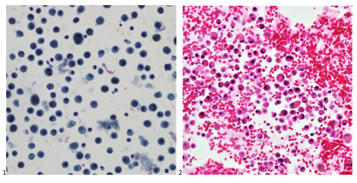
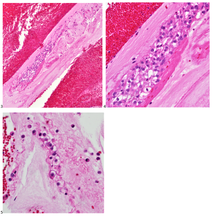
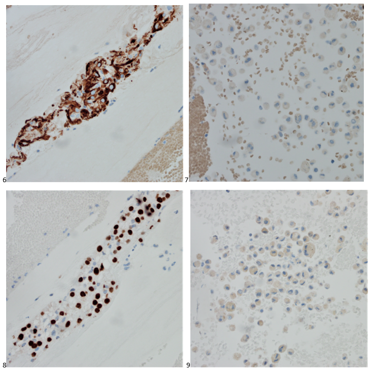
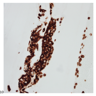
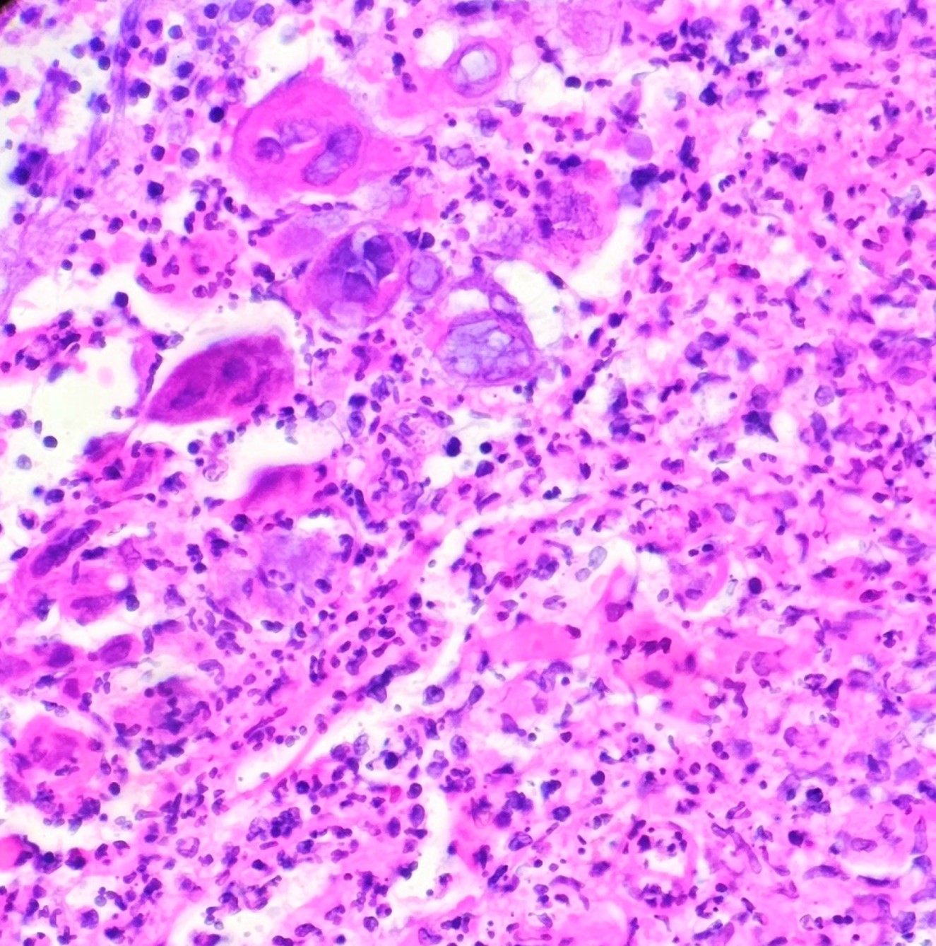
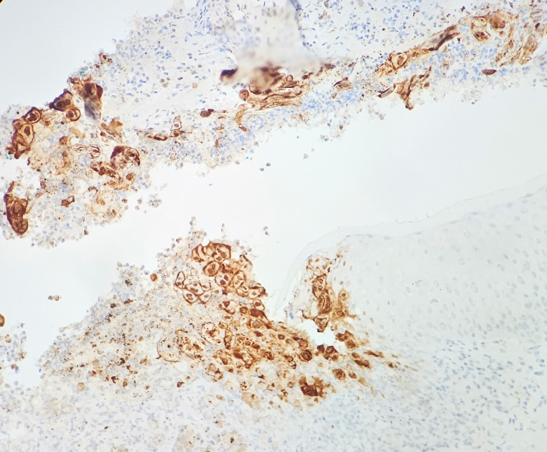
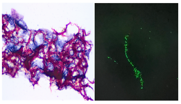
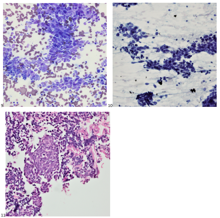
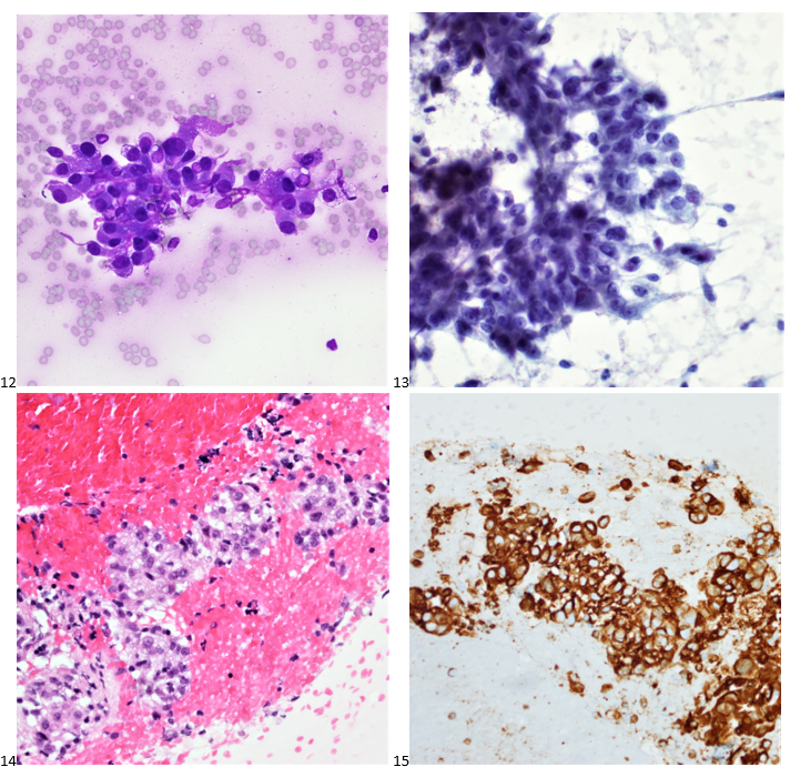
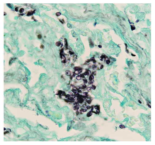
Histopathological examination of the biopsy specimen revealed granulomatous inflammation with numerous intracellular and extracellular Leishmania sp. amastigotes (Figure 2A). The amastigotes measured 2–4 µm in diameter and were oval to round with a defined nucleus and kinetoplast (Figure 2B). A diagnosis of cutaneous leishmaniasis (CL) was made, and the patient was referred to the infectious disease (ID) clinic for further evaluation and management. The patient was prescribed miltefosine for 28 days with planned follow up with ID, dermatology and ear nose and throat (ENT) groups. At a subsequent follow up visit two months later, her lesions have visibly improved, and she continues to be followed outpatient (Figure 1B).

Figure 1. Clinical presentation of a young woman with Leishmaniasis. Photographs of the patient’s left cheek at initial presentation (A) and following 28 days of miltefosine treatment (B).
Figure 2. Histopathology of Leishmania in the patient’s biopsy from the left cheek (H&E). A) A dense dermal infiltrate composed of histiocytes and lymphocytes with numerous forms consistent with amastigotes of Leishmania (orange boxes) within histiocytes (400x magnification). B) High magnification of amastigote-containing histiocytes with observable kinetoplasts (orange arrows) (1000x magnification, oil immersion).

Discussion

Leishmaniasis is a vector-borne disease transmitted by sandflies. The disease is caused by obligate intracellular protozoan parasites of the genus Leishmania. Human infection is caused by 21 of 30 known species that infect mammals in the Eastern and Western hemispheres. Examples of common species causing disease in the Eastern hemisphere include the L. donovani complex, L. tropica, L. major, and L. aethiopica whereasthe most common species found in the Western hemisphere include L. mexicana complex, L. braziliensis, and the subgenus Viannia.1 The different species are morphologically indistinguishable and can only be differentiated by isoenzyme analysis, molecular methods, or monoclonal antibodies.2

Leishmaniasis presents as a diverse range of diseases depending on the species associated with the infection. Visceral Leishmaniasis (VL) is associated with L. donovani complex in the Eastern hemisphere and L. chagasi in the Western, can be life threatening, and is characterized by fever, weight loss, hepatosplenomegaly, and pancytopenia.1 More than 90 percent of the world’s cases of VL are in India, Bangladesh, Nepal, Sudan, and Brazil.2 Worldwide, it has an estimated annual incidence of 0.7–1.0 million cases.3 The disease primarily affects the skin and can result in disfiguring lesions.

CL is the most common manifestation observed worldwide. L. major, L. tropica and L. aethiopica are common causes of CL in the East and L. braziliensis and L. mexicana are common causes of CL in the West.1 The clinical presentation of cutaneous leishmaniasis can vary depending on the Leishmania species involved, the immune status of the host, and the local immune response at the site of infection. The incubation period of cutaneous leishmaniasis can range from several days to months, with disease presenting as solitary or multifocal lesions. Affected areas may be ulcerated or nodular.4 This patient’s presentation was more nodular in appearance. Additionally, L. braziliensis can cause mucocutaneous leishmaniasis frequently months to years after spontaneously healed CL, which can erode the nasal septum, palate, or other mucosal structures.2

Serological tests can detect the presence of antibodies against Leishmania but cannot be used as a surrogate for treatment success because antibodies can continue to circulate following successful treatment.5 Histopathological examination of a skin biopsy is the gold standard for the diagnosis of cutaneous leishmaniasis. The presence of intracellular and extracellular amastigotes in a granulomatous inflammatory infiltrate is highly suggestive of the disease.6 Treatment of leishmaniasis depends on the presentation and disease severity. Topical therapy, such as paromomycin ointment, can be used for CL. Systemic therapy (such as what this patient received) is indicated in the setting of immunosuppression, or if the lesions are large, multifocal, affect the joints, hands, or feet. Various antiparasitic agents are available for the treatment of leishmaniasis, including pentavalent antimonials. amphotericin B, miltefosine, and paromomycin.6 In some cases, surgical intervention may be required to remove larger ulcers or nodules.

References

1.         Garcia, L. Diagnostic Medical Parasitology, 6th edition. “Leishmaniasis”. ASM Press. 2016. Chapter 27, p778-793.     

2. CDC – DPDx – Leishmaniasis. Published January 18, 2019. Accessed February 20, 2023. https://www.cdc.gov/dpdx/leishmaniasis/index.html

3. Burza S, Croft SL, Boelaert M. Leishmaniasis. Lancet. 2018;392(10151):951-970. doi:10.1016/S0140-6736(18)31204-2

4.         Reithinger R, Dujardin JC, Louzir H, Pirmez C, Alexander B, Brooker S. Cutaneous leishmaniasis. Lancet Infect Dis. 2007;7(9):581-596. doi:10.1016/S1473-3099(07)70209-8

5.         Aronson NE, Joya CA. Cutaneous Leishmaniasis: Updates in Diagnosis and Management. Infect Dis Clin North Am. 2019;33(1):101-117. doi:10.1016/j.idc.2018.10.004

6.         Handler MZ, Patel PA, Kapila R, Al-Qubati Y, Schwartz RA. Cutaneous and mucocutaneous leishmaniasis: Differential diagnosis, diagnosis, histopathology, and management. J Am Acad Dermatol. 2015;73(6):911-926; 927-928. doi:10.1016/j.jaad.2014.09.014

L. Jonathan He is a fourth year AP/CP resident at UT Southwestern Medical Center in Dallas, Texas.

-Dominick Cavuoti, DO is a professor in the Department of Pathology who practices Medical Microbiology, Infectious Diseases Pathology and Cytology.

-Andrew Clark, PhD, D(ABMM) is an Assistant Professor at UT Southwestern Medical Center in the Department of Pathology, and Associate Director of the Clements University Hospital microbiology laboratory. He completed a CPEP-accredited postdoctoral fellowship in Medical and Public Health Microbiology at National Institutes of Health, and is interested in antimicrobial susceptibility and anaerobe pathophysiology.

-Clare McCormick-Baw, MD, PhD is an Assistant Professor of Clinical Microbiology at UT Southwestern in Dallas, Texas. She has a passion for teaching about laboratory medicine in general and the best uses of the microbiology lab in particular.

A Traveler from Southern America with Nightly Fevers and Dysuria

Case Presentation

A 26 year old male presented to the emergency department (ED) with 1 week of malaise, high nightly fevers, abdominal pain, dark urine, and dysuria. He had no past medical history and was not taking any medications. He had been living in shelters since arriving in the US approximately 1 month ago after several months traveling through South and Central America from Venezuela. On review of systems, he also reported 3-4 days of constipation and a single episode of non-bilious, non-bloody vomit 2 days ago, accompanied by nausea. He denied mosquito bites, bloody or dark stools. On physical exam, patient appeared thin, mildly jaundiced, and had LUQ/LLQ moderate tenderness to palpation without guarding and splenomegaly. Given his travel history and presenting symptoms, the infectious disease service was consulted, and blood was drawn in the ED for further analysis. CBC and CMP were significant for normal WBC (10.21 x 10³/mcL), normocytic anemia (hemoglobin 11.3 gm/dL, MCV 80.4 femtoliters), thrombocytopenia (48 x 103/mcL), hyperbilirubinemia (total bilirubin 2.4 mg/dL, indirect bilirubin 1.7 mg/dL), and slightly elevated AST (46 units/L). Thin blood smear showed relatively enlarged erythrocytes infected with multiple ring forms and trophozoites with ameboid cytoplasm, consistent with Plasmodium vivax. Parasitemia was calculated to be 0.4%. The patient was diagnosed with uncomplicated malaria and started on artemether-lumefantrine for 3 days. He was found to a severe deficiency (41 units/1012 RBC) of glucose-6-phosphate dehydrogenase (G6PD) and therefore, at high risk of hemolysis if administered primaquine. Given his complex social situation, the patient decided to monitor himself for symptoms of relapse and defer starting primaquine for treatment of the liver stages of P. vivax. He was discharged to a local shelter with plans to follow-up closely with the outpatient infectious disease department, with strict precautions to return to the ED urgently for recurrent symptoms.

Figure 1. Thick (left) and thin (right) peripheral blood smears from patient reveal large infected red blood cells and “amoeboid” forms suggestive of P. vivax. Photo taken by Dr. Zoon Tariq, PGY3, Department of Pathology at The George Washington University School of Medicine and Health Sciences.

Discussion

Malaria should be suspected when a patient presents with a febrile illness and a travel history within a malaria-endemic region. Diagnosis of P vivax can be made through microscopic examination of blood smears, immunochromatographic rapid diagnostic tests (RDTs) and nucleic acid detection through amplification techniques.1 Examination of a thick blood smear allows efficient screening for malaria parasites, while a thin blood smear allows for species identification since parasite morphology is more clearly visualized.2 Upon examination of thin blood smears, infections by P. vivax and P. ovale may appear indistinguishable as both species infect immature, enlarged erythrocytes (1.25-2x normal), can be visualized at any stage in peripheral blood (ring, trophozoite, schizont, and gametocyte) and because Schuffner’s dots are a common morphologic feature during most stages. Defining characteristics of P. vivax include the presence of a large, ameboid trophozoite cytoplasm, and fine Schuffner’s dots and schizonts with >12 merozoites. Of note, preparation with Giemsa stain over Wright stain is preferred for demonstration of Schuffner’s dots.2 Immunochromatographic RDTs detect parasite-specific antigens (e.g., Plasmodium lactate dehydrogenase, Plasmodium specific aldolase) in a finger-prick blood sample. These tests are commercially available and relatively simple to perform and interpret, making them a useful tool for resource-limited regions.1 Nucleic acid amplification-based tools (e.g., PCR, loop-mediated isothermal amplification) are not routinely used for clinical management of malaria but do have diagnostic advantages over light microscopy and RDTs.3 PCRs are highly sensitive, can detect mixed infections even at low parasite densities, and are useful for epidemiological studies such as drug resistance identification.1

Malaria is a potentially fatal, but preventable and treatable, disease caused by infection of erythrocytes with protozoan parasites of the Plasmodium genus. Parasites are transmitted by the bites of infected female Anopheles mosquitos. Five species of Plasmodium (P. falciparum, P. vivax, P. ovale, P. malariae, P. knowlesi) infect humans. While each species of Plasmodium has unique characteristics, all species follow a similar life cycle. Erythrocyte lysis and release of merozoites cause release of pyrogens and production of inflammatory cytokines such as IL-1 and TNF-alpha, resulting in the symptomatic presentation of Plasmodium sp. infection, termed malaria. An important distinction between P. vivax and P. ovale and other Plasmodium species is that P. vivax/ovale may remain dormant in the liver (“hypnozoites”) and may resume intrahepatic replication, causing relapse of malaria weeks to years later.4 According to the WHO Malaria Report 2021, approximately half the world’s population lives in areas at risk of malaria infection. P. vivax is found predominately in Asia, Latin America, and some parts of Africa. It is the most common infective species in Latin America, accounting for an estimated 71.5% of cases in 2021.

The typical incubation time between transmission of parasites and onset of disease is approximately 14 days. P. vivax parasitesprimarily infect immature erythrocytes, representing 1-2% of the cell population. Synchronous replication and rupture of infected erythrocytes leads to the hallmark clinical presentation of cyclical severe fever and chills. Classically, P. vivax and P. ovale present with “tertian” malarial paroxysms, with fever and chills occurring every 48 hours. Patients may also complain of fatigue, malaise, headache, diaphoresis, abdominal pain, myalgias, dark urine, nausea, and vomiting. Additional clinical features and complications include anemia, jaundice, splenomegaly, and hepatomegaly. Severe infection presents with hemodynamic instability, pulmonary edema, coagulopathy, organ failure, neurological dysfunction, and potentially death.4, 5

Chloroquine or artemisinin-based combination therapy (ACT) are both effective treatments against uncomplicated, non-falciparum malaria (P. vivax, P. ovale, P. malariae, P. knowlesi).In a large systematic review, ACTs were found to be at least equivalent to chloroquine when treating the blood stage of P. vivax infection.6 ACTs are the drug of choice for non-falciparuminfections in countries where chloroquine resistance has developed, notably New Guinea and Indonesia.7To prevent relapse caused by hypnozoites of P. vivax/ovale, initial treatment is followed by administration of primaquine. Before treatment initiation with primaquine, quantitative testing for glucose-6-phosphate dehydrogenase (G6PD) deficiency should be completed since this drug may induce hemolysis in those who are deficient. A modified dosing schedule under close medical supervision is used for those who are G6PD deficient. Treatment of severe malaria is with at least 24 hours of intramuscular or intravenous artesunate, with an option to transition to an ACT regimen once oral therapy can be tolerated.1

Malaria caused by P. falciparum is typically more severe than malaria caused by P. vivax since P. falciparum infects erythrocytes of all ages, causing extensive hemolysis and related complications.4, 5 P. vivax malaria may also cause severe malaria and also relapses, emphasizing the importance of radical cure of hypnozoites with primaquine.8 Expedient and appropriate treatment leads to resolution of fever and parasitemia within days, with successful treatment confirmed by undetectable parasitemia on blood smear. Recurrence of a febrile illness should prompt re-evaluation and may suggest recrudescence or relapse due to failed therapy, or reinfection.

References

1. World Health Organization. (‎2023)‎. WHO guidelines for malaria, 14 March 2023. World Health Organization. https://apps.who.int/iris/handle/10665/366432.

5. Despommier DD, Griffin DO, Gwadz RW, Hotez PJ, Knirsch CA. Chapter 9: The Malarias. In: Parasitic Diseases. 7th ed. Parasites Without Borders, Inc.; 2019:93-122.

4. Ryan KJ. Chapter 51: Apicomplexa and Microsporidia. In: Sherris & Ryan’s Medical Microbiology. 8th ed. McGraw Hill; 2022. Accessed June 20, 2023. https://accessmedicine-mhmedical-com.proxygw.wrlc.org/content.aspx?bookid=3107&sectionid=260929904

2. DPDx – Laboratory Identification of Parasites of Public Health Concern: Malaria. Centers for Disease Control and Prevention. Updated: October 6, 2020. Accessed: June 20, 2023. https://www.cdc.gov/dpdx/malaria/index.html

3. World Health Organization. Malaria Policy Advisory Group: Meeting Report of the Evidence Review Group on Malaria Diagnosis in Low Transmission Settings. Geneva, Switzerland: WHO Headquarters, 2013. Accessed June 20, 2023. https://www.who.int/publications/m/item/meeting-report-of-the-evidence-review-group-on-malaria-diagnosis-in-low-transmission-settings

6. Gogtay N, Kannan S, Thatte UM, Olliaro PL, Sinclair D. Artemisinin-based combination therapy for treating uncomplicated Plasmodium vivax malaria. Cochrane Database Syst Rev. 2013;2013(10):CD008492. Published 2013 Oct 25. doi:10.1002/14651858.CD008492.pub3

7. Price RN, von Seidlein L, Valecha N, Nosten F, Baird JK, White NJ. Global extent of chloroquine-resistant Plasmodium vivax: a systematic review and meta-analysis. Lancet Infect Dis. 2014;14(10):982-991. doi:10.1016/S1473-3099(14)70855-2

8. Dini S, Douglas NM, Poespoprodjo JR, et al. The risk of morbidity and mortality following recurrent malaria in Papua, Indonesia: a retrospective cohort study. BMC Med. 2020;18(1):28. Published 2020 Feb 20. doi:10.1186/s12916-020-1497-0

-Cleo Whiting is a fourth-year medical student at George Washington School of Medicine and Health Sciences. Her research interests include infectious disease, autoimmune disease, and dermatopathology.

-Rebecca Yee, PhD, D(ABMM), M(ASCP)CM is the Chief of Microbiology, Director of Clinical Microbiology and Molecular Microbiology Laboratory at the George Washington University Hospital. Her interests include bacteriology, antimicrobial resistance, and development of infectious disease diagnostics.

A Diamond in the Mucin

A 35 year old male and current every day smoker transferred his care to our genitourinary (GU) clinic after undergoing a partial nephrectomy for clear cell renal cell carcinoma. A PET scan was performed, and a 5 millimeter FDG-avid right upper lobe (RUL) lung nodule was identified. While the mediastinal and hilar lymph nodes were mildly enlarged on the chest CT, they did not demonstrate FDG-avidity on the PET scan. The patient was referred to pulmonary for a workup of his subcentimeter lung nodule. Given the patient’s age and clinical history, the care team and the patient decided to pursue a lung biopsy via robotic-assisted bronchoscopy and subsequent lymph node sampling via endobronchial ultrasound (EBUS).

After the timeout, the patient was intubated, and the pulmonologist performed an airway inspection. Upon entering the right upper lobe, the pulmonologist noticed blood streaking along the bronchial wall. A right upper lobe bronchoalveolar lavage (BAL) was collected and sent for culture and cytology. Following the trachea and lung survey, the respiratory team began the robotic-assisted bronchoscopy. After confirming the correct location with both radial EBUS and fluoroscopy, multiple fine needle aspirates (FNAs) were collected from the right upper lobe lung nodule. Only benign bronchial cells were identified on rapid onsite evaluation (ROSE). A right upper lobe bronchial brushing was attempted and sent directly to cytology. Forceps were used to obtain transbronchial biopsies from the radiologically-suspicious area. The team then switched over to linear EBUS for the lymph node sampling portion of the procedure. Lymph nodes were sampled at the following stations: 11L, Level 7, and 4R. On ROSE, only lymphocytes and anthracotic pigment were identified for all three lymph nodes.

Upon e, the cytologist brought the specimens back to the laboratory for accessioning and processing. The following morning, the same cytologist screened all cytology specimens and the respective cell blocks associated with the case. The lymph nodes were signed out as negative for malignancy, with lymphocytes and anthracotic pigment present. The RUL FNA was inadequate for diagnosis as the material consisted mainly of bronchial cells and alveolar macrophages. The FNA findings were correlated with the transbronchial biopsy, which was signed out as benign pulmonary parenchyma with hemorrhage, fibrin clot, and respiratory bronchiolitis. Similarly, the bronchial brushing was negative as only benign bronchial cells were identified. In screening the BAL, there was an overabundance of hemosiderin-laden alveolar macrophages on the smear, cytospin, and SurePath liquid-based preparations (Image 1). When screening the cell block slides, abundant macrophages were also identified (Image 2).

Images 1-2. Lung, Right Upper Lobe, Bronchoalveolar Lavage 1: Pap-stained SurePath Liquid-based Prep; 2: H&E Cell Block Section (400X).

However, in the cell block sections, the cytologist also identified cells stranded within areas of mucin (Image 3). It may have been easy to dismiss these as macrophages, but contrary to the dusty hemosiderin-laden macrophages in this specimen, these cells had abundant vacuoles, and irregular nuclei with more prominent nucleoli (Images 4-5). The pleomorphic nuclei and larger vacuoles distinguished these cells from lipid-laden macrophages most often seen in aspiration pneumonia.

Images 3-5. Lung, Right Upper Lobe, Bronchoalveolar Lavage 3: H&E Cell Block section (100X); 4: H&E Cell Block section (400X); 5: H&E Cell Block section (600X).

The cytologist, marking the areas of interest, swiftly rendered a diagnosis of positive for malignant cells, favoring metastatic clear cell renal cell carcinoma. She enthusiastically described the case to the attending cytopathologist, who immediately ordered confirmatory immunostains on the unstained paraffin sections of the cell block.

With adequate controls, CD68 highlights the alveolar macrophages (not shown). The tumor cells show positive staining for AE1/AE3 and PAX-8 (Images 6-7), while macrophages show negative staining for AE1/AE3 and PAX-8 (Images 8-9). The tumor cells also show positive staining for vimentin (Image 10) and RCC (focal), and negative staining for TTF-1.

Images 6-10. Lung, Right Upper Lobe, Bronchoalveolar Lavage 6: AE1/AE3-positive tumor cells; 7: AE1/AE3-negative macrophages; 8: PAX-8-positive tumor cells; 9: PAX-8-negative macrophages; 10: Vimentin-positive tumor cells.

RUL BAL Final Diagnosis: Positive for malignant cells. Metastatic clear cell renal cell carcinoma.

When we attend a biopsy of an area clinically suspicious for metastatic renal cell carcinoma, we prepare ourselves for an overtly bloody sample. Even if the smears consist mainly of blood during ROSE, we rest assured that we will usually find tumor cells in the cell block sections. By obtaining a BAL of the blood-streaked RUL, the pulmonologist was able to pinpoint an area of lymphangitic spread. We often give little credit to BALs in cytology, especially when using concurrent techniques such as FNAs to sample a subcentimeter lung nodule without bulky disease. However, as seen in this case, sometimes it’s the only specimen that can help us render a diagnosis of metastatic cancer.

-Taryn Waraksa-Deutsch, MS, SCT(ASCP)CM, CT(IAC), has worked as a cytotechnologist at Fox Chase Cancer Center, in Philadelphia, Pennsylvania, since earning her master’s degree from Thomas Jefferson University in 2014. She is an ASCP board-certified Specialist in Cytotechnology with an additional certification by the International Academy of Cytology (IAC). She is also a 2020 ASCP 40 Under Forty Honoree.

Microbiology Case Study: Middle Aged Female Undergoing Nephrolithotomy with an Encrusted Stent

Case history

A middle-aged female with a one-year history of obstructive pyelonephrosis caused by a large nephrolith was admitted for a percutaneous nephrolithotomy and cystolitholapaxy. Previous medical history was notable for hypertension, type 2 diabetes, and peripheral vascular disease. A previously placed stent and nephrostomy tube had become encrusted, necessitating surgical intervention. CT imaging demonstrated moderate hydronephrosis, progressive encrustation of the stent, and high stone burden. Pre-operative urine cultures yielded greater than 3 organisms of unclear significance. Intraoperative findings revealed a calcified stent and large renal pelvic matrix stone which was removed and sent for culture and mineral analysis.

Laboratory identification

Intraoperative specimens were sent to the microbiology laboratory for routine culture. Gram stain of the nephrolith and associated tissue was performed, revealing gram positive coryneform rods and gram positive cocci (Image 1A). Light growth of Enterococcus faecalis was observed after 24 hours, along with a heavy amount of pinpoint, gram positive rods (Image 1B). It was noted that growth was only observed on blood agar (not chocolate agar), a feature suggestive of a lipophilic species of Corynebacterium. Colonies were visible on blood agar after prolonged incubation, and the organism was determined to catalase positive. The organism produced copious amounts of urease, leading to tube positivity within 10 minutes (Image 1C). The organism was definitively identified by MALDI-TOF MS as Corynebacterium urealyticum.

Image 1.  A) Representative Gram stain obtained from grind of nephrolith, and associated tissue submitted to the microbiology laboratory.  A mixture of coryneform rods and Gram-positive cocci can be seen.  B) Pinpoint growth of C. urealyticum on sheep’s blood agar media after 24 hours.  Extended in incubation or the addition of lipid to the culture media is needed for more robust growth.  C) Rapid urease production of C. urealyticum.  Christiansen’s urea agar slant was inoculated with C. urealyticum and photographed at different timepoints post inoculation.  Urease positivity can reliably be demonstrated within 10 minutes.

Discussion

Corynebacterium urealyticum is a lipophilic corynebacterial species frequently recovered from the urinary tracts of patients with renal or urological disease. While this organism is often associated with alkaline encrusted cystitis, it also plays an important role in renal stone formation. Due to its indolent and slow growing nature, the organism can be challenging to recover from routine urine cultures; thus, disease burden attributable to C. urealyticum is likely underestimated. Risk factors for disease include patients with urinary tract abnormalities, catheterization, prolonged hospitalization, history of immunocompromising conditions or renal transplant, and extended therapy with broad-spectrum antibiotics.1 Indeed, most contemporary clinical isolates are multidrug resistant, making antibiotic therapy of C. urealyticum challenging.

C. urealyticum is a member of the colonizing flora of both the skin and the urinary tract, and its ability to adhere to the uroepithelium is believed to mediate its ability to cause disease. The organism is frequently detected in the groin of hospitalized and institutionalized patients.2 As a member of the lipophilic corynebacteria, enhanced growth can be achieved through the addition of 0.1% Tween 80 to culture media. The impressive urease activity of C. urealyticum is central to pathogenesis as hydrolysis of urea in the GU tract leads to ammonia production and alkalinization (Image 1C). This, in turn, leads to saturation of the microenvironment with calcium phosphate and struvite crystallization which can result in sone formation.1 In settings where C. urealyticum infection may be suspected (including encrusted cystitis and encrusted pyelitis), prospective discussions with the laboratory are warranted to avoid dismissal of the organism as a diphtheroid bacilli that is a normal component of the urogenital flora. This is particularly important if the laboratory does not routinely hold cultures for longer than 24 hours.

Chronic and recurrent urinary tract infections in patients of advanced age is usually the primary presentation of C. urealyticum infection. By contrast, encrusted uropathies can develop in 4-16% of patients with C. urealyticum bacteruria and are subacute-to-chronic conditions associated with urease-producing bacteria.2 C. urealyticum is the principal cause of encrusted uropathies. This case represents a more complex case of encrusted disease leading to extensive nephrolith formation requiring surgical intervention. For urinary tract infections, vancomycin remains the antibiotic of choice for management in addition to removal of the impacted mucosal encrustations and urological consultation.3 C. urealyticum also exhibits near uniform susceptibility to linezolid. The excised nephrolith in this patient’s case was found to be composed of 90% struvite and 10% calcium phosphate. Following the procedure, imaging revealed no visible, residual stones in either the impacted kidney, ureter, or bladder. The patient is followed as an outpatient and continues to do well.

References

  1. Salem, N., Salem, L., Saber, S., Ismail, G., and Bluth, M.H. Corynebacterium urealyticum: a comprehensive review of an understated organism. Infect. Drug Resist. 2015:8 129-145.
  2. Van de Perre, E., Reichman, G., De Geyter, D., Geers, C., Wissing, K.M., and Letavernier, E. Encrusted uropathy: a Comprehensive Overview – to the Bottom of the Crust. Front. Med. 2021. 7:609024
  3. Kim, R. and Reboli, A.C. Chapter 205: Other Coryneform Bacteria, Arcanobacterium haemolyticum, and Rhodococci in Mandell, Douglas and Bennett’s Principals and Practice of Infectious Diseases 9th Ed. Elsevier, Philadelphia, PA. Pgs: 2532-2542.

-Andrew Clark, PhD, D(ABMM) is an Assistant Professor at UT Southwestern Medical Center in the Department of Pathology, and Associate Director of the Clements University Hospital microbiology laboratory. He completed a CPEP-accredited postdoctoral fellowship in Medical and Public Health Microbiology at National Institutes of Health, and is interested in antimicrobial susceptibility and anaerobe pathophysiology.


-Clare McCormick-Baw, MD, PhD is an Assistant Professor of Clinical Microbiology at UT Southwestern in Dallas, Texas. She has a passion for teaching about laboratory medicine in general and the best uses of the microbiology lab in particular.

Microbiology Case Study: An Unusual Case of Herpes Reactivation

Case history

A 37-year-old female with a past medical history of psoriasis and common variable immunodeficiency disorder (CVID) presented to her dermatologist for an ulceration on her right buttock following a camping trip about 1 month ago. She thought that she had been bitten by a bug, for the lesion became extremely pruritic and painful. The patient was self-treating the area with over-the-counter antibiotic ointment and an anti-itch cream, but the symptoms persisted. At the time, the dermatologist was also treating a lower extremity dermatophyte infection, and the antibiotic cream and anti-itch cream were discontinued and replaced with clobetasol 0.05% ointment for potential allergic dermatitis. The patient returned to the dermatologist about a month later as the site was becoming increasingly inflamed and painful. The patient had also started experiencing night sweats and fever, so she was transferred and further evaluated in the emergency department. In the ED, the differential included soft tissue infection, cellulitis, or abscess of a fungal, viral, or bacterial etiology. Labs showed evidence of inflammation with an elevated ESR and CRP. A punch biopsy was performed and pathologic examination showed an ulcer bed with prominent acute inflammatory cell infiltrate and necrosis. The infected squamous epithelium showed the3 Ms findings (Molding, Margination, Multinucleation) consistent with herpetic infection (figure 1). The diagnosis was confirmed with HSV complex IHC (figure 2) and PCR testing of the lesion came back positive for HSV-2. Of note, the patient did have a history of genital herpes; however, she was not having a typical flare, and she had been treated with a 10-day course of valacyclovir 2 weeks prior to her ED visit. The gram stain showed no evidence of neutrophils, squamous epithelial cells, or organisms, but bacterial cultures came back positive for MRSA.

Figure1. H&E section showing mixed acute and chronic inflammation with squamous cells
showing herpes viral cytopathic effect (400x magnification)
Figure 2. HSV complex (HSV1 and 2) IHC staining virally infected epithelial cells (200x).

Discussion

Herpes simplex virus (HSV) is a large, double-stranded DNA virus from the Herpesviridae family.1,2 HSV-2 is generally considered a sexually transmitted infection because it can be transmitted by contact with infected genital secretions.2,3 The viral particles within these secretions can enter epithelial cells and begin replicating, causing the characteristic intranuclear inclusions and multinucleated giant cells that can be seen under the microscope.2 When the virus infects the cells in this manner, it can also cause these infected cells to separate from each other and form grouped vesicles filled with these cell remnants.2 The virus infects nerve endings and then travels backwards to the sacral ganglia, and it can remain latent there permanently, giving it the ability to recur during the infected person’s lifetime.1 A patient can initially present with symptoms of dysuria, lymphadenopathy, fever, headaches, and myalgias, but more than half of patients may not know they have genital herpes.1,3 Recurrences can present with symptoms of tingling, burning, itching, and pain in the nerve’s distribution pattern, similar to the pain and pruritis in our presented case.2 When there is a suspected HSV infection, PCR for HSV DNA is generally the best diagnostic tool, and it is faster and more sensitive than viral culture.1,2 A patient with known herpes infection should be treated with antivirals; however, the lesions should also self-resolve within 3 weeks if the patient is not treated.1

One abnormality in our presented case was that the patient’s reactivation of genital herpes was on the buttocks. A repeat infection with HSV at a site other than genitalia is more common when the primary infection also occurred at a site other than the genitalia.4 Infections occurring at non-genital sites such as the buttocks can also occur due to self-inoculation, which may have been the case in our patient.4 Additionally, repeat infections with HSV in non-genital sites are more common when the initial infection was with HSV1, but our patient’s PCR showed the presence of HSV-2 DNA.4 One explanation for this phenomenon is that HSV-2 recurrences can occur on the buttocks due to the retrograde transport to the root ganglia in the areas that correspond to these dermatomes.4

Another abnormality in our presented case involves the patient’s persistent infection despite treatment with a course of valacyclovir for 10 days. Generally, an initial herpes infection self-resolves in a matter of weeks, and a recurrent episode will self-resolve in a matter of days, usually less than ten.1,2 It is unusual that her infection persisted despite therapy, but the patient does have a medical history significant for CVID. Patients with weakened immune systems can take longer to fight off herpes infections even if they are taking antivirals.2 Additionally, there is a theory that herpes buttocks infections last longer than in other regions due to the greater travel distance along the nerves as well as a higher concentration of nerve endings in this region.4 The patient in our case also had tissue cultures that were positive for MRSA, meaning she had a concomitant bacterial and viral infection of the buttock region, and treatment with an antiviral would not be sufficient to eradicate her coinfection.

References

1.  Johnston C, Corey L. Current Concepts for Genital Herpes Simplex Virus Infection: Diagnostics and Pathogenesis of Genital Tract Shedding. Clin Microbiol Rev. Jan 2016;29(1):149-61. doi:10.1128/cmr.00043-15

2.  Gupta R, Warren T, Wald A. Genital herpes. Lancet. Dec 22 2007;370(9605):2127-37. doi:10.1016/s0140-6736(07)61908-4

3.  Groves MJ. Genital Herpes: A Review. Am Fam Physician. Jun 1 2016;93(11):928-34.

4.  Benedetti JK, Zeh J, Selke S, Corey L. Frequency and reactivation of nongenital lesions among patients with genital herpes simplex virus. Am J Med. Mar 1995;98(3):237-42. doi:10.1016/s0002-9343(99)80369-6

-Lillian Acree is a fourth-year medical student at the Medical College of Georgia. She is interested in head and neck pathology.

-Hasan Samra, MD, is the Director of Clinical Microbiology at Augusta University and an Assistant Professor at the Medical College of Georgia.

Case Studies in Hematology: Hemoglobin S Beta Thalassemia Compound Heterozygosity in a 61 Year Old Female

A 61 year old Black woman with a diagnosis of sickle cell beta thalassemia presented to the ER with fatigue, dyspnea, and back and leg pain with some swelling in the hands and feet. Splenomegaly was noted on exam. The patient has a history of moderate to severe symptomatic anemia and is being followed by a hematologist. Her baseline Hgb is 9-10 g/dL. Her treatment plan includes Hydroxyurea, 500 mg daily, and transfusions, as needed. Her last sickle cell crisis was 2 years ago. CBC was ordered. Hgb on admission was 6.1 g/dL. Her RBC morphology showed polychromasia, target cells, sickle cells, anisocytosis, and numerous nucleated RBC forms.

The patient was admitted to the hospital. Type and crossmatch for 2 units of packed red blood cells was ordered. CT imaging was performed and revealed severe osteopenia and vertebrae deformities consistent with her history of sickle cell disease. Chest CT showed hypoinflated lungs and areas of consolidation in the lower lobes consistent with acute chest syndrome due to sickle cell beta thalassemia. She was transfused with 2 units of pRBCs and treated for sickle cell crisis. The patient remained stable and was discharged 3 days later.

Table 1. CBC results on ER admission
Figure 1. Peripheral blood smear on admission. Patient with sickle cell/beta thalassemia shows sickle cells, target cells, nucleated red cells, anisocytosis, poikilocytosis, polychromasia. (Mercy Medical Center, Baltimore, Md)

Sickle cell anemia (HbSS) and thalassemia are the world’s most common single gene disorders. Both are inherited in an autosomal recessive manner, and result in hemolytic anemias. But what happens when you inherit one gene for sickle cell and one gene for beta-Thalassemia (β-thalassemia)?

Sickle cell disease is caused by mutations in the HBB gene that provide instructions for making beta-globin. Sickle cell anemia is a hemoglobinopathy, a qualitative defect in the structure of globin chains, resulting in the production of abnormal hemoglobin. Normal adult Hemoglobin A has 2 α chains and 2 β chains (α2β2). Hb S results from the substitution of valine for glutamic acid at position 6 of the β globin chain. The resultant Hb S has reduced solubility at low oxygen tensions. Patients with sickle cell anemia have a moderate to severe chronic hemolytic anemia with recurrent painful sickle cell crisis.

Sickle cell disease is inherited in an autosomal recessive pattern from parents who have at least one mutated gene. Anyone with a sickle cell gene can pass this gene on to their children. Sickle cell anemia (HbSS) is the homozygous expression of a sickle gene from both parents and is the world’s most common inherited hematological disease. A heterozygote inherits a sickle gene from only one parent. This person is a carrier of sickle cell (HbSs), often referred to as sickle cell trait. HbSs persons do not generally exhibit symptoms or may exhibit only a mild anemia. However, under stressful conditions, such as at high altitudes, they may experience vaso-occlusive sickle crisis.

While hemoglobinopathies are a qualitative defect due to structural changes in the normal amino acid sequence of globin, thalassemias result from an imbalance in the synthesis of the globin chains that make up the hemoglobin molecule. Thalassemias involve the rate of globin chain synthesis leading to a quantitative defect. Thalassemia is divided into α-thalassemias and β-thalassemia. α-thalassemias involve genes for the α chains on chromosome 16. In α-thalassemia, the deletions involve the α1 and/or the α2 globin genes and result in decreased production of α chains. β-thalassemias mainly affect β chain production. They are disorders of reduced globin chain production from the globin chain cluster on chromosome 11.

(Since this case involves a known diagnosis with a compound heterozygous state involving a β-thalassemia gene mutation, the discussion of α-thalassemia has been limited here. Watch for a case involving α-thalassemia in a future blog!)

Beta thalassemia occurs when the beta globin chains are either produced inadequately or not at all. There are many mutations in and around the β globin gene that result in decreased β chain production. Mutations that result in the complete absence of β chain production are designated as β0. In the most severe form of β-thalassemia the patient is homozygous β00 and does not produce any β chains. Without β chains there is no Hb A (α2β2). β+ is used as the designation for any mutations of the β globin gene that cause a partial deficiency of β chains (5-30% decrease) and therefore result in a decrease in production of Hb A. The βsilent designation is used for carrier state gene mutations that result in only a mildly decreased β chain production. The degree of decrease in the β chain production is related to the degree of anemia and the severity of clinical disease.

Thalassemia, like sickle cell anemia, is a hereditary anemia inherited in the autosomal recessive manner. β-thalassemia is divided into categories based on clinical severity of disease. In β-thalassemia major a child inherits a copy of a β-thalassemia gene mutation from both parents. There are various mutations that cause genes with these mutations and different variants may be inherited from each parent. A person with thalassemia major may be homozygous β+/ β+, homozygous β0/ β0, or the compound heterozygous state β+/ β0. Hb A is only produced in patients with the β+ mutation. β-thalassemia major patients have the most severe hemolytic anemia and symptoms. β-thalassemia intermedia is characterized as homozygous βsilent or heterozygous βsilent with β+ or β0 and mild to moderate disease. β-thalassemia minor, also called β-thalassemia trait or carrier state, presents with mild but asymptomatic hemolytic anemia. These patients are heterozygous with normal β globin and have slightly decreased Hb A.

In people with sickle cell disease, at least one of the beta globin is replaced with hemoglobin S. In homozygous sickle cell anemia, both beta globin subunits in hemoglobin are replaced with hemoglobin S. In compound sickle cell diseases, one beta globin is replaced with hemoglobin S and the other beta globin is replaced with a different abnormal variant. Examples of this are Hb SC disease, and Hb SD syndrome. Compound heterozygosity is the inheritance of two different mutated genes that share the same locus. If mutations that produce hemoglobin S and beta thalassemia occur together, individuals have hemoglobin S-beta thalassemia disease. (sickle cell beta-thalassemia, Hb S β thal or sickle-β-thal). Sickle cell beta thalassemia patients have hemoglobin S (α2β26Glu→Val) and either β0 or β+.

When a qualitative hemoglobinopathy is inherited with a quantitative disorder of hemoglobin synthesis, the severity of the compound disorder is dependent on the β gene mutation. Patients with β0 produce no Hb A and have moderate to severe symptoms comparable to that of Hb SS patients. β+ patients will produce some β chains and therefore have some Hb A and milder or no symptoms.

Figure 2. Peripheral Blood smear on day 3. Sickle cell forms, polychromasia, target cells. nucleated RBCs. (Mercy Medical Center, Baltimore, Md)

Newborn screening can diagnose β0-thalassemia at birth by detecting a complete absence of hemoglobin A. However, it is not possible to make a definitive diagnosis of β+-thalassemia in the newborn because newborns have Hb F, and the reduced amount of hemoglobin A overlaps the range for normal babies. In adults with Hb S – β thal the amount of Hb S is variable. There is some Hb A in β+ patients but no Hb A detected in β0. Hb A2 and Hb F are increased. In addition to hemoglobin electrophoresis, molecular testing may also aid in the diagnosis by identifying genetic mutations. Beta globin gene sequencing can identify beta thalassemia alleles that are caused by point mutations in the beta globin gene. As well, structural variants of the beta globin gene such as Hb S can be identified with this technique. This can lead to a better understanding and clinical management of the disease.

Case Study, continued: This patient inherited a Hb S gene from one parent and a β-thal gene from the other, resulting in sickle cell beta thalassemia. This compound heterozygosity affects red blood cells both by the production of structurally abnormal hemoglobin, and by the decreased synthesis of beta globin chains. Clinical manifestations depend on the amount of beta globin chain production. Symptoms may include anemia, vascular occlusion, acute episodes of pain, acute chest syndrome, pulmonary hypertension, sepsis, ischemic brain injury, splenic sequestration crisis and splenomegaly.

Hemoglobin electrophoresis was sent out to a reference lab and results are shown in Table 2. Based on the Hemoglobin electrophoresis, is this patient Hb S- β0-thal or Hb S- β+-thal?

Table 2. Hemoglobin pattern and concentrations of a S/betathalassemia patient

Hemoglobin electrophoresis results for Hb S beta thalassemia patients are expected to show 60-90% Hb S and 10-30% Hg F. This patient’s Hgb S at 74% is within this range. This result reflexed a sickle solubility test, which was positive. As well, the elevated Hb F and Hb A2 are consistent with this diagnosis. It was noted in the discussion above that Hb S- β+-thal mutations cause a decrease of 5-30% in beta chains and therefore a decrease in Hb A. This patient’s Hb S is greater than the Hb A and her Hb A concentration is 14.8%, which is consistent with this diagnosis. Hb S- β0 mutations produce no Hb A. In this case there is some Hb A on electrophoresis but not as much as would be expected in a β+-thal mutation. Also, of note it that this patient was recently transfused with 2 units of pRBCs. Interpretations of hemoglobin electrophoresis assume that the patient has not been transfused in the last 3 months. The Hb A in this patient can be explained by these recent transfusions. Therefore, it can be concluded that the hemoglobin pattern and concentrations are consistent with transfusion of a Hb S beta0 thalassemia patient. The β0 mutation is also consistent with this patient’s moderately severe symptomatic anemia.

References

  • Keohane, Elaine, et al. Rodak’s Hematology, Clinical Principles and Application, 5th ed, Elsevier, 2016
  • McKenzie, Shirlyn. Clinical laboratory Hematology. Pearson Prentice Hall, 2004.
  • McGann PT, Nero AC, Ware RE. Clinical Features of β-Thalassemia and Sickle Cell Disease. Adv Exp Med Biol. 2017;1013:1-26. doi: 10.1007/978-1-4939-7299-9_1. PMID: 29127675.
  • Origa R. Beta-Thalassemia. 2000 Sep 28 [Updated 2021 Feb 4]. In: Adam MP, Mirzaa GM, Pagon RA, et al.,editors. GeneReviews® [Internet]. Seattle (WA): University of Washington, Seattle; 1993-2023. Available from: https://www.ncbi.nlm.nih.gov/books/NBK1426/
  • Turgeon, Mary Louis. Clinical Hematology Theory and Procedures, 6th ed, Jones and Bartlett Learning, 2017.
Socha-small

-Becky Socha, MS, MLS(ASCP)CMBBCM graduated from Merrimack College in N. Andover, Massachusetts with a BS in Medical Technology and completed her MS in Clinical Laboratory Sciences at the University of Massachusetts, Lowell. She has worked as a Medical Technologist for over 40 years and has taught as an adjunct faculty member at Merrimack College, UMass Lowell and Stevenson University for over 20 years.  She has worked in all areas of the clinical laboratory, but has a special interest in Hematology and Blood Banking. She currently works at Mercy Medical Center in Baltimore, Md. When she’s not busy being a mad scientist, she can be found outside riding her bicycle.

Microbiology Case Study: Cryptococcal Meningitis In Immunocompromised Patient

Case History

A 47-year-old male originally from Dominican Republic, with a recent diagnosis of acquired immunodeficiency syndrome (AIDS) and diffused large B cell lymphoma (DLBCL), was admitted because of seizures and a rapidly increasing left neck mass. ​MRI of the brain showed a 2.5 x 1.2 cm (about 0.47 in) lesion in the left inferior parietal lobe – (1.4×0.7cm) in the right frontal lobe, plus multiple scattered bilateral lesions. Because of this, he underwent craniotomy/craniectomy for possible resection. A biopsy was taken from the right temple lesion and sent for aerobic, anaerobic, fungal, mycobacterial culture, surgical pathology and Toxoplasma PCR (Polymerase Chain Reaction). ​

Gram stains, KOH prep, acid-fast stains, and Toxoplasma PCR of the tissue were all negative. Aerobic and anaerobic cultures did not show any growth.

Histopathology slides (GMS and H&E stains in Fig A and B) show budding yeasts morphologically consistent with Cryptococcus. Mucicarmine stain was also positive. Lumber puncture was performed the next day and Cryptococcal antigen was positive, with a titer of 1:640. Interestingly, the CSF culture and Gram stain did not reveal any organisms.

Figure A. H&E shows encapsulated variably sized transparent/gray color yeasts with thin walls. Black arrows show organisms.
Figure B. GMS stains highlight very faint staining of capsule (black arrow). Yellow arrow highlights background inflammatory cells.

Discussion

Among several species of Cryptococcus, C. neoformans and Cryptococcus gattii are pathogenic, with C. neoformans causing meningitis in immunocompromised patients worldwide whereas C. gattii has a preference for immunocompetent individuals.1 Cryptococcal disease remains a major opportunistic infection and a leading cause of mortality in patients infected with HIV in much of the developing world. Most HIV-related meningitis cases are caused by Cryptococcus neoformans.2

Cryptococci are found in soil, due to contamination with pigeon droppings. The infection occurs through inhalation, with or without symptoms of pneumonia, with subsequent dissemination to the central nervous system (CNS) via blood. Imaging findings are often unspecific or negative. CT or MRI examination of the central nervous system is performed to rule out alternative diagnoses. The diagnosis of ‘meningitis’ is made with a lumbar puncture, which typically shows lymphocytosis, an increased protein and decreased glucose concentration. Few neutrophil granulocytes are often found in CSF. This is likely because neutrophil migration is inhibited by specific polysaccharides that are part of the cryptococcal capsule.3

Cryptococci can be seen directly in the sediment of centrifuged CSF stained with India ink. The sensitivity and specificity of India ink is poor; therefore, CSF Gram stain and culture, multiplex meningitis/encephalitis PCR, and lateral flow antigen (LFA) tests have replaced the use of India ink. The cryptococcal lateral flow antigen test should be performed in CSF and serum, in addition to Multiplex ME PCR panel, and is a preferred test because of high sensitivity (93-100%) and specificity (93-98%).4

High organism burden at baseline (indicated by quantitative CSF culture or CSF antigen titre) and abnormal mental status are the most important predictors of death, while high opening pressures and a poor inflammatory response in the CSF have also been associated with poor outcome.5

On H&E it has the characteristic appearance ofencapsulated variably sized yeasts (2-20 microns) with thin walls which can be highlighted with the GMS stain. Although the presence of a capsule differentiates Cryptococcus from Histoplasma capsulatum and Blastomyces dermatitidis with the H&E or mucicarmine stain, additional confirmation can be made with Fontana-Masson stainin the absence of capsules.6 Since both C.neoformans and C. gattii produce melanin, the pathology report by FM silver or H&E/GMS stain cannot further distinguish these two closely resembled species.

Occasionally, cryptococcal meningitis cases with sterile CSF culture and/or negative Cryptococcal CSF antigen are observed in HIV individuals, regardless of the CD4 counts.7,8 However, serum Cryptococcal antigen and blood culture may be positive in those individuals.7 In our case, the diagnosis of Cryptococcal meningitis was made by the pathology report and positive CSF Cryptococcal antigen.

-Fnu Sapna is a 2ndyear AP/CP pathology resident in the Department of Pathology at Montefiore Medical Center in Bronx, NY. She completed her Medical education at Chandka Medical College in Pakistan. Her interests are putting efforts to improve screening guidelines for diagnosis of preventable gynecological and breast cancers.

-Phyu Thwe, Ph.D, D(ABMM), MLS(ASCP)CM is Associate Director of Infectious Disease Testing Laboratory at Montefiore Medical Center, Bronx, NY. She completed her medical and public health microbiology fellowship in University of Texas Medical Branch (UTMB), Galveston, TX. Her interests includes appropriate test utilization, diagnostic stewardship, development of molecular infectious disease testing, and extrapulmonary tuberculosis.

One year of Chronic Cough after Past Resolution of Viral and Bacterial Respiratory Infections

Case History

A 74-year-old female from El Salvador with a medical history of hypertension, diabetes, osteoarthritis presented with persistent productive cough. The patient states that the symptoms started on November 2022 which was believed to be associated with ongoing COVID-19 infection. Despite recovery from COVID-19 infection, significant productive cough still remained. A visit to her primary care doctor revealed streptococcal throat infection and despite completing a course of Zithromax antibiotics, a subsequent chest x-ray revealed potential right upper lobe pneumonia and she reported productive cough with occasional blood streaks. Blood work up in the Emergency Department revealed leukocytosis (20.22 x10e3/mcL) with neutrophilia (14.56 x10e3/mcL neutrophils) and monocytosis (3.03 x10e3/mcL monocytes). A chest x-ray showed bilateral reticular and airspace opacities with an air-fluid level containing opacity overlying the left mid lung, likely representing a cavitary lesion. A follow up with a computerized tomography of the chest identified innumerable randomly distributed pulmonary nodules or cavities with upper lobe predominance. The largest cavity measures approximately 6.6 cm with air-fluid levels and debris. Additionally, a small pericardial effusion with thickened pericardium was also noted. A sputum sample was submitted for Acid-fast bacilli (AFB) culture and molecular testing. AFB stain was positive for acid-fast bacilli (Figure 1). The GeneXpert MTB/RIF assay detected M. tuberculosis with no rifampin resistance marker. Growth on the Lowenstein-Jensen agar after two weeks showed buff colored rough and dry colonies and was confirmed as M. tuberculosis on the MALDI-TOF.

Figure 1. Visualization of acid-fast bacilli directly from patient specimen. Using acid-fast stain, cording was observed (Figure 1A, left) and using fluorescent staining, fluorescent rods were observed (Figure 1B, right).

Discussion

Mycobacteria are aerobic, nonmotile, thin rod shape, non-spore forming bacilli that possess mycolic acid in its cell wall giving its acid-fast stain characteristics. Tuberculosis (TB) is an airborne disease caused by Mycobacterium tuberculosis (MTB). TB remains to be the leading cause of death from a single infectious agent worldwide. According to the CDC a total of 1.6 million people died from TB in 2021 (including 187, 000 people with HIV) 1,2. The M. tuberculosis complex (MTBC) includes M. tuberculosis, M. bovis, M. bovis, Bacille Calmette-Guérin strain (BCG), M. caprae, M. pinnipedii, M. mungi,M. africanum, M. microti, and M. canettii 3. M. tuberculosis produces cord factor, a glycolipid which is also known as trehalose dimycolate, that causes the bacteria to grow in parallel strands and that appeared like cord, or rope when cultured in liquid media (Figure 1A). The cord factor is present in the outer envelope and protects the bacteria from the host response 4.

The pathogenesis of human tuberculosis involves a complex interaction between host immune system and bacterial factors 4. M. tuberculosis is carried in airborne particles generated by infected individuals. The droplet nuclei traverse the mouth or nasal passages, upper respiratory tract, and bronchi to reach the alveoli of the lungs. The bacteria are then phagocytosed by alveolar macrophages and can inhibit maturation of phagosome and block formation of phagolysosome, allowing its unchecked replication in the macrophage which results in bacteria proliferation in the alveolar macrophage and air spaces. In immunocompetent hosts, the immune response (via TLR2, TLR9, Th-1 and IFN- ꝩ cascades) may contain the infection before significant tissue destruction or systemic illness 1-4. However, in the immunosuppressed hosts, the primary infection results in a broad clinical spectrum such as meningitis, miliary tuberculosis and extrapulmonary granulomas. Post-primary/secondary tuberculosis (reactivation TB) usually begins months to years after the establishment of systemic immunity in primary TB mostly in a period when the host immune response is weakened, following exogenous or a large inoculum of virulent bacilli overwhelming the host immunity system. Extrapulmonary manifestations will develop based on the organ system affected 5.HIV infection is the greatest risk factor for reactivation of TB as the virus causes functional abnormalities in CD4+ T cells and CD8+ T cells which confer protection against active TB. Other risk factors that promote the reactivation TB include aging, malnutrition, diabetes mellitus, renal failure, cancer and immunosuppressive therapy 5. The disease typically affects the lungs (pulmonary TB) but can affect other sites as regional lymph nodes, apex of the lung, larynx, kidneys, brain, bone, joints and pleura 1,2,5.

Diagnostic tests for TB detect either the bacteria or host immune response. Specimens recommended for diagnosis of mycobacterial infection are sputum, bronchial brushing/washings /biopsies, gastric aspirates (children) urine, blood, CSF, BM, body fluids, stool (only in HIV) 5,8. Specimens from sputum and other nonsterile sites should be liquefied with N-acetyl-L-cysteine and decontaminated with NaOH and for gastric aspirate neutralized with buffer 6-8. For diagnostic purposes, all persons suspected of having TB disease at any site should have at least three consecutive sputum specimens collected in 8 to 24 hours with at least one being an early morning one for AFB smear and culture 1,8.

The organisms can be visualized under a microscope using two principal methods: carbolfuchsin staining (e.g., Acid-fast stain), or using a fluorochrome (auramine-rhodamine and auramine-O stains) procedure (Figure 1B). Microscopy is the most rapid diagnostic method for the detection of tubercle bacilli but is less sensitive; it requires a minimum of 10,000 bacilli/mL of sputum to produce a positive result. Culture is the gold standard and more sensitive method for the detection of tubercle bacilli and is necessary for performing antimicrobial drug drug-susceptibility testing and genotyping. 6,9 However culture requires 3–6 weeks for growth which delays the initiation of anti-tuberculosis drug therapy. Two types of solid media are used for mycobacterial culture: egg based (Löwenstein-Jensen) and agar based (Middlebrook 7H10, 7H11, and selective 7H11). Colony morphology of Mycobacterium tuberculosis on solid media are dry, rough, raised, wrinkled, off white to buff colored. M. tuberculosis is commonly positive for niacin, nitrate reduction test, pyrazinamidase test, but negative for 68C catalase, tween 80 hydrolysis, and 5% NaCl tolerance. Molecular techniques such as nucleic acid amplification tests revolutionized tuberculosis diagnosis since M. tuberculosis nucleic acid material can be detected directly from specimen in less than 2 hours. Two commercial NAATs for the detection of M. tuberculosis complex are available in the United States: The Amplified MTD (Mycobacterium Tuberculosis Direct) test (Hologic, Marlborough, MA) and the Xpert MTB/RIF (Cepheid, Sunnyvale, CA).

Common serological approaches for detection of M. tuberculosis are the Tuberculin skin test (TST) or IFN- ꝩ release assays (IGRA’s) 1,9,10. The tuberculin skin test is performed by intracutaneous injection of purified protein derivative of M. tuberculosis, which induces a visible and palpable induration that peaks in 48 to 72 hours. A false-positive tuberculin skin test may result on individuals with prior vaccination with BCG (Bacillus Calmette-Guerin), an attenuated strain of M. bovis. BCG immunization does not affect the test result of IGRA assay. The IGRA are blood tests that measure a person’s immune reactivity to M. tuberculosis. Both T-Spot and QuantiFERON can aid in diagnosis M. tuberculosis but do not differentiate latent infection from tuberculosis disease. IGRAs are in vitro tests that measure the IFN- γ production by T cells responding to stimulation with specific TB antigens ESAT-6, TB7.7, and CFP-10, which are not present in the M. bovis strains. Results can be interpreted both qualitatively (positive, negative, or indeterminate) and quantitatively.

The regimen currently recommended for treatment of TB is isoniazid, rifampin, ethambutol, and pyrazinamide. The initial M. tuberculosis isolate should be tested for resistance to first-line medication. Second-line drug susceptibility testing should be limited to specimens from patients who have prior TB disease treatment, contact with a patient with known TB drug resistance or positive cultures after more than 3 months of treatment 10. Multidrug-resistant TB (MDR TB) disease is defined as resistance to isoniazid and rifampin, and Extensively drug-resistant TB (XDR TB) is characterized with resistance to isoniazid and rifampin, any fluoroquinolone, and at least one of three injectable second-line drugs (i.e., amikacin, kanamycin, or capreomycin). The duration of therapy depends on the drugs used, the drug susceptibility test results, and the patient’s response to therapy. Most patients are started with a 6-month regimen plan. A difficult challenge to M. tuberculosis treatment is patient compliance with lengthened therapy. Without treatment mortality rate for tuberculosis is more than 50% 11.

References

  1. World Health Organization, Global TB Programme. Global tuberculosis report 2022. 2022 November 19. https://www.who.int/publications/i/item/9789240061729
  2. Kamholz, S. L. 1996. Pleural tuberculosis, p. 483-491. In W. N. Rom and S. Garay (ed.), Tuberculosis. Little, Brown and Co., Boston, Mass.
  3. Yanti, B., et al. The role of Mycobacterium tuberculosis complex species on apoptosis and necroptosis state of macrophages derived from active pulmonary tuberculosis patients. BMC Res Notes. 2020; 13: 415. doi: 10.1186/s13104-020-05256-2
  4. Smith, Issar. Mycobacterium tuberculosis Pathogenesis and Molecular Determinants of Virulence Clin Microbiol Rev. 2003 Jul; 16(3): 463–496. doi: 10.1128/CMR.16.3.463-496.2003
  5. Wells, C.D, et al. HIV infection and multidrug-resistant tuberculosis: the perfect storm. J Infect Dis. 2007 Aug 15;196 Suppl 1:S86-107. doi: 10.1086/518665.
  6. Dunn, J.J., Starke, J.R., Revell, P.A. Laboratory Diagnosis of Mycobacterium tuberculosis Infection and Disease in Children. J Clin Microbiol 2016 Jun;54(6):1434-1441. doi: 10.1128/JCM.03043-15.
  7. Parashar D, Kabra S, Lodha R, Singh V, Mukherjee A, Arya T, Grewal H, Singh S. 2013. Does neutralization of gastric aspirates from children with suspected intrathoracic tuberculosis affect mycobacterial yields on MGIT culture? J Clin Microbiol 51:1753–1756.
  8. Clinical and Laboratory Standards Institute. 2008. Laboratory detection and identification of mycobacteria; approved guideline—1st edition. CLSI document M48-A. Clinical and Laboratory Standards Institute, Wayne, PA.
  1. Pai M, Nicol MP, Boehme CC. Tuberculosis Diagnostics: State of the Art and Future Directions. Microbiol Spectr 2016; 4.
  2. Dheda, K. et al. The epidemiology, pathogenesis, transmission, diagnosis, and management of multidrug-resistant, extensively drug-resistant, and incurable tuberculosis. Lancet Respir Med. 2017 Mar 15;S2213-2600(17)30079-6.
  3. World Health Organization. Tuberculosis Fact Sheet. 21 April 2023. https://www.who.int/news-room/fact-sheets/detail/tuberculosis

-Dr. Carla Ayala-Soriano was born and raised in Bayamon, Puerto Rico. She attended Universidad Autonoma de Guadalajara School of Medicine where she received her doctorate degree. She completed a Bachelor of Science in Biology at the University of Puerto Rico. She spent an additional year completing a Post Bachelor Certificate in Cytotechnology. Her academic interests include Cytopathology and Gynecologic Pathology. In her spare time, Dr. Ayala-Soriano enjoys cooking, traveling, listen to music, and outdoor activities. She is pursuing AP/CP training.

-Rebecca Yee, PhD, D(ABMM), M(ASCP)CM is the Chief of Microbiology, Director of Clinical Microbiology and Molecular Microbiology Laboratory at the George Washington University Hospital. Her interests include bacteriology, antimicrobial resistance, and development of infectious disease diagnostics.

Thyroid Tales, Part 2

Typically, our patients present to the endocrinology clinic after their thyroid nodules are incidentally found on staging or surveillance after being diagnosed with a primary cancer in another part of the body. Based on TI-RADS criteria, the clinician either monitors the nodule or refers the patient to radiology for a thyroid FNA. When we hear “thyroid nodule,” we rarely assume anything other than thyroid tissue. Whether the imaging favors benign or suggests a high risk of malignancy, we prepare ourselves to assess the FNA smears for follicular cells (and all the levels of atypia), colloid, macrophages, Hurthle cells, lymphocytes, etc. While we must keep an open mind, we are always caught off guard when we see anything other than thyroid-related cells. So as promised in the first edition of this post, here are four thyroid FNA cases with unsuspecting findings.

Case 1

A 47-year-old male presented with throat dysphagia and odynophagia. CT scan revealed a destructive mass within the thyroid gland with compression and invasion of the thyroid cartilage and seemed contiguous with a large pharyngeal mass, spanning approximately 8 centimeters. A follow-up PET scan noted multiple hypermetabolic thyroid masses within both lobes, direct invasion of the subglottic trachea and upper esophagus, and mediastinal lymphadenopathy.

FNA passes were obtained from the right lobe of the palpable thyroid mass.

Images 1-2. Thyroid, Right Lobe, FNA 1: DQ-stained smear; 2: Pap-stained smear.

Smears (Images 1 & 2) revealed poorly differentiated neoplastic cells, follicular cells, and colloid (not visualized). No features of papillary thyroid carcinoma, medullary carcinoma, or Hurthle cell neoplasm/carcinoma were identified.

Immunohistochemical stains performed on cell block sections showed the poorly differentiated neoplastic cells to be negative for thyroglobulin, TTF-1, and calcitonin; Follicular cells, which may probably be differentiated neoplasm, were positive for thyroglobulin, TTF-1, and negative for calcitonin. Unfortunately, the scant cellularity in the cell block specimen precluded additional stains. Giant cell and spindle cell features were not identified in this specimen. Morphological features are compatible with poorly differentiated carcinoma of the thyroid gland; however, metastasis from other sites cannot be excluded.

The patient then underwent a total laryngectomy and thyroidectomy (Images 3 & 4) and level IV neck dissection, bilateral modified radical neck dissection, and tracheostomy with reconstruction performed. The patient then underwent adjuvant radiation followed by palliative re-irradiation and chemotherapy after abnormal activity was noted throughout the neck. Treatment was discontinued due to severe disease progression.

Images 3-4. Thyroid, Thyroidectomy: 3: H&E section (200X); 4: H&E section (600X).

Final diagnosis: Poorly differentiated thyroid carcinoma with squamous differentiation arising in association with differentiated follicle derived carcinoma cells.


Case 2

A 68-year-old female presented with a 3.7 centimeter left lobe-filling thyroid nodule and a history of melanoma of the left anterior tibial region that was excised a decade prior. During that time, a sentinel lymph node biopsy identified microscopic metastasis. Seven years after her initial diagnosis, the patient underwent an excision of a right upper quadrant subcutaneous nodule, demonstrating metastatic melanoma. Three months after that excision, the patient had a low anterior resection of a rectosigmoid metastasis. A breast lesion was then identified five months later, and the patient underwent a mastectomy for melanoma involving the breast. Six months after her mastectomy, the patient had a segmental resection and excision of a left posterior thigh nodule, at which point she was enrolled in a clinical trial. The next month, four additional subcutaneous nodules were excised on the left thigh, calf, and arm. After 2 years of relatively stable disease, the patient underwent a partial gastrectomy, partial small bowel resection, and left lower extremity mass for recurrent melanoma. The last PET avid area to biopsy was the left-lobed thyroid nodule. Under ultrasound guidance, multiple FNA passes of the solid and hypervascular thyroid nodule. The smears (Images 5 & 6) and cell block (Image 7) featuring single cells with eccentric nuclei and prominent nuclei are presented below.

Images 5-8. Thyroid, Left Lobe, FNA 5: DQ-stained smear; 6: Pap-stained smear; 7: H&E Cell Block section (600X); 8: A103-positive.

Immunostains were performed on cell block sections, and the neoplastic cells are positive for A103 (Image 8), HMB45 (scattered cells), and SOX-10, while negative for CD45, TTF-1, thyroglobulin, and calcitonin.

The patient began treatment with temozolomide and completed 33 cycles of pembrolizumab. Her most recent metastasis demonstrated extensive tumor cell necrosis, and disease progression has slowed tremendously.

Final diagnosis: Melanoma.


Case 3

After developing sudden shortness of breath and chest tightness, a 40-year-old female patient presented to the emergency department. A large mediastinal mass compressing the heart and central structures in the chest was identified on CT scan. Two thyroid nodules were also noted during that time. The patient underwent a mediastinal biopsy, which demonstrated small cell lung cancer, and the patient underwent thoracic radiation and six cycles of chemotherapy, as well as whole brain radiation. Two years later, the patient established care with endocrinology for her 1.6 centimeter solid left lobe thyroid nodule and a 1.2 cm complex thyroid nodule in the right lobe. While the right nodule was consistent with a hyperplastic nodule, the smears and cell block of the left thyroid nodule are presented below (Images 9-11).

Images 9-11. Thyroid, Left Lobe, FNA 9: DQ-stained smear; 10: Pap-stained smear; 7: H&E Cell Block section (400X).

Immunohistochemical stains performed on cell block sections demonstrate the neoplastic cells were positive for TTF-1, AE1/AE3, synaptophysin, and CD56.

The patient then completed four subsequent cycles of chemotherapy with concurrent chemoradiation and is currently on active surveillance showing no evidence of disease for over 12 months.

Final diagnosis: Metastatic small cell carcinoma.


Case 4

A 59-year-old female presented to her primary care physician for gross hematuria and fatigue. Her thyroid workup demonstrated hypothyroidism on her thyroid function panel and a 2.3 centimeter solid and hypervascular thyroid nodule in the right lobe. Her urology workup revealed a 6.7 centimeter exophytic left kidney mass, and the follow-up CT scan identified a lytic lesion in the right iliac bone. The thyroid biopsy was performed in the endocrinology clinic while she was also establishing care with the urologic oncology team the same day. The smears and cell block specimen from multiple FNA passes are presented below (Images 12-14).

Images 12-15. Thyroid, Right Lobe, FNA 12: DQ-stained smear; 13: Pap-stained smear; 14: H&E Cell Block section (400X); 15: Vimentin-positive.

Immunocytochemical stains were performed on paraffin sections of the cell block. Tumor cells show positive staining for vimentin (Image 15), focal staining for e-cadherin, and negative staining for CK7, TTF-1, thyroglobulin, CD10, and RCC.

The patient was referred to radiology for a CT-guided biopsy of the lytic bone lesion, which demonstrated similar cells. The patient had a radical left nephrectomy, followed by sunitinib. The thyroid nodule was not responding to treatment, so they patient underwent a total thyroidectomy, which showed metastatic high-grade clear cell carcinoma with sarcomatoid progression, consistent with renal primary. In some areas, the thyroid follicles were proliferating and appear atypical, probably reactive to the metastatic carcinoma. A checkpoint inhibitor was added to the patient’s therapy, but the disease continued to progress, and the patient elected for palliative care.

Final diagnosis: Poorly differentiated carcinoma, consistent with metastatic renal cell carcinoma.

That’s a wrap! Stay tuned for the next series of cytology case studies!

-Taryn Waraksa-Deutsch, MS, SCT(ASCP)CM, CT(IAC), has worked as a cytotechnologist at Fox Chase Cancer Center, in Philadelphia, Pennsylvania, since earning her master’s degree from Thomas Jefferson University in 2014. She is an ASCP board-certified Specialist in Cytotechnology with an additional certification by the International Academy of Cytology (IAC). She is also a 2020 ASCP 40 Under Forty Honoree.

Microbiology Case Study: A 21 Year Old Male with Shortness of Breath

Case History

A 21-year-old male with no significant past medical history presented to the emergency department for acute hypoxemic respiratory failure. He also reported a productive cough with green sputum and unintentional 30 lb weight loss over the past 5 months. On presentation, his oxygen saturation was in the 80s on room air and initial labs were significant for lactic acidosis and a reactive HIV Ag/Ab. His HIV viral load was 1,359,029 copies/mL with a CD4 count of 37 cells/mm3 . Imaging included a chest X-ray and chest CT, which revealed left sided pneumothorax and diffuse ground glass opacities with scattered areas of consolidation. Based on his clinical presentation and CD4 counts <200 cells/mm3, the patient was started on trimethoprim/sulfamethoxazole (TMP/SMX) for PCP prophylaxis. A bronchoalveolar lavage was performed but had no growth on bacterial and fungal cultures. On hospital day 5, the left-sided pneumothorax was persistent despite multiple chest tube placements and a right-sided pneumothorax had formed, and the patient underwent video-assisted thoracoscopic surgery and pleurodesis. During the procedure, a ruptured bleb was found at the apex of the upper lobe of the left lung and a limited apical wedge resection was performed. Pathologic examination of the resection was significant for foamy exudate-filled alveoli on H&E staining.  GMS stain revealed cup-shaped organisms confirming the presence of Pneumocystis jirovecii. The patient was continued on TMP/SMX for 21 days total and weaned off supplemental oxygen with follow-up scheduled at an HIV clinic.

Figure 1. Hematoxylin and Eosin lung tissue sections show alveolar spaces filled with
 pink, foamy amorphous material (20x and 100x magnification)
Figure 2. GMS stain show alveolar spaces filled with Fungi 4 – 6 microns, cup / boat shaped cysts (100x magnification)

Discussion

Pneumocystis jirovecii pneumonia (PCP pneumonia) is a life-threatening infection found in immunocompromised patients, with approximately a third of patients affected being HIV-positive.1 Although the incidence of infection in HIV-positive patients is declining with modern therapies, it is still commonly seen in undiagnosed HIV-positive patients who present late in the course of the disease.2

Clinically, PCP pneumonia is characterized by dyspnea, tachypnea, cough, and fever in an immunocompromised patient. Chest imaging features include bilateral interstitial infiltrates and a “ground glass” appearance on CT. Because Pneumocystis is extremely challenging to culture, diagnosis relies on these clinical findings with confirmation by staining or PCR testing of bronchoalveolar lavage fluid or lung biopsy.3 Stains that can be used to identify PCP pneumonia include Gromori-methenamine silver (GMS) stain, calcofluor white (CW) stain, Toluidine Blue O (TBO) stain, with GMS and CW stains having the highest sensitivity.3,4 On GMS stain, the cyst wall of Pneumocystis will appear black with a “crushed ping-pong ball” or crescent shaped appearance.

Treatment with TMP/SMX should be started in patients with suspected PCP pneumonia while work-up is pending. Corticosteroids can also be added to the treatment regimen in patients with more severe respiratory symptoms (5). After completion of 21 days of therapy, a lower-dose of TMP/SMX should be continued in HIV-positive patients with CD4+ counts less than 200 for prophylaxis.5

References

  1. Roux, Antoine, et al. “Pneumocystis Jirovecii Pneumonia in Patients with or without AIDS, France.” Emerging Infectious Diseases, vol. 20, no. 9, 2014, pp. 1490–1497, https://doi.org/10.3201/eid2009.131668.
  2. White, P. Lewis, et al. “Pneumocystis Jirovecii Pneumonia: Epidemiology, Clinical Manifestation and Diagnosis.” Current Fungal Infection Reports, vol. 13, no. 4, 2019, pp. 260–273, https://doi.org/10.1007/s12281-019-00349-3.
  3. Bateman, Marjorie, et al. “Diagnosing Pneumocystis Jirovecii Pneumonia: A Review of Current Methods and Novel Approaches.” Medical Mycology, vol. 58, no. 8, 2020, pp. 1015–1028, https://doi.org/10.1093/mmy/myaa024.
  4. Procop, G. W., et al. “Detection of Pneumocystis Jiroveci in Respiratory Specimens by Four Staining Methods.” Journal of Clinical Microbiology, vol. 42, no. 7, 2004, pp. 3333-3335, https://doi.org/10.1128/jcm.42.7.3333-3335.2004.
  5. Vilar, et al. “The Management of Pneumocystis Carinii Pneumonia.” British Journal of Clinical Pharmacology, vol. 47, no. 6, 1999, pp. 605–609, https://doi.org/10.1046/j.1365-2125.1999.00966.x.

-Alice Ann Lever is a fourth-year medical student at the Medical College of Georgia. She is interested in hematopathology and surgical pathology.

-Hasan Samra, MD, is the Director of Clinical Microbiology at Augusta University and an Assistant Professor at the Medical College of Georgia.