Microbiology Case Study: An 11 Year Old with Abdominal Pain

Case History

An 11 year-old patient with a history of a relapsed lymphoma presented to the hematology/oncology clinic with worsening abdominal pain. The patient was recently started on metronidazole to treat a C. difficile infection. In the clinic, the patient was found to be hypotensive, hypoxemic and pancytopenic. Blood cultures were drawn and the patient was admitted directly to the pediatric ICU and started on empiric antibiotics.

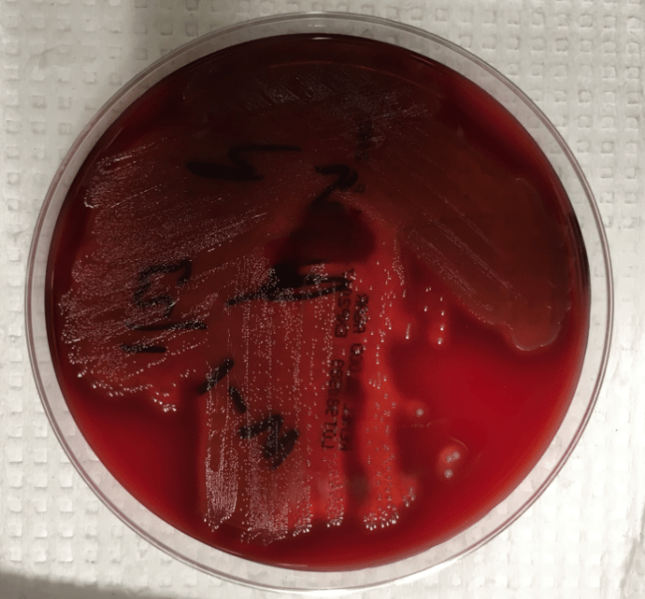
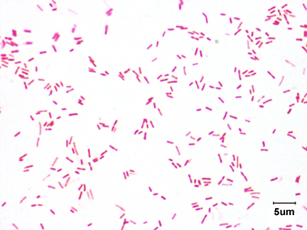
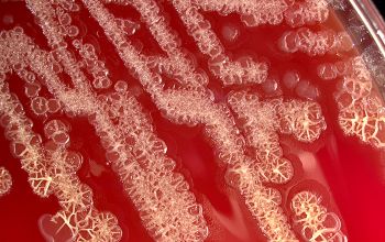
The blood cultures turned positive with Gram-positive cocci, which went on to produce small, gray, alpha-hemolytic colonies on the blood agar plate (Image 1). The colonies were catalase negative and PYR negative. The isolate was analyzed by a Bruker MALDI-TOF mass spectrometer and was identified as Streptococcus lutetiensis (score 2.19). Susceptibility testing revealed the isolate susceptible to ceftriaxone, penicillin, and vancomycin.

streplut

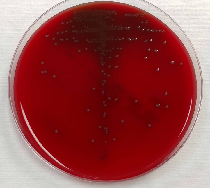
Image 1. 5% sheep blood agar growing small, grey, alpha-hemolytic colonies


Discussion

S. lutetiensis is part of the complex of organisms previously identified as the Streptococcus bovis group. This group of organisms, which possess the Lancefield Group D antigen, has undergone considerable reclassification schemes as phenotypes and genotypes have been investigated. The original biochemical classification schemes were based on their ability to ferment mannitol as well as the presence or absence of beta-glucuronidase activity. Early observations of the DNA properties from these organisms, such as %-GC base content and DNA-DNA hybridizations, identified six unique clusters of Group D streptococci [1]. One cluster group (cluster group #4) had heterogeneous biochemical phenotypes. A subcluster of this cluster group #4 was separated from the other members of the cluster based upon esculin hydrolysis. This subcluster would later go on to be named S. infantarius, so named as several isolates originated from the feces of human infants [2].

Further DNA-DNA hybridizations and ribotyping analysis led to the declaration of two S. infantarius subspecies: subsp. infantarius and subsp. coli [3]. The 16S rRNA ribotyping was problematic, however, as several species in this genus are 97-99% sequence identical.

In an attempt to address some of the limitations of relying on the 16S rRNA gene, one group analyzed the features of the conserved gene encoding the manganese-dependent superoxide dismutase gene (sodA). They observed substantial differences between S. infantarius subsp. infantarius and S. infantarius subsp. coli [4]. Thus, for the latter organism, a new species of streptococci was proposed: S. lutetiensis. It was named for Lutetia, a historical name for the city of Paris [4].

The species designation S. lutetiensis was not widely accepted, however. Based on further DNA-DNA hybridization experiments and the prior studies of the 16S rRNA, others have rejected the species name “S. lutetiensis” and maintain that it is a subspecies of S. infantarius as previously described [5].

So which name is correct? There appears to be no clear consensus about the designation of these streptococci, whether it is S. infantarius subsp. coli or whether it is another species altogether as S. lutetiensis. The Judicial Commission of the International Committee on Systematic Bacteriology reportedly met to discuss the name changes, however no resolution appears to have been determined [6]. Both names are seen in the literature as well as the names for reference organisms.

The important clinical aspect to recognize is that this organism, as well as the S. bovis group in general, can be a cause of bacteremia, endocarditis, and meningitis in children. Treatment with beta-lactam antibiotics is generally sufficient to cover these organisms.

References

  1. Farrow, J., et al., Taxonomic Studies on Streptococcus bovis and Streptococcus equinus: Description of Streptococcus alactolyticus sp. nov. and Streptococcus saccharolyticus sp nov. System. Appl. Microbiol, 1984. 5: p. 467-482.
  2. Bouvet, A., et al., Streptococcus infantarius sp. nov. related to Streptococcus bovis and Streptococcus equinus. Adv Exp Med Biol, 1997. 418: p. 393-5.
  3. Schlegel, L., et al., Streptococcus infantarius sp. nov., Streptococcus infantarius subsp. infantarius subsp. nov. and Streptococcus infantarius subsp. coli subsp. nov., isolated from humans and food. Int J Syst Evol Microbiol, 2000. 50 Pt 4: p. 1425-34.
  4. Poyart, C., G. Quesne, and P. Trieu-Cuot, Taxonomic dissection of the Streptococcus bovis group by analysis of manganese-dependent superoxide dismutase gene (sodA) sequences: reclassification of ‘Streptococcus infantarius subsp. coli’ as Streptococcus lutetiensis sp. nov. and of Streptococcus bovis biotype 11.2 as Streptococcus pasteurianus sp. nov. Int J Syst Evol Microbiol, 2002. 52(Pt 4): p. 1247-55.
  5. Schlegel, L., et al., Reappraisal of the taxonomy of the Streptococcus bovis/Streptococcus equinus complex and related species: description of Streptococcus gallolyticus subsp. gallolyticus subsp. nov., S. gallolyticus subsp. macedonicus subsp. nov. and S. gallolyticus subsp. pasteurianus subsp. nov. Int J Syst Evol Microbiol, 2003. 53(Pt 3): p. 631-45.
  6. Beck, M., R. Frodl, and G. Funke, Comprehensive study of strains previously designated Streptococcus bovis consecutively isolated from human blood cultures and emended description of Streptococcus gallolyticus and Streptococcus infantarius subsp. coli. J Clin Microbiol, 2008. 46(9): p. 2966-72.


IJF

-I.J. Frame MD, PhD, is a 1st year Clinical Pathology Resident at UT Southwestern Medical Center.

Erin McElvania TeKippe, PhD, D(ABMM), is the Director of Clinical Microbiology at Children’s Medical Center in Dallas Texas and an Assistant Professor of Pathology and Pediatrics at University of Texas Southwestern Medical Center.

Microbiology Case Study: A 28 Year Old Woman with Acute Onset Fever Post Delivery

Case History

A 28 year old woman at 37 weeks and 2 days presented in labor to our ED. After 22 hours, she delivered a healthy baby boy and sustained a second degree perineal laceration requiring repair. On hospital day 2, she reported feeling lightheaded, nauseous and “shaky.” She attempted to walk around the unit but became tremulous and unsteady, requiring assistance to get back into bed. Her vital signs were as follows: febrile at 38.8 C, BP 108/54, HR 104 and normal respiration rate at 12 breaths/min. On exam, she appeared pale and lethargic, and was noted to have a tender uterus on palpation. Based on her presentation and status post SVD, the diagnosis of endometritis was established. Blood cultures were obtained and within 16 hours, blood culture bottles were positive for gram-positive cocci. The patient was started on antibiotic therapy with ampicillin, gentamycin and clindamycin, and clinically improved within 36 hours.

streppyo1
Image 1. Blood culture on blood agar.
streppyo2
Image 2. Blood culture on chocolate agar.

Discussion

Streptococcus pyogenes is one of the most aggressive pathogens encountered in clinical microbiology. It is a beta hemolytic streptococcus and is notoriously associated with Streptococcal Toxic Shock Syndrome (STSS), necrotizing fasciitis, as well as more benign (yet still problematic) conditions, like Scarlet Fever, Impetigo, Rheumatic heart disease and Acute Post-streptococcal Glomerulonephritis. A gram-positive cocci, it possesses several virulence factors, including protein F, M protein (involved in antigen mimicry leading to valvular heart disease) hemolysins and exotoxins. These factors allow S. pyogenes to attach to and invade epithelial tissue, and in the case of hyalurondiase, potentially use hyaluron as a carbon food source. S. pyogenes agglutinates with Lancefield group A antisera and is pyrrolidonyl arylamidase (PYR) positive and VP, hippurate and CAMP test negative. Penicillin (PCN) remains the drug of choice in treating most S. pyogenes infections. Alternative antibiotic therapy includes macrolides and certain cephalosporins (e.g. cefixime, cefpodoxime). Vancomycin should be used in more severe infections such as sepsis or for patients with a PCN allergy.


-Christina Litsakos is a Pathology Student Fellow at University of Vermont Medical Center.

Wojewoda-small

-Christi Wojewoda, MD, is the Director of Clinical Microbiology at the University of Vermont Medical Center and an Assistant Professor at the University of Vermont.


Microbiology Case Study: A 47 Year Old Woman with Three Day History of Fever

Case History

A 47 year old Caucasian female presented to her primary care physician with a three day history of fever (Tmax 102°F), chills and generalized body aches. Her rapid influenza test was negative, but she was treated with oseltamivir for suspected viral infection. Her past medical history was significant for severe mitral regurgitation for which she had had a prosthetic valve replacement two years prior, ischemic cardiomyopathy with recent pacemaker placement one month prior and an undifferentiated connective tissue disease. Her current medications included hydroxychloroquine (Plaquenil) and warfarin. Her symptoms persisted and upon return to clinic, a urinalysis was performed and blood cultures were collected. On physical exam, the pacemaker site was erythematous and tender to palpation. She was started on doxycycline and fluconazole for a presumed urinary tract infection. After 4 days of incubation on the automated instrument, the two aerobic blood cultures bottles were positive and the patient was admitted to the hospital for further workup and therapy.

Laboratory Identification

Microscopic examination from the positive blood culture bottle revealed slender, beaded Gram positive bacilli (Figure 1). No definitive branching was identified. Given the morphology on Gram stain, a Kinyoun stain was performed and revealed red-purple, beaded acid fast bacilli which were consistent with a Mycobacterium spp. (Figure 2). A Mycobacterial Growth Indicator Tube (MGIT), a Lowenstein Jensen slate and blood & chocolate agars were inoculated with specimen. Given that the organism grew after 2 days, a rapidly growing Mycobacterium spp. was suspected (Figure 3). High performance liquid chromatography (HPLC) identified the organism as M. fortuitum.

mycofort1

Figure 1. Gram stain from the positive blood culture bottle showed slender, beaded Gram positive bacilli that were arranged in clumps (100x oil immersion).

mycofort2

Figure 2. Kinyoun stain of the organisms was consistent with acid fast bacilli (100x oil immersion).

mycofort3

Figure 3. Small, off-white colonies grew on chocolate agar after 3 days of incubation at 35°C in a CO2 incubator.

Discussion

M. fortuitum is a common rapid growing mycobacterial species that is ubiquitous in the environment and tap water. Most common infections due to M. fortuitum include post-traumatic or post-surgical wound infections and it can be associated with the insertion of prosthetic devices including heart valves, artificial joints and rods inserted after fractures. Of the rapid grower group (Runyon Group IV), which includes M. chelonae, M. abscessus and M. mucogenicum, it is M. fortuitum that accounts for approximately 60% of localized cutaneous skin infections and prosthetic device infections most frequently.

In the laboratory, M. fortuitum typically grows after two to five days incubation and appear as small, off-white colonies on a variety of different agars. The organism is typically slender, beaded Gram positive bacilli on Gram stain and positive for acid fast bacilli on a Ziehl-Neelsen or Kinyoun stain. As part of a traditional lab work up, M. fortuitum is arylsulfatase positive and is capable of reducing nitrates. Today a variety of methods, including HPLC, pyrosequencing, sequence analysis and matrix assisted laser desorption ionization time of flight mass spectrometry (MALDI-TOF MS), have become routine identification options. Susceptibility testing of isolates from clinically significant sites should be performed by broth microdilution and includes the following antimicrobials: amikacin, cefoxitin, ciprofloxacin, moxifloxacin, clarithromycin, doxycycline, linezolid, imipenem, meropenem, minocycline, trimethoprim-sulfamethoxazole and tobramycin.

In the case of our patient, it was discovered her pacemaker site was infected and upon further questioning it was discovered she wasn’t able to complete her antibiotic course after device placement due to nausea. A transesophageal echocardiogram showed no evidence of infective endocarditis and she was taken to the operating room for removal of the pacemaker and leads. The site was filled with pus and wound cultures obtained during surgery were consistent with M. fortuitum as well. Repeat blood cultures were negative and she was treated with intravenous amikacin and imipenem as well as oral levofloxacin for an anticipated 6-8 weeks before transitioning to oral therapy.


-Debbie Rigney Walley, MD, is a 1st year Anatomic and Clinical Pathology Resident at the University of Mississippi Medical Center.

Stempak

-Lisa Stempak, MD, is an Assistant Professor of Pathology at the University of Mississippi Medical Center in Jackson, MS. She is certified by the American Board of Pathology in Anatomic and Clinical Pathology as well as Medical Microbiology. She is the director of the Microbiology and Serology Laboratories.  Her interests include infectious disease histology, process and quality improvement and resident education. 

Microbiology Case Study: A 73 Year Old Man with Back Pain

Case History

A 73 year old man with a history of multiple back surgeries presented with bilateral lower extremity back pain of over greater than one month duration. Prior surgeries included L4/L5 fusion with pedicel screws and a decompression laminectomy one year prior to presentation. Imaging of his spine showed a fluid collection in his lumbar spine and he underwent several tissue biopsies over the course of a month which consistently showed no growth. Despite negative cultures he was treated with doxycycline and levoquin for 30 days. He was transferred to University of Vermont Medical Center (UVMMC) for IR drainage and tissue biopsy of this lumbar abscess as he continued to complain of back pain and had begun to develop bilateral lower extremity weakness. Cultures grow the organism below and close inspection revealed the presence of small feet. The organism was confirmed to be Candida albicans.

canalb1

canalb2

Discussion

Vertebral osteomyelitis due to Candida is rare, however, a review of the literature reveals that most patients have lower thoracic or lumbar spine involvement and over 80% present with >1 month of lower back pain. An elevated white blood cell count is not as sensitive as an elevated erythrocyte sedimentation rate and of all patients, less than a quarter have neurologic signs. Candida albicans was responsible for almost 2/3 of cases and the remaining cases were caused by Candia tropicalis or Candida glabrata.1 Risk factors include IV drug abuse for patients under 25 years old; for elderly patients a central venous catheter, antibiotic use and immunosuppression .1


Reference

Miller, D and Mejicano, George. Vertebral Osteomyelitis due to Candida species: Case report and review of the literature. Clinical Infectious Diseases, 2001;33:523-530.

-Agnes Balla, MD is a 3rd year anatomic and clinical pathology resident at the University of Vermont Medical Center.

Wojewoda-small

-Christi Wojewoda, MD, is the Director of Clinical Microbiology at the University of Vermont Medical Center and an Assistant Professor at the University of Vermont.


Microbiology Case Study: A 14 Year Old Cystic Fibrosis Patient

Case History

14 year old cystic fibrosis (CF) patient was admitted to the hospital for a CF exacerbation with a known rhinovirus infection. The patient reported congestion and cough with production of greenish sputum. The following was isolated from a sputum culture.

Laboratory Culture

pseaer1


Image A. MacConkey Agar plate with mucoid Pseudomonas aeruginosa colonies.

pseaer2

Image B. Kirby-Bauer Method of antimicrobial susceptibility.

Discussion

Cystic fibrosis patients have complex polymicrobial respiratory flora. Routine cultures may reveal different bacterial species that can contribute to the difficulty in treating and preventing infections in these patients. Staphylococcus aureus, Haemophilus influenza and Pseudomonas aeruginosa are main contributors to the infections found in CF patients.

In this case, we isolated a mucoid Pseudomonas aeruginosa (Image A). Pseudomonas aeruginosa is an oxidase-positive, gram negative rod-shaped bacterium that is commonly found in the environment. It is considered a severe and frequent pathogen in patients with cystic fibrosis. In chronic infections, it is thought that P. aeruginosa can undergo a “mucoid switch” where the bacterium can acquire mutations that lead to the mucoid phenotype. The phenotype is so impressive that excess polysaccharide will often drip onto the lid of the plate when stored upside down during incubation.

One of the key features of mucoid strains of P. aeruginosa is their ability to form biofilms. Biofilms consist of a matrix of polysaccharide, protein and DNA. This provides not only a protective barrier from antibiotics and the immune system, but also may contribute to the growth of other bacteria within the microenvironment.

With all the excess polysaccharide, it can be difficult to standardize the inoculum of mucoid isolates of P. aeruginosa which is an essential starting point for the microbroth dilution method of antimicrobial susceptibility testing. For this reason, susceptibility testing of mucoid isolates is often performed by Kirby-Bauer (KB) disk diffusion method. The KB method is a test of antimicrobial susceptibility that is based on the zone of inhibition surrounding disks that contain antimicrobial drugs (Image B). The strain of mucoid P. aeruginosa isolated in this case was found to be susceptible to aztreonam, ceftazidime, piperacillin/tazobactam and resistant to amikacin, cefepime, ciprofloxacin, gentamicin, levofloxacin, meropenem and tobramycin, by the Kirby Bauer method.

Case Follow Up

The patient was ultimately treated with IV piperacillin/tazobactam, as well as with chest physical therapy and hypertonic saline inhalation. They clinically improved and were ultimately discharged home after a 2 week hospital stay.

References

The mucoid switch in Pseudomonas aeruginosa represses quorum sensing systems and leads to complex changes to stationary phase virulence factor regulation. Ben Ryall, Marta Carrara, James EA Zlosnik, Volker Behrends, Xiaoyun Lee, Zhen Wong, Kathryn E. Lougheed, Huw D. Williams. PLOS ONE, May 2014, Vol. 9, Iss. 5, Pages 1-11.

Pseudomonas aeruginosa biofilms in cystic fibrosis. Niels Høiby, Oana Ciofu, and Thomas Bjarnsholt. Future Microbiology, November 2010, Vol. 5, No. 11, Pages 1663-1674.

Insights into Cystic Fibrosis Polymicrobial Consortia: The Role of Species Interactions in Biofilm Development, Phenotype, and Response to In-Use Antibiotics. Magalhaes AP, Lopes SP, Pereira MO. Frontiers in Microbiology, January 13, 2017, Vol. 7, Article 2146.

Koneman’s Color Atlas and Textbook of Diagnostic Microbiology. Gary W. Procop, Deirdre L. Church, Geraldine S. Hall, William M. Janda, Elmer W. Koneman, Paul C. Schreckenberger; Gail L. Woods. Seventh Edition. 2017. Pages 343, 1110.


mw

-Megan B. Wachsmann, MD, MSCS, is a 4th year Anatomic and Clinical Pathology Resident and Chief Resident at UT Southwestern Medical Center.

Erin McElvania TeKippe, PhD, D(ABMM), is the Director of Clinical Microbiology at Children’s Medical Center in Dallas Texas and an Assistant Professor of Pathology and Pediatrics at University of Texas Southwestern Medical Center.

Microbiology Case Study: A Bartholin Gland Cyst Culture

Case History

A swab from a bartholin gland cyst was submitted to the microbiology lab with no other clinical history. Culture produced cream-colored, mucoid colonies on blood and chocolate agar after 48 hours of incubation at 35 C (image below). The culture grew a gram negative rod that did not grow on MacConkey agar, a distinguishing characteristic for Weeksella virosa. This ID was confirmed by MALDI-TOF.

weeks1

Discussion

Originally described in 1970 as a nonsaccharolytic flavobacterium, Weeksella virosa is an uncommon aerobic gram negative rod isolated most commonly from urine, cervix and vaginal specimens. There have been case reports, however, of isolation from blood and spinal fluid. In rare instances, the bacterium has been associated with pneumonia, bacteremia, peritonitis and UTIs, more often in patients with comorbidities including end stage renal disease, diabetes, and liver disease.

In one study of 100 patients, the organism was isolated in 2% of high vaginal swabs of the female genital tract from both symptomatic and asymptomatic females. In a separate study involving women with a high risk of STDs, the incidence climbed to 15%.

The characteristic yellow tinge of the colonies is secondary to a non-diffusable pigment.  The organism is oxidase positive, indole positive and catalase positive.

There are no species-specific breakpoints. However, CLSI guidelines for other non-enterobacteriacea gram negative rods can be used.  In vitro studies have found the organism to be resistant to aminoglycosides and nitrofurantoin.

Reference

Slenker, A et al. Fatal Case of Weeksella virosa Sepsis. Journal of Clinical Microbiology. P4166-4167. Dec. 2012.

-Agnes Balla, MD, is a 3rd year anatomic and clinical pathology resident at the University of Vermont Medical Center.

Wojewoda-small

-Christi Wojewoda, MD, is the Director of Clinical Microbiology at the University of Vermont Medical Center and an Assistant Professor at the University of Vermont.


Microbiology Case Study: A 10 Year Old Girl with New Onset Fever

A 10 year old girl presented to the ED with several month history of worsening CNS symptoms, including gait instability, left-sided weakness, nausea, vomiting, several witnessed seizure events and loss of consciousness. MRI revealed right frontal mass measuring 7.0 cm in greatest dimension with midline shift. The patient underwent a right-sided craniotomy with tumor resection and was closely followed in the PICU. On post-op day 3, the patient appeared pale and lethargic with a temperature of 39.9 C. Blood cultures were collected and the patient empirically started on ceftriaxone and vancomycin for meningitis coverage. Due to worsening fever and altered mental status, a lumbar puncture was performed with the following results:

  • Opening pressure: 10.5 cm
  • Appearance: slightly cloudy
  • Cells: 91% PMN
  • Gram negative rods were identified on gram smear


OLYMPUS DIGITAL CAMERA
Gram stain shows gram negative rods. Photo credit: Dr. Gary Kaiser.

pseudostut2

Photo credit: University Medical Center of Rotterdam, Dept. of Microbiology and Infectious Disease

Discussion

Pseudomonas stutzeri is an aerobic gram negative bacilli (GNB) with a single polar flagellum that is both catalase and oxidase positive. Several defining features help distinguish P. stutzeri from other Pseudomonas species, including its nutritional versatility (utilizes starch, maltose, and ethylene glycol), its inability to produce fluorescent pigment and its “coral-like” appearance on growth medium.  There are no known virulence factors for P. stutzeri. Hospital distribution rates of P. stutzeri are approximately 1-2% of Pseudomonas spp. Nosocomial infection via medical devices or solutions is the most common route of infection. P. stutzeri isolates are sensitive to beta lactam antibiotics—meropenem is commonly used.


-Christina Litsakos is a Pathology Student Fellow at University of Vermont Medical Center.

Wojewoda-small

-Christi Wojewoda, MD, is the Director of Clinical Microbiology at the University of Vermont Medical Center and an Assistant Professor at the University of Vermont.

Microbiology Case Study: A 25 Year Old Male with Fevers, Headache, and Neck Pain

Case History

A 25 year old Caucasian male with no significant past medical history presented to the emergency department (ED) with a several day history of persistent fevers, headache (pain 10/10), dizziness and neck pain. He also reported facial and hand numbness and difficulty focusing at work.  His laboratory values at that time showed a normal white blood cell count (5.3 TH/cm2), normal hemoglobin (13.8 g/dL), slightly decreased platelet count (142,000 TH/cm2) and slightly elevated liver enzymes. A computed tomography (CT) of his head showed no abnormalities, and a lumbar puncture was performed that was suggestive of viral meningitis (92% lymphocytes). After obtaining blood cultures, the patient received a dose of vancomycin and ceftriaxone and was discharged home.

Two days later he returned to the ED with complaints of worsening neck pain, photophobia, decreased appetite, and fevers reaching 105°F. He reported fevers of such intensity that he resorted to soaking himself in ice baths. On further questioning, he reported working in a microbiology lab that handles cytomegalovirus (CMV) and attenuated mycobacterium, but was unaware of any exposures or sick contacts. He has 2 dogs he rescued (one with a history of heartworm), 3 cats (one with a history of tapeworms) and a mouse. Infectious disease was consulted and a thorough workup was initiated, which included repeat blood cultures and testing for hepatitis, human immunodeficiency virus (HIV), respiratory pathogens and syphilis. He was started on cefepime initially and then later changed to meropenem and levofloxacin.

Laboratory identification

brucan1

Figure 1. Gram stain from a positive blood culture illustrating small Gram negative coccobacilli (100x, oil immersion).

The microbiology laboratory reported the blood cultures collected during his second hospital visit were positive with small gram negative coccobacilli after approximately 60 hours on the automated instrument (Figure 1). No growth was noted after 24 hours incubation at 35°C in 5% CO2 on standard media. After 48 hours, small white colonies grew on sheep blood and chocolate agars but failed to grow on MacConkey agar. Biochemical tests revealed the organism was positive for catalase, oxidase and urease. In accordance with the suspected agents of bioterrorism manual, the culture was sent to the State Department of Health for further classification. The organism was identified by PCR as Brucella spp.  Subsequently, the Centers for Disease Control and Prevention (CDC) performed species level PCR and identified the isolate as B. canis.

On further questioning, the patient denied consuming unpasteurized milk products but reported recently adopting a pregnant dog from a local shelter, who had subsequently delivered stillborn puppies of which the patient had been in close proximity. At this point, the patient’s antibiotics were switched to a 6 week course of oral doxycycline and rifampin. On follow up visits, he was doing well and symptom free. Unfortunately, the dog also tested positive for B. canis and had to be euthanized.

Discussion

Brucella spp. are common zoonoses among wildlife and domestic animals including cattle (B. abortus), pigs (B. suis), goats (B. melitensis) and dogs (B. canis) who are usually asymptomatic carriers. While rare in the United States due to vaccination of livestock, Brucella spp. is considered endemic in areas of the Middle East, Central and South America and the Indian subcontinent. Symptoms of infection generally occur during an infectious abortion in which the placenta, fetal tissues and secretions contain high levels of the bacteria which can survive in the environment under various conditions for long periods of time. Humans are usually infected due to consumption of unpasteurized milk and cheeses. High risk professions such as veterinarians and slaughterhouse workers can also be infected by direct contract with contaminated materials or inhalation of aerosolized particles. Symptoms generally appear 1 to 2 weeks after infection with remittent/undulant fever the characteristic feature of the illness, in addition to arthralgias, fatigue, weight loss and hepatomegaly.

Laboratory identification of Brucella spp. is the gold standard but can be challenging as it is a slow growing organism and can infect personnel leading to laboratory acquired infections (LAI). When small Gram negative coccobacilli are identified that fail to grow on MacConkey agar, this should alert the laboratory worker of a potential agent of bioterrorism and work up should be performed in appropriate biosafety cabinets. Brucella spp. grows as small, smooth white colonies that appear after 24 to 48 hours incubation. It is catalase, oxidase and urease positive. Automated systems and MALDI-TOF mass spectrometry are not terribly reliable or recommended for identification of this organism due potential aerosolization events. When Brucella spp. is suspected, the level A clinical laboratory (a sentinel lab) should notify and send samples to a Level B/C lab (state health department) for confirmation.  Subsequently, confirmed isolates can be forwarded to a level D lab (CDC) for speciation.

While overall, the mortality for Brucella spp. is very low, significant morbidity can result with long term non-specific symptoms and cardiac and osteoarticular complications. Good outcomes result when acute presentations are treated with combined regimens of antibiotics. The World Health Organization (WHO) recommends the use of oral doxycycline and rifampin for 6 to 8 weeks. Susceptibility testing is not recommended as resistance is rare and the concern for laboratory safety. In the case of laboratory exposures, prophylaxis with doxycycline and rifampin for 3 weeks is recommended for high risk workers. In the case of low risk employees, temperature monitoring for 6 months and serologic testing at defined time points is standard as the incubation period for Brucella can be this long.


mb

-Melissa Brents, MD, is a 4rd year Anatomic and Clinical Pathology resident at the University of Mississippi Medical Center.

Stempak

-Lisa Stempak, MD, is an Assistant Professor of Pathology at the University of Mississippi Medical Center in Jackson, MS. She is certified by the American Board of Pathology in Anatomic and Clinical Pathology as well as Medical Microbiology. She is the director of the Microbiology and Serology Laboratories.  Her interests include infectious disease histology, process and quality improvement and resident education. 


Microbiology Case Study: A 71 Year Old Man with a dehised Corneal Wound

Case History

A 71 year old man presented with a dehisced corneal wound status post corneal transplant.

Laboratory Diagnosis

Corneal scrapings from the ulcer were submitted for interpretation and two separate organisms were isolated from the blood agar plates. The first was a non-motile gram negative rod that grew on 5% horse blood agar and MaConkeys agar. The organism was oxidase negative and spot indole negative and was identified as Klebsiella pneumoniae.

The second organism grew in bright yellow-pigmented colonies on 5% horse blood agar (Image 1) but did not grow on MacConkeys. The organism was oxidase positive and spot indole positive and was identified as Chryseobacterium indologenes by MALDI.

chryseo1
Image 1.

Discussion

Chryseobacterium indologenes, previously known as Flavobacterium indologenes is a yellow pigmented, gram-negative filamentous, non-motile rod that is a non-glucose fermenter and can be found in soil, plants, foodstuffs and water sources including those found in hospitals. It produces a water-insoluble pigment, flexirubin which gives it its characteristic color. It was first isolated from a clinical specimen in 1983 however there have been more recent reports of bacteremia related to C. indologenes related to use of indwelling devices, such as a catheters.

  1. indologenes typically exhibits resistance to multiple antibiotics, however, a case series of 16 patients with C. indologenes infections, all nosocomial and in patients with comorbidities, showed no clear relationship between antibiotic susceptibility and response to treatment (1).


Reference:

  1. Lin Y-T, Jeng Y-Y, Lin M-L, Yu K-W, Wang F-D, Liu C-L. 2010. Clinical and Microbiological Characteristics of Chryseobacterium indologenes Bacteremia. J. Microbiol. Immunol. Infection.43:498-505.


-Agnes Balla, MD is a 3rd year anatomic and clinical pathology resident at the University of Vermont Medical Center.

Wojewoda-small

-Christi Wojewoda, MD, is the Director of Clinical Microbiology at the University of Vermont Medical Center and an Assistant Professor at the University of Vermont.


Microbiology Case Study: A 14 Year Old Boy with Cystic Fibrosis

Case History

A 14 year-old-boy with Cystic Fibrosis had a respiratory culture collected at his routine clinic visit. It grew abundant mixed respiratory flora, and rare Gram-negative coccobacilli. This organism grew as non-lactose fermenting colonies on MacConkey agar (Figure 1) in approximately 36 hours and was oxidase and catalase positive. The isolate was identified by MALDI-TOF MS with a score of 2.39, which is acceptable for species-level identification.

bord1

Discussion

Our isolate was identified as Bordetella bronchiseptica. Bordetella spp. are small Gram-negative rods that often appear as coccobacilli. Like other Bordetella spp., our isolate was catalase positive. Oxidase results vary across the genus, but B. bronchiseptica is oxidase positive. Some Bordetella spp. including B. pertussis and B. parapertussis, are very sensitive to metabolites and toxic substances found in many types of microbiological media. For the best chance of recovering these fastidious Bordetella spp. in culture, specialized agar such as Regan-Lowe, Bordet-Gengou, or Stainer-Scholte medium along with extended incubation periods are used. Due to the difficulty of culturing B. pertussis and B. parapertussis, these days culture is only performed at large reference laboratories or public health facilities. Testing by PCR is the current clinical practice and has greatly improved the sensitivity of detection for these fastidious organisms. In contrast, other Bordetella spp. including B. bronchiseptica are routinely recovered in culture using standard laboratory methods.

The most clinically relevant Bordetella spp. in humans are B. pertussis, the infectious agent of whooping cough, and B. parapertussis, which causes a similar illness to B. pertussis but generally symptoms are less severe. B. bronchiseptica causes respiratory infections in many animals including cats and dogs, and is the infectious agent of kennel cough.  B. bronchiseptica can cause respiratory infection in humans who acquire the bacterium primarily from their infected pets. Human infection is rare, and most likely to occur in immunocompromised patients such as those with poorly controlled HIV or Cystic Fibrosis.

B. bronchiseptica produces a β-lactamase making the organism resistant to penicillin and many cephalosporins. Most strains are resistant to trimethoprim-sulfamethoxazole as well. In contrast, strains of B. bronchiseptica are generally susceptible to β-lactam/ β-lactamase inhibitor combinations, quinolones, aminoglycosides, and tetracycline.

Our patient was not having an exacerbation at the time of specimen collection, so he continues to do well. We expect to find B. bronchiseptica in his future sputum specimens, but the pathogenicity of B. bronchiseptica in such a low amount compared to respiratory flora is unclear.

Erin McElvania TeKippe, PhD, D(ABMM), is the Director of Clinical Microbiology at Children’s Medical Center in Dallas Texas and an Assistant Professor of Pathology and Pediatrics at University of Texas Southwestern Medical Center.