Microbiology Case: Lung Nodules in a 71-year old Male Undergoing Lung Transplant Evaluation

Microbiology Case: Lung nodules in a patient undergoing lung transplant evaluation

Case History 

A 71-year-old male with a past medical history of severe interstitial lung disease (ILD) presented for lung transplant evaluation. His chest CT demonstrated findings consistent with his known diagnosis of ILD, along with a 3.6 cm focal nodular calcific density near the right costophrenic angle, and additional scattered calcified lung nodules measuring up to 3 mm. Sputum cultures were obtained to evaluate the CT findings.

Laboratory Identification

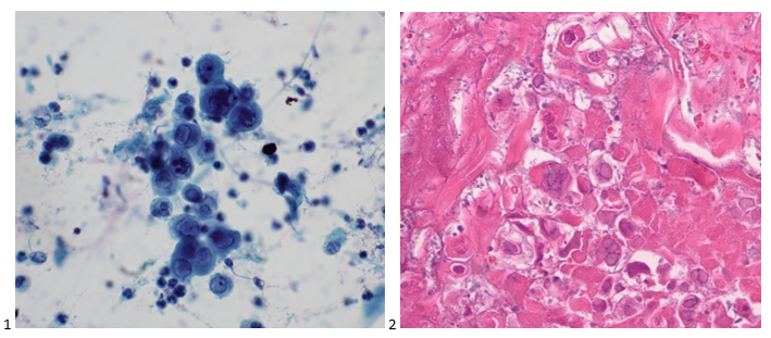
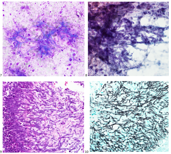
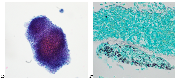
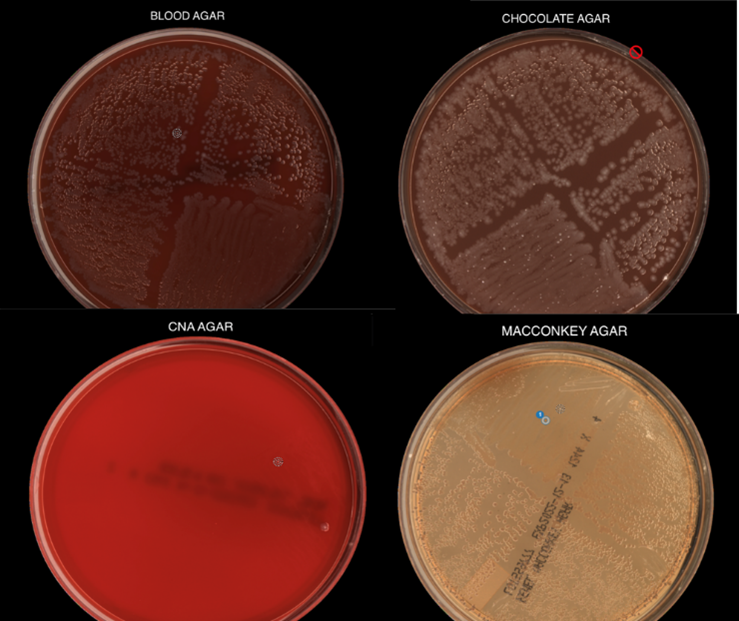
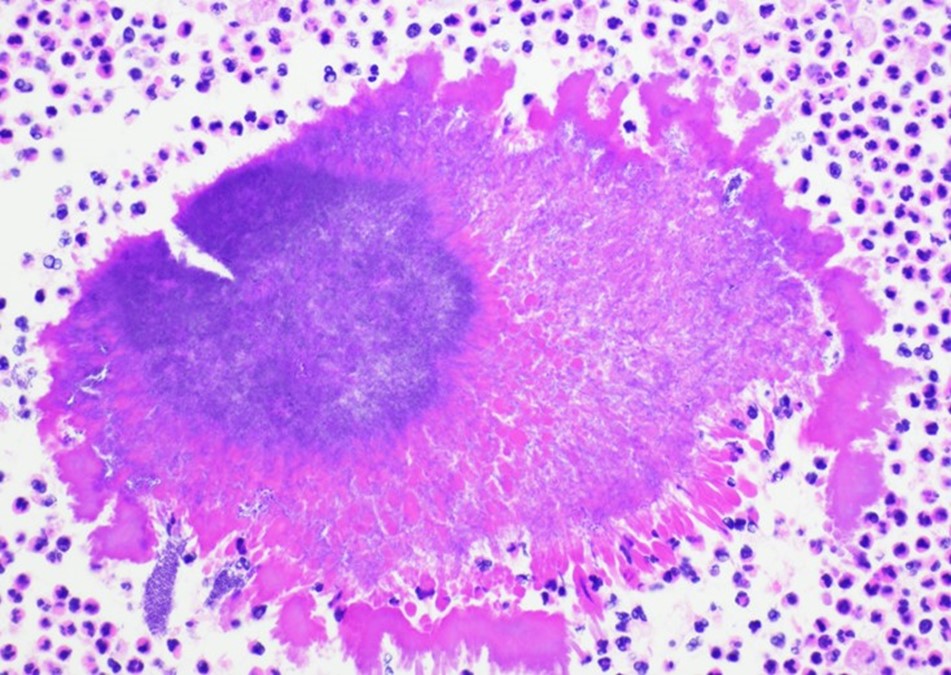
Routine bacterial, fungal, and AFB sputum testing was performed.  No AFB were recovered, and a mixture of bacteria consistent with oral flora were found on blood, chocolate and MacConkey agars.  The fungal culture grew a moderate amount of round, non-hyphenated yeast which were encapsulated, urease-positive and reacted with caffeic acid (Image 1A and 1B) consistent with Cryptococcus species.  The organism was definitively identified as Cryptococcus gattii by MALDI-TOF mass spectrometry, which was subsequently confirmed by growth and the appearance of a deep blue color on Canavanine Glycine Bromothymol Blue (CGB) agar (Image 1C).  The patient was prescribed daily fluconazole to take daily for 6 months, and underwent a successful bilateral transplant two months after treatment initiation.

Image 1.  A) Urease testing of the patient’s yeast (left, positive) versus Candida albicans (right, negative) consistent with Cryptococcus sp.  B) Caffeic acid testing of the patient’s yeast (left, positive) versus C. albicans (right, negative) consistent with Cryptococcus sp.  C) Growth of the patient’s isolate (left) versus Cryptococcus neoformans (right) on Canavanine Glycine Bromothymol Blue (CBG) agar.  The deep blue color produced by the patient’s isolate is indicative of Cryptococcus gattii.

Discussion

Cryptococci are important pathogens of both immunocompetent and immunocompromised hosts.  While Cryptococcus neoformans causes most human cryptococcal disease, Cryptococcus gattii is also a clinically important cause of infections.  Though genetically similar, these two species exhibit important differences with respect to ecological niche, pathogenesis, and epidemiology.  Historically, trees (specifically eucalyptis) have been identified as environmental reservoirs of C. gattii, while C. neoformans is associated with pigeon guano.1  Cryptococcus neoformans has a worldwide distrubtion, while C. gattii is thought to be geographically restricted to Australia, Western Canada and the Pacific Northwest of the United States, although this has become less clear in recent years.  Indeed, the patient in this case had not traveled to any of these locales; instead, he is from the southeastern United States, where the organism may be endemic.3

                There is an established association between C. gattii and infections in immunocompetent hosts, and recent evidence points to genetic and immunological factors which predispose some individuals to C. gattii infection.  Possible differences in clinical course and patient outcomes have been suggested to justify differentiation between C. gattii and C. neoformans in laboratory settings.  Importantly, routine laboratory methods such as commercial biochemical identification systems, morphological observations, and cryptococcal antigen testing lack the discriminatory power to distinguish these species.2  By contrast, MALDI-TOF MS and molecular methods, including some molecular panels performed on positive blood culture bottles, are able to discriminate between C. gattii and C. neoformans.  The use of glycine as a sole carbon and nitrogen source in the presence of canavanine allows biochemical differentiaton through the use of CGB agar.  The degredtation of L-canavanine to ammonia in the presence of glycine by C. gattii raises the pH and results in associated color change, while C. neoformans remains a yellow or green color (Image 1C).  While robust, this incubation can take up  to five days resulting in prolonged turn around times.2

Cryptococal infection begins through inhalation of the organism into the lungs, where the yeast then can dissemisate to other anatomical sites.  When compared to C. neoformans, C. gattii is less likely to present with central nervous system (CNS) involvement but exhibts a higher incidence of imaging abnormalities and mass lesions.  In pulmonary settings, C. neoformans infections commonly present with infiltrates while C. gattii infections are nodular2 as seen in this case.  Irrespective of the species involved, cryptococcal infections can be asymptomatic or classically present as a meningoencephalitis, chronic or acute pneumonia, or in a number of cutaneous manifestations.  Fluconazole is the drug of choice for management of asymptomatic or mild to moderate pulmonary infections. Despite the removal of the infected lungs, it was determined that the patient should complete the entire six month course of fluconazole due to his immunosuppression post-transplant.

References

  1. Beardsley, J., Dao, A., Keighley, C., Garnham, K., Halliday, C., Chen, S. C.-A., and Sorrell, T.C.  What’s New in Cryptococcus gattii: From Bench to Bedside and Beyond.  J. Fungi. 2023; 9, 41:1-16.
  2. Butler-Wu, S. and Limaye, A.P.  Guideline: A Quick Guide to the Significance and Laboratory Identification of Cryptococcus gattii.  American Society for Microbiology. 2011.  https://asm.org/Guideline/A-Quick-Guide-to-the-Significance-and-Laboratory-I  Accessed: February 1st, 2023.
  3. Lockhart, S., Roe, C. C., and Engelthaler, D.M.  Whole-Genome Analysis of Cryptococcus gattii, Southeastern United States.  Emerg. Infect. Dis. 2016 Jun; 22(6): 1098-1101.

-Andrew Clark, PhD, D(ABMM) is an Assistant Professor at UT Southwestern Medical Center in the Department of Pathology, and Associate Director of the Clements University Hospital microbiology laboratory. He completed a CPEP-accredited postdoctoral fellowship in Medical and Public Health Microbiology at National Institutes of Health, and is interested in antimicrobial susceptibility and anaerobe pathophysiology.

-Francesca Lee, MD, is an associate professor in the Departments of Pathology and Internal Medicine (Infectious Diseases) at UT Southwestern Medical Center


-Clare McCormick-Baw, MD, PhD is an Assistant Professor of Clinical Microbiology at UT Southwestern in Dallas, Texas. She has a passion for teaching about laboratory medicine in general and the best uses of the microbiology lab in particular.

A Series of Infectious Events

Working in a cancer center, our cytologists are well-versed in cancer morphology being able to diagnosis primary malignancies, distant metastases, and even combined metastatic disease in the same lymph node. What we don’t see as often as community hospitals are infectious diseases. However, we do have many immunocompromised patients at our institution, so the rare opportunistic infection does occur. And boy, do we get excited to pass the case around! Please find a series of infectious events embedded within this post. And unfortunately, we do not live in an area where coccidiomycosis is endemic, so beyond school, we haven’t had the pleasure of identifying those in our daily work.

Case 1. Lung, Bilateral, BAL (Bronchoalveolar Lavage)

A 73-year-old male patient was admitted to the ICU with pneumonia. The pulmonologist performed a bilateral bronchoalveolar lavage (BAL) to rule out pneumocystis pneumonia. We prepared a pap-stained smear, two cytospins, a SurePath liquid based prep, and a cell block. Two additional cytospins were sent to histology for GMS staining. While no malignant cells were identified, fragments of squamous epithelium with acute inflammation and necrosis were present. Multiple viral inclusions were identified, appearing as ground glass within the nuclei. (Image 1).  These cells present with classic 3 M features: molding, multinucleation, & margination of chromatin. The cell block also highlights viral inclusions, but demonstrates pseudohyphae and spores associated with surrounding squamous cells as well (Image 2).

Images 1-2. Lung, Bilateral, BAL. 1: SurePath LBP; 2: H&E Cell Block section (400X).

Diagnosis: Herpes Simplex Virus (HSV) and Candida.


Case 2. Lung, Left Lower Lobe, CT-guided FNA

A 72-year old male with stage IIA squamous cell carcinoma underwent a VATS right upper lobectomy and mediastinal lymph node dissection. He completed adjuvant carboplatin/gemcitabine therapy. On a surveillance CT scan, the treated area demonstrated progression as well as multiple bilateral lung nodules. To determine whether the new left lower lobe superior segment lung nodule was a metastasis or new primary, a CT-guided biopsy was performed. The smears and cell block sections were negative for malignancy but demonstrated inflammatory cells and necrotic debris, consistent with a necrotizing inflammatory process (Images 3-5). A separate pass was sent for microbiological cultures to correlate our findings. The following day, Kinyoun and GMS stains were performed on paraffin-embedded sections of the cell block. No fungal organisms were identified on GMS, but acid-fast bacilli were noted by the cytologist on the Kinyoun-stained section (Image 6).

Images 3-6. Lung, Left Lower Lobe, CT-guided FNA. 3: DQ-stained smear; 4: Pap-stained smear; 5: H&E section (100x); 6: Kinyoun stain (600x).

Diagnosis: Acid-fast bacilli (AFB), consistent with Mycobacterium Avium Complex. Isolated and confirmed by microbiology.


Case 3. Lung, Right Upper Lobe, CT-guided FNA

A 58-year-old male presented with multiple lung nodules and a brain mass. We reviewed the brain mass excision from an outside institution and agreed with the original diagnosis of anaplastic oligodendroglioma, WHO grade III with a Ki-67 proliferation index that approached 20%. EGFR was not amplified (ratio 1.2), but 1p/q19 co-deletions were noted in greater than 75% of tumor cells. To rule out primary versus metastatic disease, the patient had a CT scan-guided biopsy of right upper lobe lung mass. No malignant cells were identified in the sample; however, necrotic debris and abundant fungal hyphae were noted (Images 7-9). A portion of the sample was sent to Microbiology for culture. The following day, a GMS and PAS stains were performed on paraffin-embedded sections of the cell block which demonstrated the same fungal hyphae seen in the smears and cell block preparations (Images 10 & 11).

Images 7-11. Lung, Right Upper Lobe, CT-guided FNA. 7: DQ-stained smear; 8: Pap-stained smear; 9: H&E section (400x); 10: GMS stain (400x); 11: PAS stain (400x).

Diagnosis: Abundant fungal hyphae, consistent with Aspergillus


Case 4. Left Hilum, EBUS-FNA

A 20-year-old female patient presented with patches, pain, and inflammation on her legs, and she was diagnosed with erythema nodosum. When her swelling and pain worsened, a chest X-ray demonstrated a left hilar mass, and a subsequent CT demonstrated the mass to be encircling the left superior pulmonary artery and obstructing the pulmonary vein along with multiple peribronchial ground-glass opacities and hilar lymphadenopathy. The concern from the referring physician was thymoma versus lymphoma given her age and clinical presentation. The patient underwent an endobronchial ultrasound to assess the hilar mass and lymphadenopathy. The lymph node aspirates appeared benign, with flow cytometry supporting the cytologic diagnosis. On the left hilum FNA, there were aggregates of lymphocytes, plasma cells, and epithelioid histiocytes with caseating necrosis and fibrosis (Image 12-14). Kinyoun, PAS, and GMS stains were performed on paraffin-embedded sections of the cell block. No acid-fast bacilli were identified. Fungal organisms in the form of budding yeast were noted on GMS (Image 15) and PAS stain. The patient was prescribed a 12-week course of antifungal medication.

Images 12-15. Lung, Left Hilum, EBUS-FNA. 12: DQ-stained smear; 13: Pap-stained smear; 14: H&E section (400x); 15: GMS stain (400x)

Diagnosis: Necrotizing inflammation with fungal organisms, suggestive of Histoplasmosis.


Case 5. Lung, Right Middle Lobe, BAL (Bronchoalveolar Lavage)

A 44-year-old male patient with uncontrolled Type II diabetes and hypertension presented to pulmonary after imaging demonstrated diffuse mediastinal and hilar lymphadenopathy. The differential diagnosis was sarcoidosis versus a lymphoproliferative process. An endobronchial ultrasound was performed to evaluate the lymph nodes, all of which came back as reactive. A BAL was performed and sent for cell count, cytology, flow cytometry, and microbiology. Flow cytometry analysis demonstrated a reversed CD4:CD8 ratio, and upon further testing, the patient was determined to have HIV. Eosinophilic froth or casts were identified on the cytopreparations of the BAL (Images 16). GMS and PAS stains were performed with adequate controls, and the PAS was negative for other fungal organisms while the GMS demonstrate positive staining for what we in cytology refer to as cups or crushed ping pong balls (Image 17). He was treated with Bactrim.

Images 16-17. Lung, Right Middle Lobe, BAL. 16: Pap-stained cytospin; 17: GMS stain (600x)

Diagnosis: No malignant cells identified. Positive for Pneumocystis jirovecii.


Case 6. Lung, Right Lower Lobe, CT-guided FNA

A 68-year-old male patient with a history of a renal transplant presented with an endobronchial mass in the left lower lobe that was biopsied and diagnosed as adenocarcinoma at an outside institution. We reviewed the slides in-house and determined the original tumor to be a mucoepidermoid carcinoma. After an unsuccessful staging procedure, a mediastinoscopy was performed, and the mediastinal lymph nodes showed hyalinizing non-necrotizing granulomata, suggesting underlying sarcoidosis. No microorganisms were identified with AFB, GMS, or PAS stains. The patient did not receive adjuvant therapy following the resection of his endobronchial tumor. Seven years later, he presented to the ER for syncope and 30 lbs. weight loss in 5 months. A CT scan was performed demonstrating a thick-walled cavitary lung mass in the right lower lobe. The patient was referred to radiology for a CT-guided FNA of the RLL mass. Fibrous tissue and abundant microorganisms with a polysaccharide capsule were identified on both FNA and core biopsy (Images 18-20). The PAS, GMS, Mucicarmine (Image 21), and Fontana Masson special stains were performed on cell block sections, with proper controls, highlighting abundant microorganisms. The patient was prescribed an antifungal for his cryptococcoma (cryptococcal lung abscess).

Images 18-21. Lung, Right Lower Lobe, CT-guided FNA. 18: DQ-stained smear; 19: Pap-stained smear; 20: H&E section (600x); 21: Mucicarmine stain (400x)

Diagnosis: No malignant cells identified. Abundant microorganisms, morphologically consistent with Cryptococcus species.


If you enjoyed this special series, look out for more in the future! And feel free to recommend or request interesting cases!

-Taryn Waraksa-Deutsch, MS, SCT(ASCP)CM, CT(IAC), has worked as a cytotechnologist at Fox Chase Cancer Center, in Philadelphia, Pennsylvania, since earning her master’s degree from Thomas Jefferson University in 2014. She is an ASCP board-certified Specialist in Cytotechnology with an additional certification by the International Academy of Cytology (IAC). She is also a 2020 ASCP 40 Under Forty Honoree.

Microbiology Case Study: Traveler with Fever

Case History

An adult patient with no significant past medical history presented to the emergency room one day after arriving from a one-month stay outside of the United States. They had a fever for approximately 10 days. At the ER, their temperature measured over 102 degrees Fahrenheit. They also complained of nausea, cough, and headache. However, they denied abdominal pain, vomiting, neck pain, diarrhea, constipation, or urinary symptoms. The attending physician ordered a urinalysis, viral panel, dengue serology, malarial blood smear, urine culture, and blood culture. The urinalysis was consistent with a UTI and the patient was discharged with ceftriaxone.

The respiratory viral panel (RSV, Influenza A/B, SARS-CoV-2), malarial blood smear, urine culture, and dengue fever serology came back negative the next day. However, the Gram stain from the blood showed gram negative rods (Image 1) with concurrent bacterial growth on the blood, chocolate, and MacConkey agars. No growth was present on the CNA agar (Image 2).

Image 1. Gram stain from the positive blood culture.
Image 2. Plates demonstrating growth of this organism. Note the growth on blood agar, chocolate agar, and MacConkey agar but not CNA agar. The organism is a non-lactose fermenter.

Multiplex PCR from positive blood culture and later MALDI of the pure isolate confirmed the Salmonella organism, which was later serotyped via a Salmonella Rapid Latex Agglutination Test Kit as Salmonella typhi. Susceptibility testing revealed that the organism was susceptible to Ceftriaxone and the patient was treated accordingly.

Discussion

The Salmonella genus is divided into two species: Salmonella bongori and Salmonella enterica. The Salmonella species can be further divided into the following subspecies: enterica (group I), salamae (group II), arizonae (group IIIa), diarizonae (group IIIb), houtenae (group IV), indica (group VI). To complicate matters more, these subspecies may be further classified based on their serotype.1 Salmonella can be serotyped based on their O, H, and Vi antigens.2 As a whole, the Salmonella enterica sub enterica (group I) are gram negative rods that belong to the Enterobacteriales family. They are highly motile, facultative anaerobes that produce H2S and do not ferment lactose. We will be primarily focusing on the non-typhoidal Salmonella and typhoidal Salmonella subspecies.

The non-typhoidal Salmonella organism resides within the enteric tracts of humans and animals. They primarily cause infection via the feco-oral route via contaminated poultry, eggs, and meat products. These organisms are very sensitive to stomach acid; therefore, an abundant inoculation must take place within the gastrointestinal tract. The mucosa is invaded and becomes inflamed. The subsequent increase in prostaglandins and cAMP cause the patient to experience loose diarrhea.2 Shallow ulcerations may be present on histology. Non-typhoidal Salmonella most commonly causes gastroenteritis consisting of bloody diarrhea, fever, vomiting, and abdominal pain. Bacteremia may occur in approximately 5% of patients. In addition, sickle cell patients are at risk of developing osteomyelitis. The diagnosis is made through a stool culture and most patients can be treated symptomatically, as this is a self-limiting infection.3

On the other hand, typhoidal Salmonella may have a more serious presentation. The typhoidal Salmonella infections are caused by the Typhi and Paratyphi (A, B, or C) serotypes, with the former causing more severe illness. These serotypes reside in humans and are also transmitted via the feco-oral route. The organism reaches the basolateral side of the M cells and spread to the mesenteric lymph nodes and the blood. They also replicate within macrophages and are able to inhibit the fusion of lysosomes with phagosomes.2 Clinically, the patients present with fevers due to the bacteremia, followed by abdominal pain and the characteristic “rose spots” on the second week of infection. If not treated, patients may develop complications such as septic shock, hepatosplenomegaly, or abdominal perforations secondary to necrosis of Peyer’s patches within the GI tract. The infection is diagnosed with blood (40-80%) and stool (30-40%) cultures and must be reported to the state. Patients are parenterally treated with ceftriaxone and may be changed to other susceptible antibiotics if clinically indicated.3

There are two typhoid vaccines available: a live oral vaccine and an inactivated injection. The typhoid vaccine is recommended for certain high risk populations including travelers, those with known contact with typhoid carrier, and some laboratory workers who work routinely with this organism. The vaccine, while beneficial, does not replace proper hand and food hygiene in prevention of typhoid fever.4

References

  1. Achtman, M., Wain, J., Weill, F.-X., Nair, S., Zhou, Z., Sangal, V., Krauland, M. G., Hale, J. L., Harbottle, H., Uesbeck, A., Dougan, G., Harrison, L. H., & Brisse, S. (2012). Multilocus Sequence Typing as a Replacement for Serotyping in Salmonella enterica. PLoS Pathogens8(6), e1002776. https://doi.org/10.1371/journal.ppat.1002776
  2. Kaplan Medical. (2017). USMLE step 1 lecture notes 2017: Immunology and microbiology. Simon and Schuster.
  3. Spec, A., Escota, G. V., & Chrisler, C. (2019). Comprehensive review of infectious diseases. Elsevier.
  4. Typhoid VIS. (2019, October 30). CDC. https://www.cdc.gov/vaccines/hcp/vis/vis-statements/typhoid.html

-Ximena Wise, MD is an AP/CP pathology resident at the University of Chicago (NorthShore). She is interested in pursuing fellowships in Surgical Pathology and Gynecologic Pathology after completing her residency training.

-Paige M.K. Larkin, PhD, D(ABMM), M(ASCP)CM is the Director of Molecular Microbiology and Associate Director of Clinical Microbiology at NorthShore University HealthSystem in Evanston, IL. Her interests include mycology, mycobacteriology, point-of-care testing, and molecular diagnostics, especially next generation sequencing.

An Elderly Patient with Pyrazinamide Susceptible Mycobacterium bovis BCG Infection … Or Is It?

An elderly patient with urothelial carcinoma of the bladder was treated with intravesical Bacillus Calmette-Guerin (BCG). The patient presented nearly a year later with back pain and their laboratory tests revealed leukocytosis with neutrophilia. Magnetic Resonance Imaging (MRI) of the back showed findings suspicious for discitis/osteomyelitis of the vertebrae with epidural phegmon/abscess. The abscess fluid was sent for aerobic and anaerobic bacterial, acid-fast bacilli and fungal cultures and empiric intravenous antibiotics was commenced. Gram stain and all cultures were negative.

Their symptoms persisted and a repeat MRI 3 months later demonstrated similar findings. Decompression of the vertebrae was repeated and fluid from the disc space was sent for cultures. Again, Gram stain was negative while no growth was seen on aerobic, anaerobic and fungal cultures. However, about eight weeks after incubation, the Lowenstein-Jensen media showed rough and buff colonies (Figure 1). Matrix-assisted laser desorption/ionization-time of flight mass spectrometry (MALDI-TOF) performed on an isolate from the media confirmed the presence of Mycobacterium Tuberculosis Complex (MTBC). MALDI-TOF alone cannot distinguish between species within in the complex and thus, the result was reported as MTBC, with a comment indicating that MTBC includes M. tuberculosis and M. bovis.

Figure 1. Lowenstein-Jensen Medium with buff and rough MTBC colonies.

Upon request, phenotypic antimicrobial susceptibility testing (AST) was performed and it showed susceptibility to all primary anti-mycobacterial drugs including Pyrazinamide (PZA). Also, due to a clinical concern for M. bovis BCG infection, further species-level was required. Therefore, the isolate was sent to the Centers for Disease Prevention and Control (CDC) for species-identification/confirmation and to the State Department of Health for confirmatory AST. Results from the CDC showed M. bovis but the State Department of Health showed PZA susceptibility, inconsistent with M. bovis which is intrinsically resistant to PZA.

Discussion

M. bovis is a part of the MTBC, which includes M. tuberculosis, M. bovis and BCG strain, M. africanum, M. microti, M. orygis, M. canetti, M. caprae, M. pinnipedii, M. suricattae. M. bovis is the main cause of tuberculosis in cattle, deer, and other mammals and compared to M. tuberculosis, is a rare cause of tuberculosis in humans. There were about 59, 273 cases of tuberculosis in the U.S between 2006 and 2013 and 770-948 (1.3-1.6%) of those were due to M. bovis[1]. However, the worldwide burden is thought to be underestimated, especially in regions with considerable consumption of unpasteurized milk.

Risk factors for M. bovis infection include practices. which expose humans to mammals with M. bovis or their products. These practices include livestock farming, veterinary medicine and consumption of unpasteurized milk. Bacillus Calmette-Guérin (BCG) is a live attenuated strain of M. bovis used as tuberculosis vaccine in many areas with relatively high prevalence of tuberculosis. However, it’s also used as adjunctive therapy for non-muscle invasive bladder cancer and unfortunately, this has rarely been complicated by M. bovis BCG infection. There were about 118 cases reported between 2004 and 2015, accounting for approximately 1-5% of patients with intravesical BCG [2]. Some of the risk factors of BCG infection are traumatic catheterization, active cystitis, persistent gross hematuria following transurethral surgery, immunosuppression and age ≥70 years.

M. bovis (and M. bovis BCG) infection is indistinguishable from M. tuberculosis clinically and radiologically. However, there is a higher incidence of extrapulmonary tuberculosis and an increased risk of scrofula -infection of the lymph node(s) in proximity to the mouth and esophagus- and gastrointestinal disease.1 The laboratory workup and findings are also similar. Microscopically, primary specimen smears are screened using auramine-rhodamine stain which is the most sensitive, while carbol-fuchsin (Ziehl-Neelsen or Kinyoun) stain is used to confirm presence of growing acid-fast bacteria. MTBC is slow-growing on culture, requiring at least 7 days to form colonies on solid media. M. bovis colonies appear small and rounded, with irregular edges and a granular surface on egg-based media, and small and flat on agar media.3

MALDI-TOF which is used reliably in the workup of many bacterial infections also can’t differentiate between MTBC species. Where available, biochemical testing can be used to differentiate M. tuberculosis from M. bovis (see table 1). However, this is being replaced by newer modalities especially DNA hybridization or polymerase chain reaction (PCR)-based molecular methods such as the Region of Deletion analysis.

Species level differentiation between M. bovis and M. tuberculosis is extremely important when M. bovis is suspected because the first line drugs for treating M. tuberculosis are Rifampicin, Isoniazide, PZA and Ethambutol, and M. bovis is intrinsically resistant to PZA.1,3 The observation of this mono-resistance pattern on AST of MTBC isolate raises the suspicion for M. bovis and may warrant further workup. Importantly however, M. bovis infection cannot be excluded on the basis of an MTBC AST showing susceptibility to PZA, as this AST is difficult to perform and identifies only about 80% of M. bovis cases and approximately 7% of M. bovis cases are incorrectly reported as PZA susceptible.2 When required, isolates should be sent to a public health laboratory for M. bovis confirmation.

Table 1. Biochemical differences between M. bovis and M. tuberculosis.1

References

  1. Talbot, E. (n.d.). Mycobacterium bovis. UpToDate. Retrieved December 11, 2022, from https://www.uptodate.com/contents/mycobacterium-bovis?search=m%20bovis&source=search_result&selectedTitle=1~38&usage_type=default&display_rank=1
  2. O’Donnell, M., & Orr, P. (n.d.). Infectious complications of intravesical BCG immunotherapy. UpToDate. Retrieved December 11, 2022, from https://www.uptodate.com/contents/infectious-complications-of-intravesical-bcg-immunotherapy?search=bcg%20bladder%20cancer&source=search_result&selectedTitle=2~150&usage_type=default&display_rank=2
  3. Pfyffer, G. “Mycobacterium: General Characteristics, Laboratory Detection, and Staining Procedures.” In Manual of Clinical Microbiology, Eleventh Edition, pp. 536-569. American Society of Microbiology, 2015.

-Adesola Akinyemi, M.D., MPH, is a fourth year anatomic and clinical pathology resident and Chief resident at University of Chicago (NorthShore Program). He will be undergoing fellowship trainings in cytopathology (Northwell Health, NY) and oncologic surgical pathology (Memorial Sloan Kettering Cancer Center, NY). He is also passionate about health outcomes improvement through systems thinking and design, and other aspects of healthcare management.

Twitter: @AkinyemiDesola

-Paige M.K. Larkin, PhD, D(ABMM), M(ASCP)CM is the Director of Molecular Microbiology and Associate Director of Clinical Microbiology at NorthShore University HealthSystem in Evanston, IL. Her interests include mycology, mycobacteriology, point-of-care testing, and molecular diagnostics, especially next generation sequencing.

Microbiology Case Study: Abdominal Abscess from an Elderly Patient

An 85 year old female with past medical history of hypertension, hyperlipidemia and past surgical history of cholecystectomy presented to the emergency department (ED) with an abdominal pain in the left upper quadrant, which had been persistent for several days. Her vitals were BP:145/86 mm/Hg; pulse: 86 beats/minute; respiratory: 20/min; Temp: 98.3 °F (36.8 °C); SpO2-98%.  Her medical history revealed that she had a diagnostic laparoscopy, common bile duct exploration, and stone extraction nine months ago. Since then, the patient had a chronically draining abdominal sinus for which she underwent diagnostic laparoscopy and multiple benign peritoneal implant biopsies 5 months prior to the current event.

Examination of the LUQ revealed a fluctuant lump in the LUQ, which was close walled with no purulence or drainage. The CT abdomen demonstrated an increased infiltration of the left rectus abdominis, left anterior abdominal wall muscles, and subcutaneous tissues in the upper abdomen, with a suspicion for infectious etiology.

The patient was evaluated by general surgery for abscess at the LUQ. The abscess was drained, the fluid was sent for a bacterial culture, and the patient was started on IV vancomycin and Zosyn. Blood cultures were collected but had no growth. The pathology report of peritoneum implants and soft tissue biopsies showed focal necrotizing granulomatous inflammation but negative special stain for fungi (GMS-F) and acid-fast bacilli (AFB). The Gram stain of her abscess fluid culture was negative with a few neutrophils. However, her culture grew spready colonies on blood and chocolate agar after 4 days of incubation (Figure 1). Since the initial Gram stain was negative, Kinyon stain was performed and was positive (not shown). It was identified by Matrix-assisted laser desorption ionization Time of Flight (MALDI-ToF) as Mycobacterium fortuitum species.

Figure 1. Dry spready colonies on Chocolate agar plate.

Discussion

There has been recent evidence of an increased prevalence of Nontuberculous Mycobacterium (NTM), and it is becoming a major public health concern.1,2 NTM is a diverse group of ubiquitous, environmental, acid-fast organisms that can produce a wide range of diseases, most of which are found in skin and soft tissue infections (SSTI).3 Historically, NTM has been classified into Runyon groups based on the colony morphology, growth rate, and pigmentation.4 Identification is made with rapid molecular diagnostic technology. However, grouping the species of NTM is based on the growth rates and divided into rapidly growing mycobacteria (RGM) and slowly growing mycobacteria (SGM).

RGM includes species that grow on the media plates within 7 days and subdivided into 5 groups based on pigmentation and genetic similarity: Mycobacterium fortuitum, Mycobacterium chelonae/abscessus, Mycobacterium mucogenicum, and Mycobacterium smegmatis. Most SSTIs commonly associated with surgery and cosmetic procedures are caused by 3 RGM species: M fortuitum, M abscessus, and M chelonae. These infections are nonspecific in their clinical presentations and may present with abscesses, cellulitis, nodules, ulcers, panniculitis, draining sinus tracts, folliculitis, papules, and plaques. There is a delay in diagnosis of these infections, as mycobacterial cultures are not routinely performed on surgical wound infections or skin biopsy specimens which are essential for an accurate diagnosis, especially because the treatment varies depending on the species and its sensitivities.5

M. Fortuitum is a Gram positive, acid-fast, aerobic rod-shaped, saprophytic, rapidly growing NTM that is typically considered an opportunistic pathogen. They are widely distributed in the nature and can be isolated from soil, dust, natural surface and municipal water, wild and domestic animals, fish, hospital environment, contaminated medical instruments, and implants. Common culture media include Middlebrook 7H10 or 7H11 agar, BACTEC 12B broth and 5% sheep blood agar or chocolate agar. These organisms may not stain well with the Ziehl-Neelsen or Kinyoun method and may not be recognized readily with the fluorochrome method due to lipid rich long-chain mycolic acids in their cell walls. Because of the high mycolic acid content in the cell wall, it does not stain well by the Gram stain, which is likely the reason for the negative Gram stain results in our patient abscess culture.

It is well known that older biochemical tests are replaced by newer diagnostic methods including matrix associated laser desorption/ionization time-of-flight mass spectrometry (MALDI-TOF MS) and molecular methods, including line probe hybridization assays, as well as 16S ribosomal RNA sequencing. DNA line probe assays provide a rapid means of identification and currently there are two commercially available assays: INNO-LiPA MYCOBACTERIA v2 assay (Fujirebio Europe, Ghent, Belgium) and GenoType assay (Hain Lifescience GmbH, Nehren, Germany). However, neither of them is currently FDA-approved, and therefore, the use is largely restricted to the public health or reference laboratories in United States. Studies utilizing these lines probe assays have reported satisfactory sensitivity and specificity.6,7,8,9 Notably, a study by Fida et al., reported a case of Mycobacterium smegmatis that was misidentified as Mycobacterium fortuitum by a DNA line probe assay.

In our case, histopathology reported necrotizing granulomas with a negative AFB stain. There has been literature evidence reporting that these SSTIs cases present with a mixed suppurative-granulomatous inflammation, with only a few cases showing well-formed granulomas.10 In most of these pathological cases, mycobacterial stains, such as AFB or FITE, are negative. However, negative stains do not entirely exclude the diagnosis and hence medical management by clinicians should be based on the culture, which remains the gold standard method for identification of AFB.11

There is limited literature evidence of M fortuitum as an opportunistic pathogen causing disseminated infection especially in immunosuppressed patients or receiving steroids.12 A case report of chyluria caused by Mycobacterium fortuitum infection in a 64-year-old male, who was successfully treated with two weeks of amikacin, trimethoprim-sulfamethoxazole and levofloxacin followed by 24 weeks of levofloxacin and doxycycline.13 Another case of Mycobacterium fortuitum osteomyelitis of the cuboid bone following a penetrating plantar trauma. The patient underwent a single-stage surgery and resolved the infection after 5 months of treatment with gentamicin-/vancomycin.14 M. Fortuitum is resistant to all antituberculosis drugs but susceptible to macrolides, amikacin, doxycycline, fluoroquinolones, and trimethoprim-sulfamethoxazole. Therefore, an aggressive and prolonged NTM treatment is required to completely clear the infection and reduce the recurrence.

References

-Preeti Malik, M.D, MPH, PGY2 Pathology resident at Montefiore Medical Center.

-Phyu M. Thwe, PhD, D(ABMM), MLS(ASCP)CM is Associate Director of Infectious disease testing laboratory at Montefiore Medical Center, Bronx, NY. She completed her CPEP microbiology fellowship at the University of Texas Medical Branch in Galveston, TX. Her interest includes appropriate test utilization and extra-pulmonary tuberculosis.

MRSA Testing

Methicillin-resistant Staphylococcus aureus (MRSA) is a well-known cause of bacteremia, pneumonia, skin and soft tissue infections, and osteomyelitis, resulting in significant morbidity and mortality worldwide.1 Many testing methods (e.g. MALDI-TOF with susceptibility testing, molecular, chromogenic agar) have been developed for identification of MRSA and clinical microbiology laboratories will often use more than one. On occasion this leads to discrepant results which can be challenging to resolve and report.

How does methicillin resistance work?

Staphylococcus aureus (SA)has a peptidoglycan cell wall containing alternating N-acetylglucosamine (NAG) and N-acetylmuramic acid (NAM) molecules with peptide chains reinforced by crosslinks. Crosslinking is mediated by penicillin-binding proteins (PBPs), which are the targets of beta-lactam antibiotics such as penicillins and cephalosporins.2 In methicillin-sensitive S. aureus (MSSA), these antibiotics bind PBPs and prevent formation of crosslinks, thus disrupting cell wall synthesis. However, methicillin resistance can occur if the PBPs are altered. MRSA produces PBP homologues such as PBP2a (encoded by the mecA gene) or more rarely, PBP2c (encoded by mecC), which don’t allow beta-lactam antibiotics to bind strongly so crosslinking occurs.3,4

Image generated by author.

What tests are used to identify MRSA?

MRSA testing can be genotypic or phenotypic, but most cannot be performed directly on patient samples. With molecular testing, we can detect mecA and/or mecC, the genes most commonly responsible for methicillin resistance. However, positive molecular results on a direct specimen source (e.g., positive blood culture) cannot be definitively attributed to SAif other mecA-harboring organisms such as methicillin-resistant Staphylococcus epidermidis are also present.5

When there is a pure isolate of SA growing in culture, lateral flow assays and latex agglutination tests can be used to interrogate the presence of mecA. Both lateral flow assays and latex agglutination tests detect PBP2a using antibodies specific to this alternative penicillin-binding protein. Chromogenic agars are a modern-day biochemical test, taking advantage of specific enzymes produced by MRSA (e.g. phosphatase) which cleave chromogens in the media.6

Disk diffusion and broth/agar dilution are the standard phenotypic methods for quantitating antimicrobial resistance in SA growing in bacterial culture. Despite the name, methicillin is no longer used for testing or treatment of MRSA. Per Clinical and Laboratory Standards Institute, oxacillin-resistant and cefoxitin-resistant SA should both be reported as MRSA and considered resistant to all beta-lactam antibiotics.7

Why don’t my test results match?

Although detection of the mecA gene or its protein product PBP2a are the standard7, mixed MSSA and MRSA cultures can lead to discrepant results. Another source of genotypic-phenotypic discrepancy are mecA mutations where the gene is still present and detected, but functional PBP2a is no longer produced. PBP2c only shares ~70% homology to PBP2aand is not detected by latex agglutination assays4-5, and mecC-mediated MRSA might be resistant only to cefoxitin and not oxacillin7. Other mechanisms of MRSA resistance are still being studied and not all are included on molecular test panels.

References

  1. Turner, N.A., Sharma-Kuinkel, B.K., Maskarinec, S.A. et al. Methicillin-resistant Staphylococcus aureus: an overview of basic and clinical research. Nat Rev Microbiol 17, 203–218 (2019). https://doi.org/10.1038/s41579-018-0147-4
  2. Sawa, T., Kooguchi, K. & Moriyama, K. Molecular diversity of extended-spectrum β-lactamases and carbapenemases, and antimicrobial resistance. j intensive care 8, 13 (2020). https://doi.org/10.1186/s40560-020-0429-6
  3. Srisuknimit V, Qiao Y, Schaefer K, Kahne D, Walker S. Peptidoglycan Cross-Linking Preferences of Staphylococcus aureus Penicillin-Binding Proteins Have Implications for Treating MRSA Infections. J Am Chem Soc. 2017 Jul 26;139(29):9791-9794. doi: 10.1021/jacs.7b04881.
  4. Ballhausen B, Kriegeskorte A, Schleimer N, Peters G, Becker K. The mecA homolog mecC confers resistance against β-lactams in Staphylococcus aureus irrespective of the genetic strain background. Antimicrob Agents Chemother. 2014 Jul;58(7):3791-8. doi: 10.1128/AAC.02731-13.
  5. Lakhundi S, Zhang K. Methicillin-Resistant Staphylococcus aureus: Molecular Characterization, Evolution, and Epidemiology. Clin Microbiol Rev. 2018 Sep 12;31(4):e00020-18. doi: 10.1128/CMR.00020-18.
  6. Flayhart D, Hindler JF, Bruckner DA, et al. Multicenter evaluation of BBL CHROMagar MRSA medium for direct detection of methicillin-resistant Staphylococcus aureus from surveillance cultures of the anterior nares. J Clin Microbiol. 2005;43(11):5536-5540. doi:10.1128/JCM.43.11.5536-5540.2005
  7. CLSI Performance Standards for Antimicrobial Susceptibility Testing M100, 32nd edition. (2022) Clinical and Laboratory Standards Institute

– Angelica Moran, MD, PhD is a clinical microbiology fellow at University of Chicago Medicine and NorthShore University Healthsystem and research fellow at the Duchossois Family Institute. She is interested in translational research developing clinical laboratory diagnostics for precision medicine and the microbiome.

-Paige M.K. Larkin, PhD, D(ABMM), M(ASCP)CM is the Director of Molecular Microbiology and Associate Director of Clinical Microbiology at NorthShore University HealthSystem in Evanston, IL. Her interests include mycology, mycobacteriology, point-of-care testing, and molecular diagnostics, especially next generation sequencing.

Microbiology Case Study: A 70 Year Old with Fevers, Rigors, and Dizziness

Case Description

A 70 year old female arrived in the hospital with chief complaints of 6 days of fever, rigors, weakness, headache, and dizziness; she has a history of asthma, type 2 diabetes, supraventricular tachycardia and exercise-induced ventricular tachycardia. The patient was also seen 5 days before the current visit for abdominal pain, nausea, and fever. The abdominal pain has gone, but she has had a loss of appetite. She admitted that she sleeps with her dog in bed during that visit. No scleral icterus, rash, cough, urinary tract burning, or neck stiffness was reported on any visits.

CT scan, CBC with differential, BMP, liver function panel, Coag, blood culture, and blood parasite tests were ordered. On the CBC, the cells below were flagged for review (Figure 1).

Figure 1. A Cellavision capture of morulae inside a neutrophil.

Discussion

The round light purple dots pointed by the arrow in Figure 1 are morula indicative of Anaplasma phagocytophilum, formally named “human granulocytic anaplasmosis (HGA)”. Historically, Ehrlichia phagocytophila and Ehrlichia equi were recognized separately (Sexton & McClain, 2022). HGA is a tick-borne illness more commonly found in the northeast U.S., and the case number has continuously increased in recent years (Centers of, 2022). The tick bite is not painful, and the first symptom usually shows after about a week from the bite. Early diagnosis can be hard at the initial stage since laboratory serology tests often give negative results for the antibodies. It is essential to carefully review the clinical signs and symptoms, travel history, outdoor activity, and animal contacts (Centers of, 2022). PCR is the most sensitive and specific method of diagnosis. Blood smears can be made to confirm the parasite morphology, although patients can have leukopenia leading to decreased sensitivity.

Lab results showed critical hyponatremia (121 mmol/L) and thrombocytopenia (33 K/uL) in this case. The patient was admitted to the floor and prescribed 10 days of doxycycline.

Extreme hyponatremia related to anaplasmosis is not common, and the causing mechanism is unclear; however, all the reported cases fit the description of SIADH – syndrome of inappropriate secretion of antidiuretic hormone (Ladzinski et al., 2021).

References

  1. Centers for Disease Control and Prevention. (2022, August 15). Epidemiology and statistics. Centers for Disease Control and Prevention. Retrieved 2022, from https://www.cdc.gov/anaplasmosis/stats/index.html
  2. Ladzinski, A. T., Baker, M., Dunning, K., & Patel, P. P. (2021). Human granulocytic anaplasmosis presenting as subacute abdominal pain and hyponatremia. IDCases, 25. https://doi.org/10.1016/j.idcr.2021.e01183
  3. Sexton, D. J., & McClain, M. T. (2022, March 21). Human ehrlichiosis and anaplasmosis. UpToDate. Retrieved 2022, from https://www.uptodate.com/contents/human-ehrlichiosis-and-anaplasmosis

-Sherry Xu is a Masters Student in the Department of Pathology and Laboratory Medicine at the University of Vermont Larner College of Medicine.

-Christi Wojewoda, MD, is the Director of Clinical Microbiology at the University of Vermont Medical Center and an Associate Professor at the University of Vermont.

Microbiology Case Study: An Elderly Adult Presenting with Foodborne Illness Related to Shellfish Consumption

Case History

An adult consumed shellfish at a restaurant. Approximately 12 hours after this dinner, the patient experienced the first signs of loose stools, fever, and abdominal cramping. The patient had watery diarrhea for the next three days with 8 bouts a day. The patient did not have a fever after the first day. The patient denied blood in stool or nausea or vomiting. The patient did not have a recent travel history and denied recent antibiotic use. On the 4th day of symptoms, the patient was seen by their primary care provider. The physical exam was unremarkable except for dehydration. A stool and blood sample were obtained and aggressive hydration was recommended. Blood smear, complete blood panel, and basic metabolic panel resulted in normal. Shigella, Salmonella, Campylobacter, and Shiga-toxin-producing gene were not detected by PCR. The stool sample was set up for culture. Mucoid colonies were noticed after 12 hours on the blood agar plate. MALDI revealed Grimontia hollisae.

Discussion

The genera of Grimontia is one of the new members of the Vibrionaceae family. Grimontia hollisae, previously known as Vibrio hollisae, is currently the only known pathogenic species in the Grimontia genera. Vibrio hollisae was first described and named by Hickman et al. in 1982.1 However, based on phylogenetic and phenotypical differences V. hollisae was placed into a novel genus, named Grimontia.2 It is named after French microbiologist Patrick P. A. Grimont.

G. hollisae are halophilic, gram negative, oxidase-positive, indole-positive, ornithine-negative, and motile by a single polar flagellum.2 One of the most important features of G. hollisae is its failure to grow on thiosulfate-citrate-bile salts-sucrose (TCBS) agar, the main phenotypical difference from vibrios.2 However, it does grow well on sheep blood agar and marine agar.3 G. hollisae is generally transmitted via shellfish (mostly oysters, mussels, and prawns etc.).2 However, it can also be transmitted through infected ocean water, and other foods that are cross-contaminated with the organism.4 To date, the person-to-person spread has not been documented.4

Diagnosis of G. hollisae can be challenging since it does not grow on Vibrio-selective media (TCBS agar) or on MacConkey.5 However, the organism grows well on blood agar plate. Spot oxidase and indole tests may be helpful to rule-in a possible Vibrio or Grimontia species in suspicious cases.5 It is important that the stool sample should be collected as soon as possible in patients suspicious for vibrio gastroenteritis.5 Cary-Blair medium should be used as transport medium.5

The incubation period of G. hollisae is usually 12-24 hours (ranging between 4-96 hours).4 It primarily causes moderate to severe gastroenteritis.3 Signs and symptoms of G. hollisae gastroenteritis include fever, abdominal cramping, watery diarrhea, nausea, and vomiting. Although it is mostly self-limited, it may also cause serious conditions such as hypovolemic shock, sepsis, hepatitis, and ileus.3, 6-8 Rarely, grossly bloody stool can be seen in severe cases.9 Treatment is mostly supportive, oral hydration is preferred over intravenous in tolerating patients.

G. hollisae disease, clinically, is still considered Vibriosis.4 Janda et al. showed that among the all other causes of Vibriosis, G. hollisae comprises only 1.2% of the cases.5 In 83% of these cases, the organism was isolated from the gastrointestinal system.5 Skin and soft tissue specimens were other resources where G. hollisae was isolated.5 In the same study, it has been shown that unlike V. cholerea, V. mimicus, and V parahaemolyticus, G. hollisae has never caused an epidemic, a pandemic, or an outbreak.5 However, unfortunately, the numbers of vibriosis are in increasing trend due to rising sea surface temperature.10 Considering the record high temperatures and heat waves in recent years, it is more than a lucky guess that we may see more and more Vibriosis cases in the next years, especially in the summer seasons. As microbiologists and healthcare workers we should be aware of these organisms, their capabilities, their limits, and how to prevent the spread of them.

References

  1. Hickman FW, Farmer JJ 3rd, Hollis DG, Fanning GR, Steigerwalt AG, Weaver RE, Brenner DJ. Identification of Vibrio hollisae sp. nov. from patients with diarrhea. J Clin Microbiol. 1982 Mar;15(3):395-401. doi: 10.1128/jcm.15.3.395-401.1982. PMID: 7076812; PMCID: PMC272106.
  2. Thompson FL, Hoste B, Vandemeulebroecke K, Swings J. Reclassification of Vibrio hollisae as Grimontia hollisae gen. nov., comb. nov. Int J Syst Evol Microbiol. 2003 Sep;53(Pt 5):1615-1617. doi: 10.1099/ijs.0.02660-0. PMID: 13130058.
  3. Hinestrosa F, Madeira RG, Bourbeau PP. Severe gastroenteritis and hypovolemic shock caused by Grimontia (Vibrio) hollisae infection. J Clin Microbiol. 2007 Oct;45(10):3462-3. doi: 10.1128/JCM.01205-07. Epub 2007 Aug 17. PMID: 17704283; PMCID: PMC2045321.
  4. https://www.oregon.gov/oha/PH/DiseasesConditions/CommunicableDisease/ReportingCommunicableDisease/ReportingGuidelines/Documents/vibrio.pdf
  5. Janda JM, Newton AE, Bopp CA. Vibriosis. Clin Lab Med. 2015 Jun;35(2):273-88. doi: 10.1016/j.cll.2015.02.007. Epub 2015 Apr 9. PMID: 26004642.
  6. Edouard S, Daumas A, Branger S, Durand JM, Raoult D, Fournier PE. Grimontia hollisae, a potential agent of gastroenteritis and bacteraemia in the Mediterranean area. Eur J Clin Microbiol Infect Dis. 2009 Jun;28(6):705-7. doi: 10.1007/s10096-008-0678-0. Epub 2008 Dec 17. PMID: 19089475.
  7. Gromski MA, Relich RF, Siwiec RM. Grimontia hollisae: A Cause of Severe Ileus in a Seafood-Loving Traveler: 968. American Journal of Gastroenterology: October 2015 – Volume 110 – Issue – p S415-S416
  8. Edouard S, Daumas A, Branger S, Durand JM, Raoult D, Fournier PE. Grimontia hollisae, a potential agent of gastroenteritis and bacteraemia in the Mediterranean area. Eur J Clin Microbiol Infect Dis. 2009 Jun;28(6):705-7. doi: 10.1007/s10096-008-0678-0. Epub 2008 Dec 17. PMID: 19089475.
  9. Abbott SL, Janda JM. Severe gastroenteritis associated with Vibrio hollisae infection: report of two cases and review. Clin Infect Dis. 1994 Mar;18(3):310-2. doi: 10.1093/clinids/18.3.310. PMID: 8011809.
  10. Baker-Austin C, Trinanes J, Gonzalez-Escalona N, Martinez-Urtaza J. Non-Cholera Vibrios: The Microbial Barometer of Climate Change. Trends Microbiol. 2017 Jan;25(1):76-84. doi: 10.1016/j.tim.2016.09.008. Epub 2016 Nov 12. PMID: 27843109.

-Kadir Isidan, MS, MD is a pathology resident at University of Chicago (NorthShore). His academic interests include gastrointestinal pathology and cytopathology.

-Paige M.K. Larkin, PhD, D(ABMM), M(ASCP)CM is the Director of Molecular Microbiology and Associate Director of Clinical Microbiology at NorthShore University HealthSystem in Evanston, IL. Her interests include mycology, mycobacteriology, point-of-care testing, and molecular diagnostics, especially next generation sequencing.

Microbiology Case Report: Left Upper Quadrant Abdominal Pain in a 39 Year Old Male

A 39 year old male presented to a hospital in Dallas, TX with left upper quadrant abdominal pain, nausea, decreased appetite, and a feeling of bloating. The abdominal pain was described as a gradual onset of pain over the course of 2 to 3 weeks. He had no known weight loss, night sweats, chills, diarrhea, or recent trauma. The patient was afebrile on exam with unremarkable vital signs and reported tenderness in the left upper quadrant on palpation of the abdomen. Of note, he was admitted to the hospital 6 weeks prior with abdominal discomfort and was found to have a splenic abscess on computed tomography (CT) scan of the abdomen. There was no surgical drainage of the abscess at that time, and he was treated with two weeks of antibiotics with initial improvement in symptoms. The patient had a past medical history of 3 previous episodes of acute sigmoid diverticulitis that were each treated with bowel rest and 14 days of empiric antibiotics. After the second episode of diverticulitis, the patient had a colonoscopy with findings of colitis and 2 polyps were removed that were negative for malignancy. Following the third episode of diverticulitis, the patient had a sigmoid and partial descending colectomy about 2 years prior to the current presentation.

On admission, a CT scan of the abdomen and pelvis revealed a 3.5 x 1.9 cm air and fluid collection of the inferior border of the spleen and 5.2 x 1.6 cm fluid collection of lateral spleen. The collections were noted to be increased compared to the prior imaging 6 weeks before. Blood cultures were without growth at 5 days. A transthoracic echocardiogram showed no significant valvular abnormalities or vegetations. On hospital day 5, the patient was taken to the operating room for a laparoscopic splenectomy and left diaphragm repair. Surgical findings included a large spleen with omental adhesions and a thick rind along the spleen, which was closely adherent to the diaphragm. A portion of the colon closely adherent to the spleen was also noted. Histopathologic examination showed multifocal splenic abscesses with surrounding fibrosis on hematoxylin and eosin (H&E) stain and granules with surrounding Splendore-Hoeppli material on higher magnification (Figure 1). On Grocott-Gomori methenamine silver (GMS) stain, the granule was seen to be composed of mixed bacterial morphologies with a predominance of filamentous rods typical of Actinomyces (Figure 2). Based on histopathological examination, a diagnosis of splenic actinomycosis was rendered.

Figure 1. Granule with surrounding Splendore-Hoeppli material (H&E 400x magnification).
Figure 2. Granule with mixed bacterial morphologies (GMS 100x magnification).

Discussion

Actinomycosis is a slowly progressive infection characterized by fibrotic mass-like lesions, abscesses, granules, progression across tissue planes, and the development of sinus tracts. The incidence of actinomycosis has declined in the U.S., which is thought to be due to better oral hygiene and the organism’s susceptibility to a wide range of antibiotics.4 The clinical manifestation of actinomycosis is classified by the anatomical site of infection. This includes oral-cervicofacial, thoracic, abdominopelvic, central nervous system, musculoskeletal, and disseminated forms of disease. Oral-cervicofacial disease is the most common form and classically develops with fevers and perimandibular soft tissue swelling that may have a firm or “woody” consistency on palpation.4 Abdominopelvic disease occurs in about 20% of cases with intra-abdominal manifestations usually due to appendicitis, inciting trauma, or previous surgical procedure and pelvic disease most often due to intra-uterine contraceptive devices.1 The clinical manifestations of actinomycosis are often difficult to correctly diagnose, and the presentation and imaging findings often mimic malignancy further complicating the assessment. Diagnosis relies on consideration of the disease process and diagnostic sampling for histopathology and microbiologic studies.

Although most actinomycotic lesions are polymicrobial, species of the genus Actinomyces are the predominant etiologic agents.2 Actinomyces are a group of gram positive filamentous facultatively anaerobic or microaerophilic bacteria that are normal flora of the gastrointestinal and genitourinary tracts. The organisms typically have true branching and may appear beaded due to irregular Gram staining. Importantly, Actinomyces spp. will be negative with modified acid-fast staining, which can be used to differentiate it from Nocardia spp. The bacteria are relatively slow growing on primary culture and mature colonies may have a variety of morphologies. The classic “molar tooth” appearance is characteristic of A. israelii.3 On histopathology, actinomycotic lesions have a surrounding area of fibrosis and central suppurative inflammation with granules. The granules consist of accumulations of organisms with club-shaped ends and filamentous rods seen on special staining.4 Optimal diagnosis would consist of visualization of these features on histopathology or other direct method. Isolation of the organism can be useful but should be taken in the context of the clinical picture as the mere isolation of Actinomyces in culture does not always imply actinomycosis.

Splenic involvement of actinomycosis is an uncommon cause of the intra-abdominal disease process. In our case, the most likely etiology for splenic actinomycosis was due to the recurrent episodes of acute sigmoid diverticulitis with breaches in the mucosal barrier and direct invasion into the spleen. The surgical management in this case was splenectomy to avoid splenic rupture. Medical management involves antibiotic therapy with high-dose penicillin as first-line therapy. The treatment duration has historically been to treat with parenteral penicillin for 2 to 6 weeks and then transition to oral penicillin or amoxicillin up to a year based on clinical response.

References

  1. Bennhoff D: Actinomycosis: diagnostic and therapeutic considerations and a review of 32 cases. Laryngoscope 1984; 94: pp. 1198-1217.
  2. Blaser MJ, Dolin R, Bennett JE. Mandell, Douglas, and Bennett’s Principles and Practice of Infectious Diseases. Ninth edition. Elsevier; 2020.
  3. Pfaller, M. A., Carroll, K. C., & Jorgensen, J. H. (2015). Manual of clinical microbiology (11th edition.). ASM Press.

-Zane Conrad, MD is a medical microbiology fellow at UT Southwestern Medical Center.

-Dominick Cavuoti, DO is a professor at UT Southwestern and practices Infectious disease pathology, medical microbiology and cytology.

-Andrew Clark, PhD, D(ABMM) is an Assistant Professor at UT Southwestern Medical Center in the Department of Pathology, and Associate Director of the Clements University Hospital microbiology laboratory. He completed a CPEP-accredited postdoctoral fellowship in Medical and Public Health Microbiology at National Institutes of Health, and is interested in antimicrobial susceptibility and anaerobe pathophysiology.

-Clare McCormick-Baw, MD, PhD is an Assistant Professor of Clinical Microbiology at UT Southwestern in Dallas, Texas. She has a passion for teaching about laboratory medicine in general and the best uses of the microbiology lab in particular.

Microbiology Case Study: A 28 Year Old with Unilateral Groin Pain

Case History

A 28 year old male reported to the ED with complaints of right groin pain, nausea, and vomiting for the past five days. The patient was not taking any active home medications and reported no chronic medical conditions at the time of presentation. He reported that he and his fiancée have a 5 month old kitten but denied any scratches or bites. Physical exam showed a tender right inguinal region covering a hard, non-reducible mass with no overlying erythema or fluctuance. Due to fever and tachycardia (temperature: 38.3 ˚C/ pulse: 136 beats/min), patient met criteria for sepsis without shock. CT of the abdominal pelvis showed enlarged right inguinal lymph nodes with suspected lymphadenitis and no inguinal hernia. Patient was started on ampicillin/sulbactam, ceftriaxone to cover possible STD, and azithromycin to cover possible cat-scratch disease. STI testing was negative for trichomonas, syphilis, chlamydia, and gonorrhea. Due to suspected azithromycin allergy, doxycycline was administered instead for empiric cat scratch disease treatment. Serology studies for Bartonella henselae in addition to right inguinal lymph node biopsy (Images 1 and 2). Lymph node biopsy revealed multiple cores displaying reactive lymphoid tissue with microabscesses surrounded by palisading histiocytes concerning for cat scratch disease lymphadenitis. Serology results showed elevated IgG and IgM titers for Bartonella henselae and PCR testing for Bartonella henselae performed on the lymphoid tissue confirmed the diagnosis. Patient was discharged on doxycycline and pain management medications.

Images 1 (left) and 2 (right). Lymph node biopsy showing necrotizing stellate   granulomas with neutrophilic infiltration and necrosis.

Discussion

The majority of cat scratch disease infections are caused by Bartonella henselae, a facultative, intracellular gram negative bacillus.1,2 Bartonella henselae is usually acquired through a cat flea (Ctenocephalides felis) vector or transferred from a cat scratch or bite.1 Culture and polymerase chain reaction (PCR) have demonstrated Bartonella presence in cat saliva, gingiva, blood, claws, skin, and feces.3 Due to its fastidious nature, it is difficult to culture Bartonella henselae from samples taken from the human lymph node.4 In the past, Warthin-Starry or Steiner stains have been used to identify Bartonella henselae microscopically. 5,6,7 However, these silver stains are historically expensive, bulky, and difficult to interpret. Therefore, diagnosis typically relies on the combination of a variety of factors, including clinical, epidemiological, serological, and histological.4 PCR and serology or immunofluorescence have proven to be effective in detection of Bartonella henselae and are commonly used in the clinical setting for confirmation of diagnosis.4,8 Necrotizing stellate granulomas with neutrophil infiltration are the characteristic findings on histology (Images 1 and 2). Early histological findings are more likely to show histiocytes, follicular hyperplasia, and microabscesses bordering a thickened lymph node capsule.9

Cat scratch disease is most frequently characterized by self-limiting lymphadenopathy.1 The lymphadenopathy is usually close to the location of the cat scratch or bite and develops 1-2 weeks after exposure, although nearly a quarter of patients with cat scratch disease do not report close contact with cats.1 A papule or wheal may develop at the site of infection prior to lymphadenopathy.1 Cat scratch disease has not been documented to be transmitted between individuals.1 Fever, malaise, arthralgia and headache are other commonly reported symptoms.1 While most symptoms will resolve spontaneously, lymphadenopathy may last for weeks to months.1, Nonclassical presentations of cat scratch disease are reported in 10-15% of cases. Less common presentations that have been reported include, but are not limited to endocarditis, ophthalmic disease, central nervous system disease, hepatitis, splenitis, osteomyelitis, musculoskeletal arthropathy, and pulmonary disease. Immunocompromised patients infected with Bartonella henselae may present with widespread disease or with other diseases associated with Bartonella, including bacillary angiomatosis. While the majority of cases will resolve spontaneously, antimicrobial therapy including azithromycin can be used for treatment.1 In patients allergic to macrolides, doxycycline has proven to be effective. Pharmacologic pain management is also indicated when necessary.1

References

  1. Zangwill, K. M. (2021). Cat Scratch disease and Bartonellaceae: the known, the unknown and the curious. The Pediatric Infectious Disease Journal40(5S), S11-S15.
  2. Welch, D. F., Hensel, D. M., Pickett, D. A., San Joaquin, V. H., Robinson, A., & Slater, L. N. (1993). Bacteremia due to Rochalimaea henselae in a child: practical identification of isolates in the clinical laboratory. Journal of Clinical Microbiology31(9), 2381-2386.
  3. Lappin MR, Hawley J. Presence of Bartonella species and Rickettsia species DNA in the blood, oral cavity, skin and claw beds of cats in the United States. Vet Dermatol. 2009 Oct;20(5-6):509-14. doi: 10.1111/j.1365-3164.2009.00800.x. PMID: 20178489.
  4. Hansmann Y, DeMartino S, Piémont Y, Meyer N, Mariet P, Heller R, Christmann D, Jaulhac B. Diagnosis of cat scratch disease with detection of Bartonella henselae by PCR: a study of patients with lymph node enlargement. J Clin Microbiol. 2005 Aug;43(8):3800-6. doi: 10.1128/JCM.43.8.3800-3806.2005. PMID: 16081914; PMCID: PMC1233974.
  5. Cotter B, Maurer R, Hedinger C. Cat scratch disease: evidence for a bacterial etiology. A retrospective analysis using the Warthin-Starry stain. Virchows Arch A Pathol Anat Histopathol. 1986;410(2):103-6. doi: 10.1007/BF00713512. PMID: 2432720.

-Grant Whitebloom is a second-year medical student at the Medical College of Georgia. He is interested in Internal Medicine and its subspecialties.

-Hasan Samra, MD, is the Director of Clinical Microbiology at Augusta University and an Assistant Professor at the Medical College of Georgia.