Case History
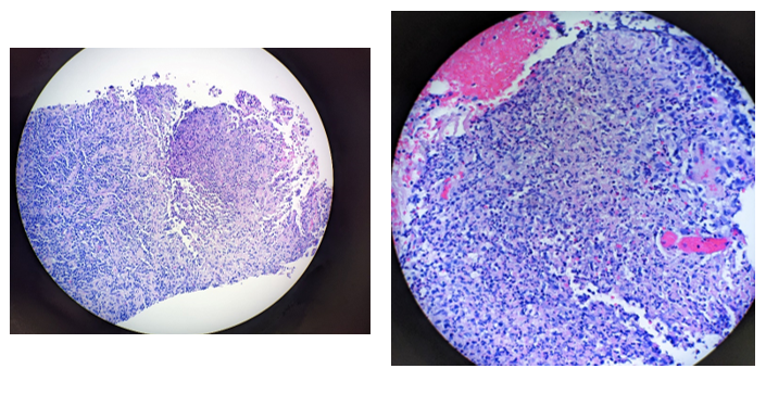
A 28 year old male reported to the ED with complaints of right groin pain, nausea, and vomiting for the past five days. The patient was not taking any active home medications and reported no chronic medical conditions at the time of presentation. He reported that he and his fiancée have a 5 month old kitten but denied any scratches or bites. Physical exam showed a tender right inguinal region covering a hard, non-reducible mass with no overlying erythema or fluctuance. Due to fever and tachycardia (temperature: 38.3 ˚C/ pulse: 136 beats/min), patient met criteria for sepsis without shock. CT of the abdominal pelvis showed enlarged right inguinal lymph nodes with suspected lymphadenitis and no inguinal hernia. Patient was started on ampicillin/sulbactam, ceftriaxone to cover possible STD, and azithromycin to cover possible cat-scratch disease. STI testing was negative for trichomonas, syphilis, chlamydia, and gonorrhea. Due to suspected azithromycin allergy, doxycycline was administered instead for empiric cat scratch disease treatment. Serology studies for Bartonella henselae in addition to right inguinal lymph node biopsy (Images 1 and 2). Lymph node biopsy revealed multiple cores displaying reactive lymphoid tissue with microabscesses surrounded by palisading histiocytes concerning for cat scratch disease lymphadenitis. Serology results showed elevated IgG and IgM titers for Bartonella henselae and PCR testing for Bartonella henselae performed on the lymphoid tissue confirmed the diagnosis. Patient was discharged on doxycycline and pain management medications.
Discussion
The majority of cat scratch disease infections are caused by Bartonella henselae, a facultative, intracellular gram negative bacillus.1,2 Bartonella henselae is usually acquired through a cat flea (Ctenocephalides felis) vector or transferred from a cat scratch or bite.1 Culture and polymerase chain reaction (PCR) have demonstrated Bartonella presence in cat saliva, gingiva, blood, claws, skin, and feces.3 Due to its fastidious nature, it is difficult to culture Bartonella henselae from samples taken from the human lymph node.4 In the past, Warthin-Starry or Steiner stains have been used to identify Bartonella henselae microscopically. 5,6,7 However, these silver stains are historically expensive, bulky, and difficult to interpret. Therefore, diagnosis typically relies on the combination of a variety of factors, including clinical, epidemiological, serological, and histological.4 PCR and serology or immunofluorescence have proven to be effective in detection of Bartonella henselae and are commonly used in the clinical setting for confirmation of diagnosis.4,8 Necrotizing stellate granulomas with neutrophil infiltration are the characteristic findings on histology (Images 1 and 2). Early histological findings are more likely to show histiocytes, follicular hyperplasia, and microabscesses bordering a thickened lymph node capsule.9
Cat scratch disease is most frequently characterized by self-limiting lymphadenopathy.1 The lymphadenopathy is usually close to the location of the cat scratch or bite and develops 1-2 weeks after exposure, although nearly a quarter of patients with cat scratch disease do not report close contact with cats.1 A papule or wheal may develop at the site of infection prior to lymphadenopathy.1 Cat scratch disease has not been documented to be transmitted between individuals.1 Fever, malaise, arthralgia and headache are other commonly reported symptoms.1 While most symptoms will resolve spontaneously, lymphadenopathy may last for weeks to months.1, Nonclassical presentations of cat scratch disease are reported in 10-15% of cases. Less common presentations that have been reported include, but are not limited to endocarditis, ophthalmic disease, central nervous system disease, hepatitis, splenitis, osteomyelitis, musculoskeletal arthropathy, and pulmonary disease. Immunocompromised patients infected with Bartonella henselae may present with widespread disease or with other diseases associated with Bartonella, including bacillary angiomatosis. While the majority of cases will resolve spontaneously, antimicrobial therapy including azithromycin can be used for treatment.1 In patients allergic to macrolides, doxycycline has proven to be effective. Pharmacologic pain management is also indicated when necessary.1
References
- Zangwill, K. M. (2021). Cat Scratch disease and Bartonellaceae: the known, the unknown and the curious. The Pediatric Infectious Disease Journal, 40(5S), S11-S15.
- Welch, D. F., Hensel, D. M., Pickett, D. A., San Joaquin, V. H., Robinson, A., & Slater, L. N. (1993). Bacteremia due to Rochalimaea henselae in a child: practical identification of isolates in the clinical laboratory. Journal of Clinical Microbiology, 31(9), 2381-2386.
- Lappin MR, Hawley J. Presence of Bartonella species and Rickettsia species DNA in the blood, oral cavity, skin and claw beds of cats in the United States. Vet Dermatol. 2009 Oct;20(5-6):509-14. doi: 10.1111/j.1365-3164.2009.00800.x. PMID: 20178489.
- Hansmann Y, DeMartino S, Piémont Y, Meyer N, Mariet P, Heller R, Christmann D, Jaulhac B. Diagnosis of cat scratch disease with detection of Bartonella henselae by PCR: a study of patients with lymph node enlargement. J Clin Microbiol. 2005 Aug;43(8):3800-6. doi: 10.1128/JCM.43.8.3800-3806.2005. PMID: 16081914; PMCID: PMC1233974.
- Cotter B, Maurer R, Hedinger C. Cat scratch disease: evidence for a bacterial etiology. A retrospective analysis using the Warthin-Starry stain. Virchows Arch A Pathol Anat Histopathol. 1986;410(2):103-6. doi: 10.1007/BF00713512. PMID: 2432720.

-Grant Whitebloom is a second-year medical student at the Medical College of Georgia. He is interested in Internal Medicine and its subspecialties.

-Hasan Samra, MD, is the Director of Clinical Microbiology at Augusta University and an Assistant Professor at the Medical College of Georgia.
