Hematology Case Study: 75 Year Old Man with Leukopenia

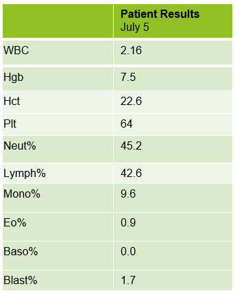
A 75 year old male first presented earlier this year with abnormal CBC results. The patient has a history of Type 2 diabetes, high blood pressure and atrial fibrillation. He was diagnosed with non-small-cell lung cancer (NSCLC) 6 years ago. His stage II NSCLC was completely removed with surgery. Surgery was followed up with adjuvant cisplatin-based chemotherapy to reduce the chance that the cancer would return. In June, he was referred to the hematology oncology department following consecutive CBCs that revealed leukopenia and thrombocytopenia. The CBC results from these specimens are shown below in Table 1.

Table 1. CBC results from a 75 year old male.

The peripheral blood sample from June was sent for flow cytometry. A leukemia/lymphoma phenotype was performed. Result comments noted proportionately decreased granulocytes with a left shift and 4% blasts. The blasts were CD34+, CD117+, HLA-DR+, CD13+ and CD33+ and were identified as myeloblasts. There were proportionately increased atypical monocytes with CD23 expression. Lymphocytes were also proportionately increased and included an increased population of CD57+, CD3+ T cells consistent with T-cell large granular (LGL) expansion. Immunophenotypic findings raised a concern for a myelodysplastic process. The hematologist discussed the findings with the patient and the patient was scheduled for a bone marrow biopsy. The procedure was performed 3 weeks later. CBC results on the day of the procedure are shown below in Table 2.

Table 2. CBC results day of bone marrow procedure. Pre-op diagnosis: Anemia, Leukopenia.

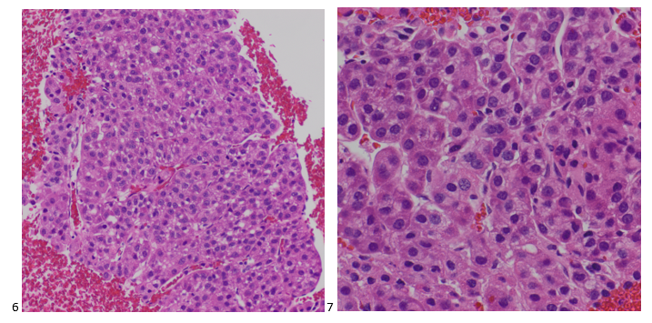
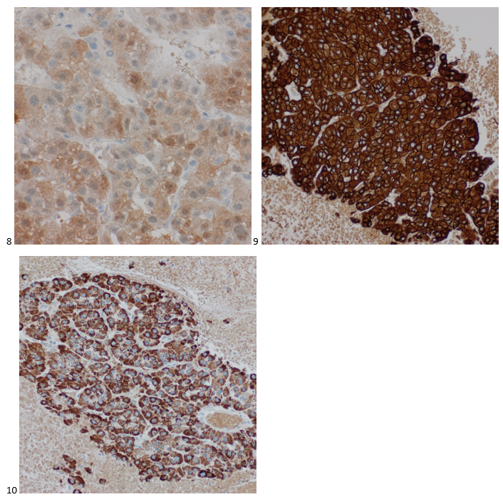
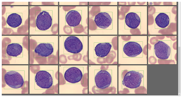
Bone marrow aspirate showed markedly increased myeloblasts (55%), consistent with acute myeloid leukemia (AML), nonacute promyelocytic leukemia (APL) type. The phenotype of the blasts was CD13+, CD33+, CD117+ and HLA-DR+. Blasts were negative for CD34. Several genomic variations were found in the specimen. These included variations in IDH2, SRSF2, STAG2 and ASXL1. Diagnosis: Increase in myeloblasts consistent with AML, nonAPL type.

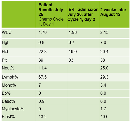
In July, 20 days after the bone marrow procedure and AML diagnosis, the patient was scheduled to begin his first cycle of Azacitidine (Vidaza). Based on his critical hemoglobin, the patient received 1 unit of packed RBCs followed by his first Vidaza injections. This Cycle 1, Day 1 chemotherapy was well tolerated, and he returned home. The following day he returned for his second treatment. His CBC showed good response to the previous day’s transfusion and his Cycle 1, Day 2 Vidaza was administered without incident. However, that evening the patient presented to the ER with nausea, vomiting and nose bleeds. The patient was admitted to the hospital and received another RBC transfusion. For the next several days the patient continued to do poorly, requiring additional RBC transfusions, and the Vidaza treatments were deferred, then discontinued. The patient had several ER visits and hospital admissions with transfusions over the next 2 weeks. During this time, we saw his blast% on his differential peak at over 60%. The patient was transferred to the palliative care team with care and comfort measures. CBC results from Cycle 1, Day 1 and subsequent CBC results are shown below. Note the sharp increase in blasts over a 2-week period.

Table 3. CBC results after chemotherapy initiated, then discontinued
Image 1. Cells classified as blasts on CellaVision

AML is the most common acute leukemia in adults. In AML with minimal differentiation, evidence of bone marrow failure is characterized by anemia, neutropenia, and thrombocytopenia. The median age for patients with AML in the US is 66-67, and those who are older than 55-65 at diagnosis often have challenges and lower odds for long term survival. These older patients tend to have poor tolerance to traditional aggressive chemotherapy because of other health issues. This patient was likely not a good candidate for strong chemotherapy because of his age and health history. In these more fragile patients, Vidaza may be used. Vidaza is a class of drug called a hypomethylating agent that works by switching off DNA methyltransferase. This switches on genes that stop the cancer cells growing and dividing. The goal is to reduce the number of abnormal blood cells and to control cell growth.

As you can see from the CBC results, the onset of this patient’s AML was very abrupt, and the disease progressed rapidly. He has several risk factors that made him more likely to be diagnosed with AML. Older age is a risk factor for AML, and AML is more common in males than females. He has a history of smoking which is a behavioral risk factor associated with AML. Additionally, patients with cancer who are treated with certain chemotherapy drugs are more likely to develop AML in the years following treatment. This patient was treated with cisplatin following lung cancer surgery. Cisplatin is an alkylating agent which has been linked to an increased risk of AML.

Also interesting is the note on the peripheral blood phenotype interpretation that a T-cell large granular lymphocyte (LGL) expansion was present. These are an increased population of CD57+, CD3+ T cells. LGL clones have been described in AML and a hallmark of this association is cytopenia, as is observed in this patient. The patient’s poor prognosis can partly be attributed to the p.Gly646TrfsTer12 alteration in the ASXL1 gene, identified in the bone marrow interpretation. This alteration is associated with decreased overall survival and poor prognosis which was observed in this patient.

I work in a hospital with a large hematology/oncology practice, and we see a lot of adult leukemia patients. Many of the patients we see regularly have Chronic Lymphocytic Leukemia (CLL). We feel like we get to know these patients, because even though we never see them, we see their CBCs every week, sometimes for many years. This was an interesting case because it reminded me of the sudden onset and rapid progression of AML. It was amazing to see the differentials change so dramatically in a matter of weeks. This patient is currently receiving care and comfort end of life measures.

References

Fattizzo, B, Bellani, V, et al. Large Granular Lymphocyte Expansion in Myeloid Diseases and Bone Marrow Failure Syndromes: Whoever Seeks Finds. Front. Oncol., Sec. Cancer Immunity and Immunotherapy. 01 October 2021.

Pratcorona M, Abbas S, Sanders MA, Koenders JE, et al.Acquired mutations in ASXL1 in acute myeloid leukemia: prevalence and prognostic value. Haematologica. 2012 Mar;97(3):388-92. doi: 10.3324/haematol.2011.051532. Epub 2011 Nov 4. PMID: 22058207; PMCID: PMC3291593.

https://www.cancer.net/cancer-types/lung-cancer-non-small-cell/types-treatment

Thomas XG, Dmoszynska A, Wierzbowska, et al. Results from a randomized phase III trial of decitabine versus supportive care or low-dose cytarabine for the treatment of older patients with newly diagnosed AML. Journal of Clinical Oncology 29:2011

Turgeon, Mary Louis. Clinical Hematology Theory and Procedures, 6th ed, Jones and Bartlett Learning, 2017.

Socha-small

-Becky Socha, MS, MLS(ASCP)CMBBCM graduated from Merrimack College in N. Andover, Massachusetts with a BS in Medical Technology and completed her MS in Clinical Laboratory Sciences at the University of Massachusetts, Lowell. She has worked as a Medical Technologist for over 40 years and has taught as an adjunct faculty member at Merrimack College, UMass Lowell and Stevenson University for over 20 years.  She has worked in all areas of the clinical laboratory, but has a special interest in Hematology and Blood Banking. She currently works at Mercy Medical Center in Baltimore, Md. When she’s not busy being a mad scientist, she can be found outside riding her bicycle.

Tumor on the Brain

Back in my Master’s program at Jefferson, I fondly remember the week we covered central nervous system (CNS) tumors. I was fascinated by the mnemonic tools we would use to identify different CNS tumors, such as “fried eggs” for oligodendrogliomas, perivascular pseudorosettes in ependymomas, and the whorling associated with meningiomas. Fortunately, for our patients, and unfortunately, for our diagnostic curiosity, we rarely see CNS tumors at my institution. Brain lesions resulting from metastatic carcinomas are typically well-identified via imaging and treated appropriately by the surgical, medical, and radiation oncology teams, but cytologists are available to screen cerebrospinal fluids (CSFs) for CNS involvement. For primary CNS tumors, however, we’re left recollecting the core memory of the second semester of our didactic phase. When a metastatic CNS tumor made its way into our lab, our cytology team swooned with excitement. (Yes, I know, but please introduce me to a lab professional who doesn’t embrace their quirks.) A 27-year-old male patient presented to radiation oncology three years after surgical debulking of a brain tumor at an outside institution. The patient, who was referred to radiation oncology at to treat the residual tumor at the original institution, did not follow up and developed an 8 centimeter recurrence a year after the initial resection. At this point, the patient experienced complete vision loss and underwent a biparietal-occipital craniectomy. A repeat brain MRI was performed a year later, and once again, a large enhancing extra-axial mass was identified along with multiple smaller masses also increasing in size. The patient received radiation after worsening difficulty with ambulation. After almost completing the planned fractions of radiation, the patient elected to stop their radiation therapy due to worsening seizures. A left neck mass was identified six months prior, and while the mass had not grown or caused pain, the patient was referred to head and neck surgical oncology for evaluation. Surveillance imaging demonstrated an enlarged left level 5A lymph node, suggestive of metastatic disease. Multiple ultrasound-guided fine needle aspiration biopsies were obtained from the lymph node, and ROSE was performed. The Diff-Quik-stained and concurrent Pap-stained smears demonstrated lesional tissue, although everything from epithelioid histiocytes to spindle cell melanoma to a renal primary were considered as a differential. Based on the location, a salivary gland primary was also a possibility for this case. The streaked cytoplasm and pseudoinclusions in both smears were concerning for a metastasis of the patient’s primary CNS tumor, but we were still hesitating to make the call.

Images 1-4. Lymph Node, Neck, Left, Level 5A, US-guided FNA. 1-2: Diff-Quik-stained smears, 3-4: Pap-stained smears.

The following morning, the H&E-stained FFPE cell block sections demonstrated the characteristic whorls expected for the patient’s primary, although the idea of metastasis was uncanny.

Images 5-6. Lymph Node, Neck, Left, Level 5A, US-guided FNA. H&E sections (6: 100x, 7: 400x).

We then used immunohistochemical studies to confirm our morphologic diagnosis. Immunostains performed on the cell block slides with adequate controls show that the tumor cells are positive for vimentin and PR (focal), while negative for AE1/AE3, EMA, CK7, CK20, TTF-1, Napsin A, p40, Pax8, synaptophysin, and S-100. The Ki-67 proliferation index fell at 18%, which is consistent with intermediate aggressive disease in a WHO Grade 2 atypical meningioma.

Images 7-8. Lymph Node, Neck, Left, Level 5A, US-guided FNA. Cell block section immunohistochemistry. 7: Vimentin-positive; 8: focally PR-positive.

The patient had next gen sequencing performed on his tissue, which demonstrated an NF-2 mutation, indicating he may benefit from MTOR inhibitors, but he elected not to pursue systemic therapy.

Where meningiomas account for 36% of primary brain tumors, atypical meningiomas comprise only 5-15% of all meningiomas (Cai et al., 202. Extracranial metastasis of atypical meningioma is a rare event, with only a few cases documented in the literature. While meningioma metastases are uncommon, a thorough collaboration between clinical impression and pathologic interpretation is necessary to ensure the possibility is not entirely excluded.

References

Cai C., Kresak J.L., Yachnis A.T. (2021) Atypical meningioma. Pathology Outlines. Retrieved October 11th, 2022, from https://www.pathologyoutlines.com/topic/cnstumoratypicalmeningioma.html.

P.S. I’d like to take this opportunity for a shameless plug. My Doctor of Health Science (DHSc) research survey is live now through November 23rd, 2022. If you’re a medical laboratory professional or pathologist, please consider contributing to our field of laboratory medicine! Click the following link to read the consent form and take the one-time anonymous survey. Thank you for your time!

https://www.surveymonkey.com/r/leadinglabs

-Taryn Waraksa-Deutsch, MS, SCT(ASCP)CM, CT(IAC), has worked as a cytotechnologist at Fox Chase Cancer Center, in Philadelphia, Pennsylvania, since earning her master’s degree from Thomas Jefferson University in 2014. She is an ASCP board-certified Specialist in Cytotechnology with an additional certification by the International Academy of Cytology (IAC). She is also a 2020 ASCP 40 Under Forty Honoree.

Microbiology Case Study: What’s with the Rash?

Case presentation

A 79 year old female with a past medical history of COPD, hypertension, diabetes, and eczema presented to the emergency department with a localized rash on the right knee (Figure 1). The rash began after gardening and persisted for three weeks.

The patient reported some itching, warmth, and tenderness but denied nausea, vomiting, fever, and diarrhea. Her vital signs were BP 175/76| Pulse 91 | Temp 98.5 °F (36.9 °C) (Oral) | Resp 20 | SpO2 96%. The remainder of her physical exam was notable: right knee skin rash. There was no induration or fluctuance or drainage. She exhibited a full range of knee motion; there was no palpable knee joint effusion (Figure 1).

Lab CBC results were unremarkable. X-Ray knee AP and lateral – right showed soft tissue prominence anterior to the patella, which suggests prepatellar edema and a fluid collection. Lyme antibody screening was negative. Two sets of blood culture bottles were sent to the microbiology laboratory. After 24 hours of incubation, aerobic bottles were positive with the organism shown in: Gram stain (Figure 2), culture growth showing alpha-hemolytic colonies (Figure 3), H2S production on the TSA agar slant (Figure 4). 

Identification by Matrix-assisted laser desorption ionization Time of flight (MALDI-ToF) revealed Erysipelothrix rusiopathiae at a score above 2.0. 

Discussion

Erysipelothrix is a non-spore-forming, catalase-negative, facultative gram positive bacillus. It is not acid-fast or motile. It is distributed worldwide and is primarily considered an animal pathogen responsible for causing erysipelas that may affect a wide range of animals. Erysipelothrix is ubiquitous in soil, food scraps, and water contaminated by infected animals.1 It can survive in the soil for several weeks. In pig feces, the survival period of this bacterium ranges from 1 to 5 months.

Erysipelothrix can also cause zoonotic infections in humans, called erysipeloid. Most human infections are acquired through occupational exposure, such as fish handlers, veterinarians, and butchers, via direct injection of the organism through abrasion or injuries. Notably, the human disease of “erysipelas” is not caused by Erysipelothrix but by Streptococcus. 

Erysipeloid typically develops at the site of infection between 2 and 7 days after exposure. E. rusiopathiae infection can be categorized as 1) localized cutaneous erythematous 2) generalized cutaneous form due to traumatic injury and skin penetration of the organism, and 3) septicemic form.2 Skin infection can sometimes progress to bacteremia, most commonly associated with endocarditis3. The implication of endocarditis in the setting of E. rusipathiae infection is associated with increased mortality rate.2,3 

E. rusiopathiae can easily be grown on routine media, including blood and chocolate agar plates, in a clinical microbiology laboratory.1 The colonies appear as small alpha-hemolytic and can resemble alpha Streptococcus species. It can also be confused with Corynebacterium species due to the similarity in Gram stain characteristics. E. rusipathiae produces H2S on the triple iron sugar media (Figure 4), which is one of the distinguishing morphologies from other Gram-positive rods, such as Listeria or Bacillus species.1 It can be identified by Matrix-assisted laser desorption ionization Time of Flight (MALDI-ToF) directly from the positive blood culture broth (using Sepsityper Kit with Bruker MALDI-Biotyper (MBT)) or from isolated colonies. 

E. rusiopathiae is generally sensitive to penicillin. It is intrinsically resistant to vancomycin and aminoglycosides.4 CLSI (Clinical Laboratory of Standard Institution) M45 ED3 recommended ampicillin or penicillin for primary testing agents.4 While antimicrobial susceptibility testing is not warranted for every case of E. rusiopathiae, it is imperative that the organism be identified due to the critical nature of infection resulting in endocarditis. Since vancomycin is typically used for broad-spectrum coverage of gram positive organisms,4 early identification of this organism and notification of clinicians is helpful for appropriate antimicrobial management.

References

  1. Jorgensen et.al., Chapter 27. Manual of Clinical Microbiology. 11th Edition.

2. Principe L, Bracco S, Mauri C, Tonolo S, Pini B, Luzzaro F. Erysipelothrix Rhusiopathiae Bacteremia without Endocarditis: Rapid Identification from Positive Blood Culture by MALDI-TOF Mass Spectrometry. A Case Report and Literature Review. Infect Dis Rep. 2016 Mar 21;8(1):6368. doi: 10.4081/idr.2016.6368. PMID: 27103974; PMCID: PMC4815943.

3. Wang T, Khan D, Mobarakai N. Erysipelothrix rhusiopathiae endocarditis. IDCases. 2020 Sep 9;22:e00958. doi: 10.1016/j.idcr.2020.e00958. PMID: 32995274; PMCID: PMC7508995.

4. CLSI. Methods for Antimicrobial Dilution and Disk Susceptibility Testing of Infrequently Isolated or Fastidious Bacteria. 3rd ed. CLSI guideline M45. Wayne, PA: Clinical and Laboratory Standards Institute; 2016.

-Azal Al-Ani, MD is a third-year AP/CP pathology resident at Montefiore Medical Center, Bronx, NY. She completed her medical school at Al-Anbar Medical College, Iraq. Her interest includes hematopathology and dermatopathology

-Phyu M. Thwe, PhD, D(ABMM), MLS(ASCP)CM is Associate Director of Infectious disease testing laboratory at Montefiore Medical Center, Bronx, NY. She completed her CPEP microbiology fellowship at the University of Texas Medical Branch in Galveston, TX. Her interest includes appropriate test utilization and extra-pulmonary tuberculosis.

Into the Badlands of Safety

During a recent trip to South Dakota, I was able to visit Badlands National Park. I am not a hiker or a camper, so I was not sure I would enjoy the park very much, but it turned out to be the highlight of our vacation. The vastness of the landscape and the unusual beauty of the rock formations cannot be captured in pictures. It is truly something that should be see in person at least once in a lifetime. While walking the trails of the park, it looked and felt like walking in an alien world. It looks strange, and there are hidden dangers- rattlesnakes, potential high heat, and crumbly walkways with sudden drop-offs.

The experience reminded me of how the laboratory must seem to visitors or workers who need to come into the department to perform various duties. The laboratory must seem like a foreign world, and indeed, there are many hidden dangers within. If I had walked blindly into Badlands National Park and not read the warning signs, would I have been bitten by a snake or could I have walked off a cliff? Of course. Do the signs in your lab adequately warn visitors of the dangers? Do visitors pay attention?

The lab staff reported a plugged floor drain under the hematology analyzer, so the facilities plumber arrived to repair it. He asked the staff if the analyzer was running, and because they were not processing any samples, they said it was not. When the plumber bent down to look at the drain, the analyzer cycled waste through the drain line which quickly splashed into the eyes and mouth of the plumber.

Warning signs are required in many labs for many reasons, but they are not sufficient for protection from the hazards in the department. Those who enter the “badlands” of the lab need to be told about the dangers, and they need as much information as possible. If someone is coming in to work on equipment, offer proper personal protective equipment. If someone will be on the floor of a biohazard lab, make sure a lab coat and gloves are in use, and lay a pad on the floor if possible. Make sure people understand proper terminology. An analyzer may not be actively running samples, but it is still “on,” and there are still potential hazards present.

It should not be assumed that people who come to work in the lab department will have general knowledge of the laboratory or of lab safety practices. It is a good practice to use a safety training checklist for vendors or others who enter the department and to go over that checklist at least annually. Couriers can be harmed by pathogens, chemicals, or dry ice. Phlebotomists who are expected to process samples should be trained in centrifuge operations, spill clean up and more. Environmental service workers and biomedical engineering staff need to understand the chemical and biohazards in the department.

Instrument service representatives have training, but not typically much of that training is focused on lab safety. Some representatives keep a reusable lab coat with them, and wear it from lab to lab, washing it at home when visibly dirty. There are some OSHA violations in those behaviors. PPE that is used on a lab cannot be removed from that lab (except for professional laundering). Lab coats used in a laboratory cannot be laundered at home. These are unsafe (and illegal) practices, but until someone notifies the representatives about them, the behaviors will continue.

When someone works in the lab “badlands” every day, it is easy to become complacent about the hazards within, or they may be well-trained and no longer consciously think about the tools they use to mitigate those hazards. That is not true for someone who may enter, someone who does not have the same background, experience, and training. Laboratorians are responsible for their safety as well, and educating those visitors about the potential dangers can keep them safe so they can go into more familiar climates with their health fully intact. 

Dan Scungio, MT(ASCP), SLS, CQA (ASQ) has over 25 years experience as a certified medical technologist. Today he is the Laboratory Safety Officer for Sentara Healthcare, a system of seven hospitals and over 20 laboratories and draw sites in the Tidewater area of Virginia. He is also known as Dan the Lab Safety Man, a lab safety consultant, educator, and trainer.

Microbiology Case Study: A 67 Year Old with Foot Pain

Case description

A 67 year old male presented at the clinic with a primary complaint of foot pain; she has a previous medical history of M. tuberculosis infection of her prosthetic joint, osteoarthritis, and leukopenia. The patient described joint pains during the check-up and mentioned that she also started to have periumbilical pain two weeks ago, along with worm-like objects in her stool. The patient was in Ethiopia for 8 months in the past year and was very active. He has had some weight loss but no change in appetite; he denies any diarrhea, skin rashes, fever, or chills. The patient consumed undercooked meat products during the time she visited Ethiopia. No abnormal neurological symptoms presented at the time of the visit.

Orders were placed for H. Pylori antigen, fecal bacteria pathogen PCR, Giardia and Cryptosporidium antigen, and Ova & Parasite exam for the patient’s GI symptoms. The Ova & Parasite exam detected the objects in Image 1.

Image 1. Patient stool sample wet mount preparation.

Discussion

The Ova & Parasite exam was reported as Taenia species. The eggs had a diameter of around 37um. An infectious disease consult was ordered and a single dose of 600mg praziquantel was prescribed for the treatment. Repeat Ova & Parasite exams are ordered for 3 days post-treatment looking for dying parasites and 1 month post-treatment to confirm the cure (no eggs).

Taenia in the Taeniidae family of tapeworms (BioLib, n.d.). Three species are commonly found and most clinically important in human infection: Taenia saginata, Taenia solium, and Taenia asiatica; most Taeniasis is asymptomatic or has mild symptoms (Centers for, 2020b).

Taenia solium, or pork tapeworm often found in pork, is the most dangerous species to humans for two reasons. First, this is the only species that can cause the neurologic symptoms by cysticercosis in brain tissue; second, this species can take humans as intermediate hosts, which means it can cause human to human transmission within the household (Schmidt et al., 2009).

Taenia asiatica also lives in pigs, primarily in the liver instead of muscle. This species has a very similar genetic, morphology, and immunology to T. saginata. It is frequently found in Asia (Schmidt et al., 2009).

Taenia saginata, or beef tapeworm, is what our patient was assumed to have in this case. The life cycle is shown below in Figure 2. The patient presented because his ankle pain started to impact his walking significantly; however, he was not seeking help for his worm-like objects in the clinic, probably due to the mildness of the symptoms. The parasite infection was brought into sight because of his travel history and stool observation. Per CDC, Eastern Europe, Russia, eastern Africa, and Latin America are the highest risk areas (Centers for, 2020a). The patient stayed for 8 months in Ethiopia in eastern Africa. Ethiopia has a relatively poor sanitation status and a high prevalence of taeniasis (Jorga, 2020). The major contributors for our infectious disease clinicians to assume this patient has T. saginata infection but not T. solium infection are: there are no neurological symptoms, and there is no pork exposure due to his religion. Visualization of the tapeworm eggs or segments is important for identification the species. In this case, many eggs were found on the wet mount slide from the patient’s stool sample.

Treatment of taeniasis is with Praziquantel. Praziquantel removes the tapeworms from the human body by detaching the worm suckers from vessel walls. The medication is safe to give to ≥1year old patients (UpToDate, 2022).

Image 2. Taeniasis life cycle. Alive Taenia eggs or gravid proglottids in the environment get ingested by farm or wild animals. Oncospheres develop in the GI tract, then hatch to the intestine wall and penetrate the wall to migrate to muscle tissue. In the muscle tissue, oncospheres develop into cysticerci (cysticercosis happens at this step). After the meat products (generally animal muscle) get ingested by humans, the cysticerci grow into adult worms in humans. Some segments/worms/eggs will be released into the environment through feces to complete the life cycle (which allows detection and diagnosis of human infections).
https://www.uptodate.com/contents/image/print?imageKey=ID%2F64879

References

BioLib: Biological library. Taenia | BioLib.cz. (n.d.). Retrieved from https://www.biolib.cz/en/taxon/id43806/

Centers for Disease Control and Prevention. (2020a, September 18). CDC – taeniasis – general information . Epidemiology & Risk Factors. Retrieved from https://www.cdc.gov/parasites/taeniasis/epi.html

Centers for Disease Control and Prevention. (2020b, September 18). CDC – taeniasis – general information . frequently asked questions. Retrieved from https://www.cdc.gov/parasites/taeniasis/gen_info/faqs.html

Jorga, E., Van Damme, I., Mideksa, B. et al. Identification of risk areas and practices for Taenia saginata taeniosis/cysticercosis in Ethiopia: a systematic review and meta-analysis. Parasites Vectors 13, 375 (2020). https://doi.org/10.1186/s13071-020-04222-y

Schmidt, G. D., & Roberts, L. S. (2009). Chapter 21 Tapeworms. In Foundations of Parasitology, eighth edition (pp. 346–351). essay, McGraw-Hill Higher Education.

UpToDate. (2022). Praziquantel: Drug information. UpToDate. Retrieved from https://www.uptodate.com/contents/table-of-contents/drug-information

-Sherry Xu is a Masters student in the department of Pathology and Laboratory Medicine at the University of Vermont Medical Center.

-Christi Wojewoda, MD, is the Director of Clinical Microbiology at the University of Vermont Medical Center and an Associate Professor at the University of Vermont.

Microbiology Case Study: A 46 Year Old with Chest Pain

Case History

A 46 year old male with a history of cystic fibrosis and bilateral lung transplant two years prior presented to the hospital with chest pain and hemoptysis. The patient was recently diagnosed with COVID-19, and a CT chest revealed multiple rounded, mass-like opacities with central cavitation. As imaging was not consistent with COVID-19 pulmonary disease and no clear risk for tuberculosis could be identified, a bronchoscopy with transbronchial biopsy was performed. Tissue and bronchiolar lavage fluid were collected and submitted to the microbiology laboratory for analysis. Viral etiologies including influenza A/B, Parainfluenza 1-3, Adenovirus, RSV and metapneumovirus were ruled out through molecular studies. Galactomannan was negative from the BAL fluid, as were fungal and mycobacterial cultures and Mycobacterium tuberculosis PCR. GMS staining of the biopsy was negative but organizing pneumonia and mononuclear infiltrate was noted. The patient had a history of recurrent multidrug-resistant Pseudomonas aeruginosa infection and was being managed with empiric ceftazidime/avibactam.

Laboratory Identification

Gram stains of both the tissue and BAL fluid were generally unremarkable. Histopathological analysis of the transbronchial tissue revealed changes suggestive of organizing pneumonia with mononuclear infiltrate (Image 2, left). Bacterial growth of a predominant organism from both the BAL and biopsy tissue was observed on plates after 48 hours on blood and chocolate agars but was absent on MacConkey agar. At 96 hours, the colonies of the organism had become mucoid, slightly pink and had coalesced (Image 1, right). Gram staining of the growth revealed short, poorly staining gram positive coccobacilli with a beaded appearance. Due to the incomplete gram staining of this isolate, modified acid-fast staining was attempted which was positive (Image 1, left). The organism was both catalase- and urease-positive. The isolate was subsequently identified by MALDI-TOF MS as Rhodococcus equi, and the patient was discharged from the hospital on imipenem and linezolid.

Image 1. (Left) Modified acid-fast (MAF) staining revealing small, MAF-positive coccobacilli (black arrowheads).  (Right) Characteristic, mucoid salmon-colored colonies of the isolate on blood agar after 96 hours incubation. ​
Image 2. (Left) Transbronchial biopsy revealing areas of histiocyte aggregation and mononuclear infiltrate (H&E, 10X magnification).  (Middle) Representative image of expanded histiocytes with small, pale-staining round forms in a background of neutrophils (H&E, 40X magnification).  (Right) Representative image of histiocytes filled with coccoid and coccobacilliary forms (GMS, 40X magnification).​

Discussion

Rhodococcus equi is a zoonotic pathogen which primarily causes infections among immunocompromised hosts. Infrequently isolated clinically, the organism is a primary pathogen of horses causing pneumonia with abscess formation in foals, often with dissemination into peripheral sites due to high organism burden. The organism is excreted in feces of infected animals, leading to contamination of soils from farms, ranches, and other agricultural environments from which the organism is either aerosolized and inhaled or acquired via direct inoculation.1 While human infections are classically associated with exposure to horses or their environment, there is a growing body of literature to suggest that many patients with microbiologically proven cases of R. equi infection lack such environmental exposures. This patient falls into the latter category, with no known exposure to livestock.

                R. equi is a member of the aerobic actinomycetes. Like Nocardia sp., the cell wall of R. equi contains mycolic acids which lead to positivity when stained with a modified acid-fast stain. The organism is a facultative, intracellular pathogen surviving within macrophages and histiocytes, leading to granulomatous inflammation, eventually leading to necrosis.2 Immunosuppression (including HIV infection or immunosuppressive therapy) is a major risk factor for R. equi infection, as most clinical cases are reported in this setting. In immunocompromised hosts, the spectrum of disease manifestations of R. equi are diverse, but most commonly (approx. 80%) include pulmonary involvement3 with upper lobe cavitary pneumonia.4 Characteristic malakoplakia (an infiltration of foamy histocytes with intracellular bacteria and basophilic inclusions name Michaelis-Gutmann bodies)1 can be associated with R. equi infection. These structures were noticeably absent in this patient’s case despite the observed histocyte aggregation and mononuclear infiltrate (Image 2, center, left).

R. equi pneumonia among solid organ transplant recipients, such as the patient in this case is associated with low overall morbidity and mortality, but require protracted antibiotic therapy regimens.1 Susceptibility testing is warranted to guide therapy of R. equi due to unpredictable resistance patterns among isolates. This patient’s isolate was revealed to be susceptible to amoxicillin/clavulanate, ceftriaxone, imipenem, ciprofloxacin, moxifloxacin, clarithromycin, amikacin, tobramycin, minocycline, trimethoprim/sulfamethoxazole, vancomycin, linezolid, and rifampin. The patient was discharged on imipenem/linezolid. At follow-up, the patient had clinically improved with a resolution of symptoms, but his radiologic abnormalities persisted and thus remains on oral therapy with moxifloxacin and minocycline.

References

Yamshchikov, AV, Schuetz, A, and Lyon, GM. Rhodococcus equi infection. 2010. Lancet Infect. Dis. 10:350-359.

Prescott, JF. Rhodococcus equi: an Animal and Human Pathogen. 1991. Clin. Microbiol. Rev. 4(1):20-34.

Weinstock, DM, and Brown, AE. Rhodococcus equi: an emerging pathogen. 2002. Clin. Infect. Dis. 34:1379-1385.

Mutaner, L, et. al. Radiologic featuresof Rhodococcus equi pneumonia in AIDS. Eur. J. Radiology. 1997. 66-70.

-Andrew Clark, PhD, D(ABMM) is an Assistant Professor at UT Southwestern Medical Center in the Department of Pathology, and Associate Director of the Clements University Hospital microbiology laboratory. He completed a CPEP-accredited postdoctoral fellowship in Medical and Public Health Microbiology at National Institutes of Health, and is interested in antimicrobial susceptibility and anaerobe pathophysiology.

-Dominick Cavuoti is a Professor in the Department of Pathology at UT Southwestern Medical Center. Dr. Cavuoti is a board certified AP/CP who is a practicing Clinical Microbiologist, Infectious Disease pathologist and Cytopathologist.


-Clare McCormick-Baw, MD, PhD is an Assistant Professor of Clinical Microbiology at UT Southwestern in Dallas, Texas. She has a passion for teaching about laboratory medicine in general and the best uses of the microbiology lab in particular.

Microbiology Case Study: A 17 Year Old with Chest Pain

Case History

A 17 year old female who presented to the emergency department with complaints of fever, vomiting, diarrhea, and chest pain for the past two weeks. She also reported an unintentional weight loss of 20 lbs. Her medical history consisted of essential hypertension for which she was previously on medication, however had been discontinued two years ago due to normal blood pressure. The patient reported that she is sexually active with one male partner and denied use of protection. She denied any other sexual partners or any prior history of sexually transmitted infections. Her urine NAAT testing was positive for chlamydia, but negative for gonorrhea. Blood cultures collected at the time of admission resulted in growth of gram-negative diplococci on day 2 of admission (Image 1) and colony growth on chocolate agar (Image 2). The organism was positive for both catalase and oxidase and identified by matrix-assisted light desorption ionization- time of flight (MALDI-TOF) as Neisseria gonorrhoeae. Due to her chest pain complaints and QT prolongation on EKG, a trans-thoracic echo was performed that demonstrated a large aortic root abscess suggestive of infective endocarditis. Ceftriaxone was started as treatment for her gram-negative endocarditis, and she was emergently transferred to another facility where an aortic valve replacement and patch aortoplasty were performed.

Image 1. Gram stain of the blood culture showing gram negative diplococci.
Image 2. Neisseria gonorrhoeae on chocolate agar producing small gray-white colonies.

Discussion

Neisseria gonorrhoeae is a fastidious, oxidase positive, gram negative diplococcus, commonly transmitted through sexual contact.2,3 Neisseria uniquely grows on chocolate agar and VPN/Thayer Martin agar, and has virulence factors such as pilli that attach to mucosal surfaces, and many antigenic variations that make it a highly resistant organism prone to reinfection.

In the laboratory, N. gonorrhea grows well on chocolate agar after 24-48 hours of incubation (Image 2) with less robust or no growth on blood agar. It is positive for both catalase and oxidase. Traditionally, sugar fermentation was used to differentiate Neisseria species from one another, but more ore rapid identification methods (MALDI-TOF and PCR) are being increasingly used in most clinical laboratories

In men, Neisseria usually ascends the genitourinary tract to cause prostatitis. In women, the infection can disseminate to cause pelvic inflammatory disease, which can cause scarring in the fallopian tubes, resulting in infertility. Neisseria also can present as an asymmetric polyarthritis, most commonly to the knees. The main treatment of Neisseria gonorrhea is ceftriaxone. Gentamicin is an acceptable alternative in patients with severe cephalosporins allergy.

This case involves a rare presentation of infective endocarditis caused by disseminated gonorrhea infection. Previous reported cases of gonococcal endocarditis1,4 reported ad subacute presentation in around 2-4 weeks with generalized fatigue, fevers, arthritis, rash, renal dysfunction, and new cardiac murmurs. Because it can present without preceding genitourinary symptoms, disseminated gonorrheal can be difficult to recognize. The infection is usually aggressive, forming large vegetations and rapid valve destruction, despite antibiotic treatment. Most commonly it involves the aortic valve, as seen in the case presented above, but can also involve the mitral and tricuspid valves in some cases. The damage usually requires valve replacement surgery in addition to antimicrobial therapy.5,6 Lastly, this case demonstrates the limitations of the urine NAAT to diagnose gonorrhea specifically in females and/or asymptomatic patients due the possible presence of inhibitors and the need for further testing if clinical suspicion remains.7

References

  1. Said M, Tirthani E. Gonococcal Infective Endocarditis Returns. Cureus. 2021 Sep 14;13(9):e17955. doi: 10.7759/cureus.17955. PMID: 34660143; PMCID: PMC8515499.
  2. Ryan, K. J., Ray, G., and Sherris, J. C. (2004). Sherris Medical Microbiology: An introduction to Infectious Diseases, 4th edition. McGraw-Hill Medical.
  3. Centers for Disease Control and Prevention. Gonorrhea. Available from: https://www.cdc.gov/std/gonorrhea/stdfact-gonorrhea-detailed.htm. Last updated 2021 July 22; cited on 2022 March 21.
  4. Fenech, Marylou, et al. “Neisseria Gonorrhoeae Infective Endocarditis.” BMJ Case Reports, BMJ Specialist Journals, 1 May 2022
  5. Thompson EC, Brantley D. Gonoccocal endocarditis. J Natl Med Assoc. 1996 Jun;88(6):353-6. PMID: 8691495; PMCID: PMC2608094.
  6. Nie S, Wu Y, Huang L, Pincus D, Tang YW, Lu X. Gonococcal endocarditis: a case report and literature review. Eur J Clin Microbiol Infect Dis. 2014 Jan;33(1):23-7. doi: 10.1007/s10096-013-1921-x. Epub 2013 Jul 16. PMID: 23856883.
  7. Whiley DM, Tapsall JW, Sloots TP. Nucleic acid amplification testing for Neisseria gonorrhoeae: an ongoing challenge. J Mol Diagn. 2006 Feb;8(1):3-15. doi: 10.2353/jmoldx.2006.050045. PMID: 16436629; PMCID: PMC1871692.

-Olivia Piscano is a second-year medical student at the Medical College of Georgia. She is currently interested in Internal Medicine, Pediatrics, and Infectious Disease.

-Hasan Samra, MD, is the Director of Clinical Microbiology at Augusta University and an Assistant Professor at the Medical College of Georgia.

Microbiology Case Study: 57 year old Female with Altered Mental Status and Declining Health

Case Description

A 57 year old female presents to the emergency department with altered mental status, decreased appetite and chest, abdominal and pelvic pain. She has a complex medical history including end-stage renal disease, cardiovascular disease with pacemaker placement, and recurrent ascites. Her physical exam was notable for hypotension (65/52), hypothermia (31.7°C), and abdominal distention. A CT of her abdomen and pelvis revealed marked ascites, and bloodwork indicated leukocytosis (12.81, ref 4.22-10.33), elevated lactate (4.1, ref 0.5-2.2), and acidemia (7.27). Given the concern for septic shock, an infectious workup was initiated. A diagnostic paracentesis was undertaken which revealed rare yeast forms by Gram stain (Figure 1). Routine and fungal cultures of the ascites fluid grew yeast which was identified as Cryptococcus neoformans by matrix-assisted laser desorption/ionization-time-of-flight mass spectrometry (MALDI-TOF MS). Bacterial blood cultures turned positive after 3 days and Cryptococcus neoformans was identified (Figure 2). Interestingly, serum cryptococcal antigen tests were negative.

The patient was treated with amphotericin B, vancomycin, and meropenem. Despite intervention, the patient’s clinical condition continued to deteriorate, leading to multiple organ failure. The patient was transitioned to comfort care and expired soon thereafter.

Figure 1. Gram stain of peritoneal fluid (ascites) demonstrating two yeasts with surrounding capsule and host cells (100x objective, oil immersion).
Figure 2.  Bacterial blood culture with encapsulated yeasts on Gram stain (100x objective, oil immersion).

Discussion

This case highlights two significant points: 1) atypical presentation of a cryptococcal infection and 2) the value of complimentary approaches in the diagnostic workup of an infectious etiology.

Cryptococciare environmental fungi with worldwide distribution, classily causing opportunistic central nervous system infection in patients with either uncontrolled HIV or other significantly immunocompromising conditions. In this patient population, a staggering 70-90% of cryptococcal infections manifest as meningitis, with a one-year mortality rate as high as 70% in some regions.1 Meningitis caused by Cryptococcus sp. accounts for nearly 1 in 7 HIV-related deaths worldwide.2 This case and others,3 serve as an important reminder that cryptococcal disease may present in different ways, including peritonitis in a patient without significant immunosuppression.

Several methods are available to aid in the identification of Cryptococcus sp.4 Direct microscopic evaluation of Cryptococcus in fluid and tissue samples reveals round, narrow-based budding yeast of variable size (2-20 um). The capsule of Cryptococcus is classically seen with India ink, though recent work has shown that Gram stain is as effective.5 In our patient, direct microscopy of the peritoneal fluid provided the first clue that Cryptococcus was the causative agent. Other useful stains used for histopathological analysis include mucicarmine and Fontana-Masson, which stain the capsule and melanin, respectively. In culture, strains of Cryptococcus sp. typically elaborate a robust capsule leading to the formation of mucoid colonies. However, acapsular strains have also been identified.

Biochemical hallmarks of Cryptococcus sp. include the production of urease and phenoloxidase leading to the formation of melanin which is absorbed into the cell wall. Phenoloxidase activity is exploited for diagnostic purposes as it leads to melanized pigmentation in the presence of caffeic acid, such as in either a caffeic acid disk test or on Bird Seed agar.

Non-culture-based methods for the diagnosis of cryptococcal infections include detection of cryptococcal capsular antigens by either ELISA, latex agglutination, or a lateral flow immunoassay (LFA). The LFA is a cost-effective test with rapid turnaround time which exhibits strong agreement with other antigen detection methods. While the LFA has proven useful in a variety of settings,6 our patient’s LFA was negative, underlining the importance of using orthogonal methods in parallel to identify microbes. In cases where patients are infected with an acapsular strain of Cryptococcus, the antigen testing will be negative since the antigenic target (i.e the capsule) is missing. An important consideration when assessing discrepancies between Gram stain/culture and ancillary immunoassay testing is “prozone effect”. Prozone, or Hook effect, is a phenomenon where overwhelming amounts of analyte impairs immunocomplex formation, causing a lack of analyte detection (i.e false-negative test). To account for prozone, a serial dilution series of the sample with repeat testing should be performed to ensure accurate correlation.

References

  1. World Health Organization. Guidelines for the diagnosis, prevention, and management of cryptococcal disease in HIV-infected adults, adolescents and children, March 2018: supplement to the 2016 consolidated guidelines of the use of antiretroviral drugs for treating and preventing HIV infection.
  2. Rajasingham R, Smith RM, Park BJ, Jarvis JN, Govender NP, Chiller TM, Denning DW, Loyse A, Boulware DR. Global burden of disease of HIV-associated cryptococcal meningitis: an updated analysis. The Lancet infectious diseases. 2017 Aug 1;17(8):873-81.
  3. El-Kersh K, Rawasia WF, Chaddha U, Guardiola J. Rarity revisited: cryptococcal peritonitis. Case Reports. 2013 Jul 10;2013:bcr2013009099.
  4. Mais DD. Quick compendium of clinical pathology. American Society for Clinical Pathology Press; 2018.
  5. Coovadia YM, Mahomed S, Dorasamy A, Chang C. A comparative evaluation of the Gram stain and India ink stain for the rapid diagnosis of cryptococcal meningitis in HIV infected patients in Durban: brief report. Southern African Journal of Infectious Diseases. 2015 Jan 1;30(2):61-3.
  6. Perfect JR, Bicanic T. Cryptococcosis diagnosis and treatment: What do we know now. Fungal Genetics and Biology. 2015 May 1;78:49-54.

AUTHORS

-Andrew T. Nelson, MD, PhD, is a Clinical Pathology resident at UT Southwestern Medical Center in his second year. He has an interest in Clinical Chemistry.

-Andrew Clark, PhD, D(ABMM) is an Assistant Professor at UT Southwestern Medical Center in the Department of Pathology, and Associate Director of the Clements University Hospital microbiology laboratory. He completed a CPEP-accredited postdoctoral fellowship in Medical and Public Health Microbiology at National Institutes of Health, and is interested in antimicrobial susceptibility and anaerobe pathophysiology.

-Clare McCormick-Baw, MD, PhD is an Assistant Professor of Clinical Microbiology at UT Southwestern in Dallas, Texas. She has a passion for teaching about laboratory medicine in general and the best uses of the microbiology lab in particular.

An Adult Patient Presents with Mild Penile Irritation and Discharge

Patient History

An adult male presented to the primary care office with mild penile irritation and discharge without fever, dysuria, or other lesions. He is sexually active and reported recent unprotected sex with multiple partners. He is on pre-exposure prophylaxis for HIV and tested non-reactive for HIV, HCV, and syphilis antibodies. Chlamydia and gonorrhea were detected in his urine, rectal, and throat specimens by PCR. The lab paged the director to review and verify the results. Is it possible to be positive for both chlamydia and gonorrhea?

Discussion

In the United States, chlamydia and gonorrhea are the most commonly reported sexually transmitted bacterial infections. While most cases of chlamydia and gonorrhea are sexually transmitted, neonates can become infected by perinatal transmission.1,2,3 To prevent long-term complications in women, all sexually active women aged <25 years and older women with increased risk of infection should get tested annually for chlamydia and gonorrhea. All pregnant women <25 years old or are considered high risk should be screened at the first prenatal visit and in the third trimester or at the time of delivery for both organisms. CDC recommends screening genital and extragenital sites at least annually for all sexually active MSM at risk for infection.4

Chlamydia trachomatis (C. trachomatis) is a gramvnegative, obligate, aerobic, coccoid or rod shape bacteria that does not grow in routine culture. C. trachomatis cannot synthesize ATP and humans are the only known natural host for C. trachomatis.4 Neisseria gonorrhoeae (N. gonorrhoeae) is a Gram-negative, facultatively intracellular, obligate aerobe diplococci. While this organism can be grown in culture, sensitivity is lower compared to routine molecular methods. Co-infection is common, with an estimated 10–40% of patients with gonorrhea also infected with chlamydia, and the data also suggested an interplay between these two pathogens. 5,6,7 Patients with chlamydia and gonorrhea co-infection can have increased gonococcal bacterial load, which might facilitate gonorrhea transmission compared with a single infection. Chlamydia can evade the host immune response by preventing neutrophil extracellular traps (NETs) production, which can help gonorrhea to establish intracellular infection.8 Studies in mice suggest that C. trachomatis induces changes in the genital tract immune environment, making it a more permissive environment for N. gonorrhoeae.9

Appropriate specimens include self- or clinician-collected vaginal swab, endocervical swab, urethral swab, and first catch urine. For chlamydial and gonococcal infection diagnosis, CDC recommends testing by nucleic acid amplification tests (NAATs). NAATs are more sensitive and specific compared to other methods. FDA has approved NAATs for urogenital specimens and only particular platforms are approved for rectal and oropharyngeal specimens. C. trachomatis does not grow in routine culture and diagnosis at this time relies solely on NAAT. For N. gonorrhoeae, culture and antibiotic susceptibility should be evaluated in case of suspected treatment failure. Our lab uses Abbott Real-time CT/NG assay, which is currently FDA approved for testing urogenital specimens only.

CDC recommends treating chlamydia with a seven-day course of doxycycline with sexual abstinence until treatment completion/resolution of symptoms. Azithromycin or levofloxacin can be used as alternatives. For gonorrhea, a single ceftriaxone intramuscular injection is recommended, and gentamicin with azithromycin can be used in case of cephalosporin allergy. Unfortunately, for pharyngeal gonorrhea, there is no reliable alternative available for ceftriaxone allergy. Sexual partner evaluation, testing, and presumptive treatment are recommended, along with patient treatment.10 In cases where the chlamydial infection has not been ruled out, patients should also receive anti-chlamydial therapy. A test-of-cure (follow-up testing) for gonorrhea is required in throat infections only after 14 days of the treatment.10

References:

  1. Kreisel KM, Spicknall IH, Gargano JW, Lewis FM, Lewis RM, Markowitz LE, Roberts H, Satcher Johnson A, Song R, St. Cyr SB, Weston EJ, Torrone EA, Weinstock HS. Sexually transmitted infections among US women and men: Prevalence and incidence estimates, 2018. Sex Transm Dis 2021; in press.
  2. CDC. Sexually Transmitted Disease Surveillance, 2020. Atlanta, GA: Department of Health and Human Services; April 2022.
  3. https://www.cdc.gov/std/chlamydia/stdfact-chlamydia-detailed.
  4. https://www.cdc.gov/std/treatment-guidelines/msm.
  5. Creighton S, Tenant-Flowers M, Taylor CB, Miller R, Low N. Coinfection with gonorrhea and chlamydia: how much is there and what does it mean? Int J STD AIDS. 2003; 14:109–13.
  6. Althaus CL, Turner KM, Mercer CH, Auguste P, Roberts TE, Bell G, Herzog SA, Cassell JA, Edmunds WJ, White PJ, Ward H, Low N. Effectiveness and cost-effectiveness of traditional and new partner notification technologies for curable sexually transmitted infections: observational study, systematic reviews and mathematical modelling. Health Technol Assess. 2014 Jan;18(2):1-100, vii-viii. doi: 10.3310/hta18020. PMID: 24411488; PMCID: PMC4780998.
  7. Creighton S. Gonorrhoea. BMJ Clin Evid. 2014:2014.
  8. Rajeeve K, Das S, Prusty BK, Rudel T. Chlamydia trachomatis paralyses neutrophils to evade the host innate immune response. Nat Microbiol. 2018 Jul;3(7):824-835. doi: 10.1038/s41564-018-0182-y. Epub 2018 Jun 25. PMID: 29946164.
  9. Vonck RA, Darville T, O’Connell CM, Jerse AE. Chlamydial infection increases gonococcal colonization in a novel murine coinfection model. Infect Immun. 2011 Apr;79(4):1566-77. doi: 10.1128/IAI.01155-10. Epub 2011 Jan 18. PMID: 21245268; PMCID: PMC3067530.
  10. St. Cyr S, Barbee L, Workowski KA, et al. Update to CDC’s Treatment Guidelines for Gonococcal Infection, 2020. MMWR Morb Mortal Wkly Rep 2020;69:1911–1916. DOI: http://dx.doi.org/10.15585/mmwr.mm6950a6external icon

-Payu Raval, MD is a 2nd year anatomic and clinical pathology resident at University of Chicago (NorthShore). Her academic interests include hematology, molecular, and surgical pathology.

-Paige M.K. Larkin, PhD, D(ABMM), M(ASCP)CM is the Director of Molecular Microbiology and Associate Director of Clinical Microbiology at NorthShore University HealthSystem in Evanston, IL. Her interests include mycology, mycobacteriology, point-of-care testing, and molecular diagnostics, especially next generation sequencing.

Triaging Times

As a clinical instructor and lead cytologist at my institution, I like to remind our newer cytologists and cytology students of the importance of being prepared for FNA biopsies so they develop good habits or best practices as they become more experienced. This level of preparation helps to create a culture of ongoing learning and improvement, which is necessary for the laboratory. In my experience, I’ve met some cytologists who prefer to go into a case blind, with the mindset that knowing the patient’s clinical history in advance muddies their knowledge, skills, and abilities, limiting their mindset by excluding the possibility of other diagnoses. While diving into the unknown might seem exciting, it is also a hindrance and could result in errors, especially when the clinical history helps us triage the patient’s sample. For example, knowing that the patient has a history of lymphoma or that the presentation state includes bulky lymphadenopathy prompts us to collect additional needle passes to send for flow cytometry analysis. Another concern is not knowing whether the patient has a history of breast, gastric, or esophageal cancer, and consequently processing the specimen routinely, which may result in an extended cold ischemic time. This delay in fixation along with insufficient formalin fixation can yield false negatives on ER/PR IHC in breast cancers and HER2 FISH in breast, gastric, and esophageal cancers, which could restrict the use of hormone therapies, such as tamoxifen and aromatase inhibitors for hormone receptor-positive (HR+) cancers, or trastuzumab for HER2+ cancers. I cannot overemphasize the importance of familiarizing yourself with clinical history and communicating case specifics while you act as a mediator between clinician and pathologist.

Whether the clinical history impacts the pre-analytical phase, such as specimen collection (limiting cold ischemic time or collecting additional needle passes for ancillary studies) or the analytical phase, as such processing (formalin fixation) and diagnosis (selecting an appropriate immunoprofile), we must remain vigilant and proactive in laboratory medicine. In this case, knowing the patient’s clinical history was of the utmost significance as it helped to reduce the number of immunostains and ancillary studies necessary to make the diagnosis. Using morphologic criteria in tandem with the patient’s clinical history narrowed the differential diagnoses to just two possible types of cancer, presented below.

A 59 year old male patient presented to the emergency room after an automobile accident. On imaging, the X-ray and CT scan identified a left humerus mass and fracture, and bloodwork was performed. His medical record was sparse and uneventful with no recent visits or encounters. To build a more comprehensive wellness profile and prepare for surgery, he was also offered a one-time screening for Hepatitis C, as an adult who was born between 1945 and 1965.

The left humerus mass was biopsied via CT-scan guidance and two passes were obtained. The Diff-Quik stained smears demonstrate large polygonal cells, some with abundant, granular cytoplasm and some isolated cells with naked nuclei. Vessels also appear to traverse some of the cell groups.

Images 1-2: Bone, Humerus, Left, CT-guided FNA. Diff-Quik-stained smears.

The Pap-stained smears also demonstrate polygonal cells with granular cytoplasm, nuclei with coarse chromatin, and prominent nucleoli. An interesting feature frequently identified in this case is the intranuclear inclusions, and in hindsight, a focus on these may have further reduced the number of immunostains performed.

Images 3-5: Bone, Humerus, Left, CT-guided FNA. Pap-stained smears.

The H&E-stained cell block sections show trabeculae with endothelial wrapping around the cell cords. While renal cell carcinoma was listed as a differential diagnosis due to its telltale oncocytic cytoplasm and vascularity, hepatocellular carcinoma was favored.

Images 6-7: Bone, Humerus, Left, CT-guided FNA. H&E sections (6: 100x, 7: 400x).

Immunostains were performed using proper positive and negative controls on the cell block sections, and the tumor cells show positive staining for Arginase, cam5.2, and Hepar1, while negative staining for CK7 and PAX8 (not shown).

Images 8-10: Bone, Humerus, Left, CT-guided FNA. Cell block section immunohistochemistry. 8: Arginase-positive; 9: cam5.2-positive; 10: Hepar1-positive.

Fortunately, before ordering immunostains, both our cytologist and pathologist working on the case peered into the patient’s medical record and noticed that he had recent bloodwork which demonstrated a positive Hepatitis C screening. This diagnosis was as recent as the identification of his humerus mass. Had it not been for his car accident, I can’t imagine how long he would have gone undiagnosed for both hepatitis and metastatic hepatocellular carcinoma. Incidental findings save lives, folks.

Granted, in settings of unknown primaries with widespread metastatic disease, such as carcinomatosis, an extensive workup is almost always inevitable. Narrowing down possible etiology based on information such as gender, age, and environmental or occupational exposure can help, but that doesn’t always yield a definitive answer as time- or cost-effectively as possible. In this case, that one clue of untreated Hepatitis C was all the cytopathology team needed. A rarity, sure, but as we are asked to do more personalized tests with less material, think of the patient’s specimen as a puzzle and keep your eye out for a clue both under the microscope and behind the computer. You never know what you might find that reduces errors and unnecessary testing while efficiently leading to a definitive diagnosis.

-Taryn Waraksa-Deutsch, MS, SCT(ASCP)CM, CT(IAC), has worked as a cytotechnologist at Fox Chase Cancer Center, in Philadelphia, Pennsylvania, since earning her master’s degree from Thomas Jefferson University in 2014. She is an ASCP board-certified Specialist in Cytotechnology with an additional certification by the International Academy of Cytology (IAC). She is also a 2020 ASCP 40 Under Forty Honoree.