The Importance of Mentors

Hello again readers. It’s been a while as I took some time off from blogging. But I hope to update you every once in a while when I can. So, I’m currently in my final year of residency and have been serving as chief since April 1st. My chief term ends before the end of the year to provide time to focus on studying for the boards (we take our boards in mid-May).

When I reminisce about residency, I can’t believe that almost four years have flown by so quickly. I remember arriving in Chicago for a 2 week boot camp we had prior to our start date on July 1st just like it was yesterday. It was nice to be back in a familiar city (The University of Chicago is my alma mater), although many things had changed in the two decades since I had last been a college student in the Windy City.

Thinking about it now, I really appreciate all the thought and hard work that my director of surgical pathology, Dr. Elizabeth Wiley, had put into organizing this boot camp to ease us into the transition to residency (and surgical pathology). We learned Rokitansky method of autopsy dissection on 3 pig blocks that she personally picked up from the butcher’s for us and later had to complete a competency exam by ourselves on a 4th pig block. We learned to cut frozen sections on various tissues from our pig blocks (and of course, had a competency exam on that as well). We learned to gross uteri and prostates on ground turkey versions (complete with chickpea leiomyomas) that she and her fellows had made for us. We had weekly online exams on histology (we had a slide scanner which I now appreciate that not everyone has one) and special didactics on surgical pathology topics we don’t see much during general sign-out (dermatopathology, neuropathology, hepatopathology, and nephropathology) in addition to the usual goings-on during a surgical pathology rotation. We eventually had online modules on surgical pathology as well. And of course, we had three months straight (I hear its five months now) of learning to gross with our awesome fellows. I now appreciate more deeply just how innovative and dedicated Dr. Wiley was to our surgical pathology education. And even though I ultimately chose to pursue fellowships in hematopathology and molecular genetic pathology, the foundation in surgical pathology that was established during that boot camp still helps and influences me now.

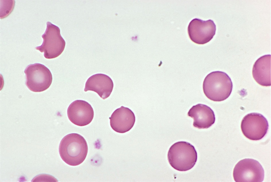
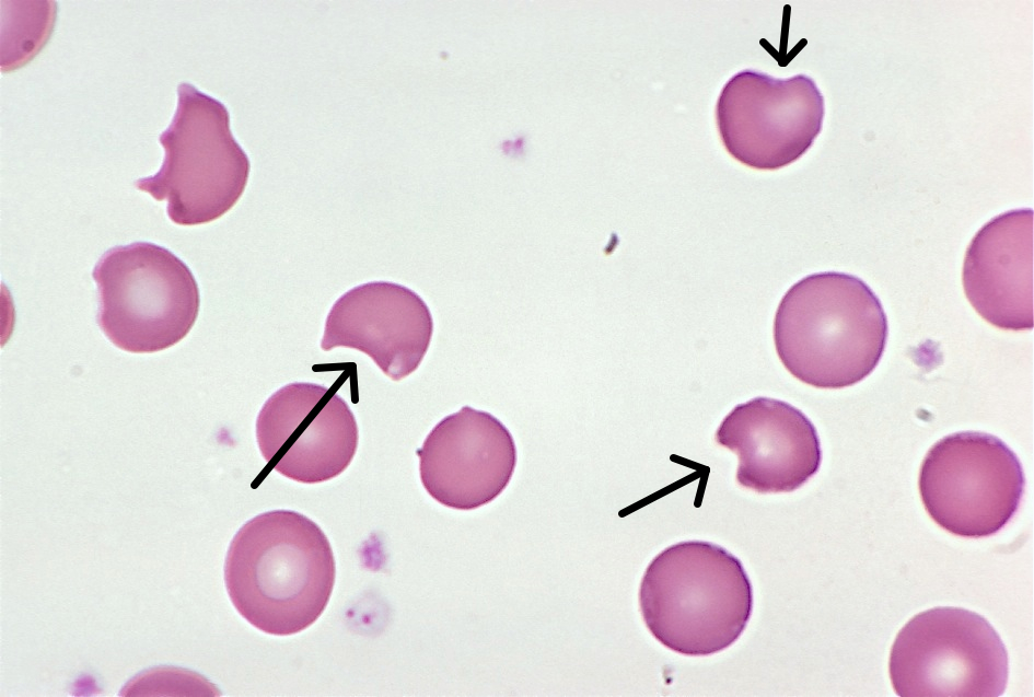
During my first year, I was also fortunate to have hematopathology at Jesse Brown VA Medical Center with Dr. John Kennedy. He is a hematopathologist who was trained as a morphologist before the heavy reliance on flow cytometry and IHC. And he taught me to love the morphology of blood cells especially with respect to lymphomas. I had enjoyed my hematology sub-I at the NIH when I was a medical student but had originally entered residency thinking that I’d pursue molecular genetic pathology and clinical microbiology. But a great mentor can really open your mind to a different path and that is what Dr. Kennedy did for me. The second time I rotated with him, he was away for much of the rotation. But I loved the opportunity that I was given to take care of the hematopathology service in his absence. I looked at all the daily cases and performed path reviews and counts on peripheral blood smears and body fluids and the surgical pathology attending at the end of the day would review and sign-out my work. This experience of graduated responsibility helped me to decide to pursue hematopathology and it was nice to have someone believe in me and my abilities. I was also fortunate that I was in a city where the Lymphoma Foundation also held quarterly inter-program Lymphoma Rounds which I was able to attend.

So my advice is to identify mentors early on in your residency. You may not be able to see the ripple effect they have on your life until later on but I promise that they will touch your lives in an indelible manner that will help shape the pathologist you will become later on in life.

This week I’m at the ASCP Annual Meeting and will write about those experiences in a future blog, but for now, I’d like to take this opportunity to personally thank my mentors. I hope that I take your lessons and make you proud as a future physician-scientist with a public health (molecular epidemiology aka biomarker discovery which was one of my areas of specialization during my MPH) focus. You have touched my life in ways that I may not always be able to articulate but do acknowledge and appreciate.


Chung

-Betty Chung, DO, MPH, MA is a fourth year resident physician at Rutgers – Robert Wood Johnson University Hospital in New Brunswick, NJ.

You Make the Diagnosis: a 32-Year-Old with Mild Jaundice

A 32-year-old male presents with mild jaundice and flank pain. He recently developed sinusitis, which was treated with trimethoprim-sulfamethoxazole, but is otherwise healthy. His hemoglobin is 10.2 g/dL (13.5-17.5), MCV is 90 μm3 (80-100), and total bilirubin is 3.4 mg/dL (0.2-1.5). A representative field from his blood smear is shown here. What is the most likely diagnosis?

g6pd1

  1. Aplastic anemia
  2. Iron-deficiency anemia
  3. Glucose-6-phosphate dehydrogenase deficiency
  4. Autoimmune hemolytic anemia
  5. Megaloblastic anemia

The answer is glucose-6-phosphate dehydrogenase (G6PD) deficiency. G6PD deficiency is an X-linked recessive disorder in which patients produce decreased amounts of G6PD, a red blood cell enzyme involved in detoxifying free radicals.

When a patient with G6PD deficiency is exposed to an oxidant stress (which can be anything from an illness to ingestion of certain foods or drugs), the resulting reactive oxygen species attack structures within the red cell.

Globin chains are particularly vulnerable to oxidant damage. They become denatured and stick to the inside of the red cell membrane, forming inclusions called Heinz bodies, which are visible on crystal violet staining. Heinz bodies are removed by macrophages in the spleen, leaving visible “bites” in the red cells. Several bite cells are visible in this patient’s blood smear (arrows).

g6pd2

Most episodes of hemolysis in patients with G6PD deficiency resolve on their own after the offending substance is removed.

Krafts

-Kristine Krafts, MD, is an Assistant Professor of Pathology at the University of Minnesota School of Medicine and School of Dentistry and the founder of the educational website Pathology Student.

You Make the Diagnosis: A 62 Year Old Female with Hemoptysis and Fatigue

A 62-year-old female presents with hemoptysis and fatigue. A large mass is found adjacent to the right main bronchus. A representative field is shown here. What is the diagnosis?

smallcell

  1. Invasive adenocarcinoma
  2. Squamous cell carcinoma
  3. Adenocarcinoma in situ (formerly bronchoalveolar carcinoma)
  4. Small cell carcinoma
  5. Large cell carcinoma


The diagnosis in this case is small cell carcinoma. Also called “oat cells,” the malignant cells in this tumor tend to be round to somewhat elongated in shape, with the typical “salt-and-pepper” chromatin of neuroendocrine tumor cells. The cells are often so closely apposed that their nuclear contours show a characteristic “molding” effect. Small cell carcinoma is a fast growing tumor (note the large mitotic figure at 4 o’clock) with a poor prognosis.


Krafts

-Kristine Krafts, MD, is an Assistant Professor of Pathology at the University of Minnesota School of Medicine and School of Dentistry and the founder of the educational website Pathology Student.


Microbiology Case Study: 38-Year-Old with Intermittent Straight Catheterization

Case History:

A 38 year old man with history of secondary progressive multiple sclerosis complicated by neurogenic bladder requiring intermittent straight catheterization presented with fevers, chills, and weakness. Clinical evaluation revealed hypotension and leukocytosis concerning for sepsis. His urinalysis was positive for 3+ blood, 1+ leukocyte esterase and nitrites. Urine was sent to our laboratory for culture and grew bacteria with the below gram stain and colony morphology.

Gram stain showing gram negative bacilli.
Sheep blood agar growing nonhemolytic grey-yellow mucoid bacterial colonies.
MacConkey agar growing mucoid lactose fermenting (pink) bacterial colonies.


Laboratory identification:

Klebsiella and Raoultella both are gram negative bacilli that form lactose fermenting, yellow, mucoid bacterial colonies. The bacterial colonies appear mucoid because of the bacteria’s polysaccharide capsule. Distinguishing these two types of bacteria requires molecular analysis as their morphology can be identical. In our initial identification process, only one type of bacteria was detected on the urine culture. Given the gram stain and colony morphology, our differential included Klebsiella, Enterobacter, and Roaultella. The bacteria was verified as Roaultella species using mass spectrometry. However, our laboratory also received two sets of blood cultures which showed the same morphology as above but the bacteria was identified as K. oxytoca. This prompted a review of the urine culture in which a subtle second bacterial colony morphology was seen and confirmed as K. oxytoca.

Discussion:

In summary, our patient had a urinary tract infection caused by Roaultella and Klebsiella which was the source of the patient’s sepsis. Only the Klebsiella was detected in his blood stream. Raoultella and Klebsiella are both gram negative, oxidase negative, non-motile, capsulated, facultative anaerobic bacilli within the Enterobacteriaceae family. Raoultella was initially classified within the Klebsiella genus, but reclassified based on comparative analysis of the 16S rRNA gene and rpoB gene which encodes the β subunit of bacterial RNA polymerase. Members of the Raoultella genus include R. electrica, R. terrigena, R. planticola, and R. ornithinolytica and are found in the environment, specifically in plants, soil, and water. Raoultella species are rare in human disease but have been documented to cause bacteremia, urinary tract infection, conjunctivitis and cholecystitis.

Klebsiella causes human disease much more frequently than Roaultella. Klebsiella are part of human oropharynx and gastrointestinal flora that act as opportunistic pathogens. Mode of transmission may occur endogenously or through person to person spread, hence nosocomial infections are common. K. oxytoca and K. pneumoniae are the most frequently implicated species in human disease and may cause a wide spectrum of infections such as urinary tract infections, respiratory infections, enteritis, meningitis and bacteremia.

For our patient, we reported antibiotic sensitivities that were susceptible to both types of bacteria. The patient was treated with ceftriaxone and then transitioned to cefpodoxime with resolution of his urosepsis. In general, Raoultella’s susceptibility to antimicrobial agents are not well studied and should be based on the antibiotic sensitivities of each strain. In contrast, Klebsiella is well documented to have increasing antimicrobial resistance. Most clinical strains are resistant to ampicillin, carbenicillin and ticarcillin as well as extended spectrum beta-lactam drugs. Resistance to beta-lactam drugs is the result of Klebsiella bacteria that harbor plasmids which produce beta-lactamase enzymes.


-Jill Miller, MD is a 2nd year anatomic and clinical pathology resident at the University of Vermont Medical Center.

Wojewoda-small

-Christi Wojewoda, MD, is the Director of Clinical Microbiology at the University of Vermont Medical Center and an Assistant Professor at the University of Vermont.


Microbiology Case Study–35-Year-Old with a Puncture Wound

Case History:

A 35 year old healthy man presented with a puncture wound of the left hand. He was bit by his neighbor’s dog. Fluid expressed from the wound was sent to the microbiology laboratory with the following gram stain and colony morphology.

past1
Gram stain showing small gram negative coccobacilli.
Sheep blood agar with smooth, gray, non-hemolytic bacterial colonies.


Laboratory Identification:

Bacterial colonies, as shown above, grew on sheep blood and chocolate agars. No growth was present on CNA or MacConkey agars which in combination with the gram stain indicated a type of fastidious gram negative bacteria. Additionally, the bacterial colonies were oxidase positive, catalase positive and indole positive. These findings were consistent with Pasteurella species and confirmed to be P. multocida by mass spectrometry.

Discussion:

Pasteurella species are nonmotile, gram-negative, facultative anaerobic coccobacilli or rods. P. multocida is the most common species involved in human disease. P. multocida is part of the normal oropharyngeal flora in multiple animals including cats, dogs, cattle, horses, rodents and other animals. This bacteria has been reported in up to 70-90% cats and 40-60% of dogs. Humans who have frequent exposure to animals may also harbor P. multocida as part of their normal flora.

Pasteurella are opportunistic pathogens that require mechanical disruption of host barriers. The vast majority of P. multicida related diseases are wound infections and/or cellulitis as a result of a cat bite or scratch. Transmission of Pasteurella also occurs in partially healing wound or regions of poor skin integrity that are licked by a cat or dog. Pasteurella infections may be complicated by septic arthritis and osteomyelitis if wounds are deep and inoculate the underlying soft tissue. Patients who are immunocompromised may develop bacteremia with more widespread infections. The majority of Pasteurella species are susceptible to penicillin, cephalosporins and tetracycline. Our laboratory does not report antibiotic sensitivities for P. multocida for a few reasons: Pasteurella is rarely resistant to penicillin, and bite wounds are generally mixed infections.


-Jill Miller, MD is a 2nd year anatomic and clinical pathology resident at the University of Vermont Medical Center.

Wojewoda-small

-Christi Wojewoda, MD, is the Director of Clinical Microbiology at the University of Vermont Medical Center and an Assistant Professor at the University of Vermont.

Make the Diagnosis–A 58 Year Old Male with Abdominal Pain

A 58-year-old male presents with abdominal pain and bloody diarrhea which had a gradual onset over the last 3 weeks. He has lost 8 pounds during this period of time. A cecal biopsy is performed, and a representative section is shown here. What is the most likely diagnosis?

amoebiasis1

  1. Amoebiasis
  2. Ascariasis
  3. Candidiasis
  4. Cryptosporidiosis
  5. Giardiasis

The diagnosis in this case is amoebiasis. The most common cause of amebic enterocolitis is Entamoeba histolytica, an organism spread through ingestion of cysts in contaminated food or water. The trophozoite forms are round, and frequently show intracytoplasmic red cells.

Amoeba with intracytoplasmic red cells

Occasionally, the organisms invade other organs, such as the liver, lung, and heart. Most patients may be treated on an outpatient basis, unless there is severe colitis or extraintestinal disease.

The organisms frequently invade through the mucosa and into the submucosa, often with lateral extension.

"Flask-shaped" ulcer in amoebiasis
“Flask-shaped” ulcer in amoebiasis

Occasionally, the organisms invade other organs, such as the liver, lung, and heart. Most patients may be treated on an outpatient basis, unless there is severe colitis or extraintestinal disease.

Krafts

-Kristine Krafts, MD, is an Assistant Professor of Pathology at the University of Minnesota School of Medicine and School of Dentistry and the founder of the educational website Pathology Student.

Microbiology Case Study–Upper Thigh Pain

An 85 year old man presented with right medial upper thigh pain and swelling.  Imaging revealed a large pseudoaneurysm in the right superficial femoral artery with evidence of rupture. The patient was taken to the operating room for placement of a gortex stent graft. His postoperative course was complicated by development of a large hematoma at the surgical site. Incision and drainage of the hematoma was surgically performed and fluid from the hematoma was sent to the microbiology laboratory.

Gram stain with multiple gram negative bacilli.
White-grey bacterial colonies growing on blood agar plate.
Grey semi-translucent non-lactose fermenting colonies growing on MacConkey agar.


Laboratory Identification:

The gram stain and plates confirmed the bacteria were non-lactose fermenting, non-hemolytic gram negative bacilli which is consistent Salmonella. Salmonella species was confirmed by mass spectrometry. Another feature helpful in the identification of Salmonella is its ability to produce hydrogen sulfide. Although not performed in this case, Salmonella will produce colonies with black centers when grown on Xylose lysine deoxycholate agar (selective agar that has thiosulfate which Salmonella metabolize to hydrogen sulfide).

The bacterial isolates of Salmonella were forwarded to the public health laboratories where serotype is determined based on serologic reactions to O and H antigens. The O antigen is the most external component of the lipopolysacccharide of gram negative bacteria and the H antigen is the antigenic determinant that makes up the flagellar subunits . This Salmonella species was identified to be S. enteritidis. Two sets of the patient’s blood cultures also grew S. enteritidis.

Discussion:

Salmonella are motile, gram negative bacilli that are widely disseminated in nature. Various animals such as turtles, lizards, snakes and birds are associated with Salmonella. Salmonella may infect humans via ingestion of contaminated food products that are typically of poultry or dairy origin. Person to person transmission may also occur by fecal-oral route. Salmonella has multiple virulence factors that allow it to evade the immune system. One of its virulence factors is the polysaccharide capsule that surrounds the O antigen. The O antigen is highly immunogenic and shielding the O antigen prevents its recognition by antibodies. Additionally, Salmonella can periodically change its H antigen as another protective mechanism against antibodies.


Jill Miller, MD is a 2nd year anatomic and clinical pathology resident at the University of Vermont Medical Center.

Wojewoda-small

Christi Wojewoda, MD, is certified by the American Board of Pathology in AP/CP and Medical Microbiology. She is currently the Director of Clinical Microbiology at the University of Vermont Medical Center and an Assistant Professor at the University of Vermont.


The Importance of Truly Internalizing Feedback and Learning From It

Recently, I’ve felt a shift in the timeline. Part of this I can attribute to having less time to myself as I ease into chief resident duties. Time I would’ve spent doing (or postponing) other activities is now relegated to this new role. But this increased demand on my time is not the only factor. Time feels like it is more rapidly passing with each year of residency, and more accelerated as of late.

Taking the annual RISE this time of year also contributes to this. I’m reminded that I should have reached some invisible bar on the meter stick in terms of my knowledge base and hope that I am commensurate with where I should be at this point in my residency. Because sooner than I may feel comfortable with, I will be expected to be “competent” enough to serve as a junior attending during my fellowships. And even though I’ve put it off until a later date, I know that I should also start composing a study plan soon for my boards because time is short between now and graduation.

Lately, probably because it was my most recent rotation, I’m reminded of my surgpath fellows during my PGY-1 telling me that I would learn the most from my cases, both AP and CP. Even though I was listening, I don’t think that I quite understood the depth of those words until my third year. During residency, we often don’t have much time to think because our service duties occupy much of that time. And the desire and need for sleep occupies much of the remainder of the time. But the light bulb moment has gone off so to speak in terms of what they meant by “learning from my cases” – be deliberate and start early.

For much of my first two years, as I’ve previously written, I’ve had a love/hate relationship with surgpath. Maybe those words are too strong, because I neither loved nor hated it, more like was ambivalent toward it. I naturally gravitated toward those subspecialties (obviously not surgpath) that I felt more comfortable with because of my previous training and interests – we all do.

But now I find that grossing is more meaningful and less of a chore to get through for me because I truly understand now how important it is that I do it well – be able to identify the important lesions and sections (90-95% of the diagnosis is off the gross, after all), cut thin and deliberate sections that look like “sushi” as one resident described my grossing, and understand how the sections I provide contribute to staging in the case of cancers. I understand these aspects better now because my grossing skill was called into question during my 2nd year. Since then, I’ve put a good amount of effort into correcting any deficiencies. Even the rotation director who originally brought up this issue, joked about the disasters of my first day on surgpath at his hospital at every end-of-the-rotation evaluation I had since then. His method of feedback may have been dramatic at the time but he really did provide me with a defining moment that changed my outlook and approach and for that I am grateful.

But it’s necessary to be deliberate and start early whatever rotation you’re on. Even though I read about the diagnoses for most of my big resection cases or at least did a quick pathology outlines search each time, I really wish that I would’ve spent even more time really reading up on those cases besides the cursory skim to come up with a diagnosis earlier in my residency. These days, I try to read a little every day, whether it be from a textbook or a journal article. And I’ve found that my knowledge, understanding, and skills improve at a faster rate. But I do wish that I had started this process from my PGY-1 so that I wouldn’t feel like I’m behind where I should be in terms of being ready for boards…so that I didn’t feel like I’m going to have to cram like I used to during college and med school for boards or wonder how to retain info that I learned two years ago on a rotation I haven’t had since PGY-1.

So really listen to the feedback from those more experienced then you. It probably took them longer than they would’ve liked to get to that light bulb moment. That is probably why they are making it a point to bring up that pearl of wisdom to you that they should’ve and wished they could’ve known then.


Chung

-Betty Chung, DO, MPH, MA is a third year resident physician at Rutgers – Robert Wood Johnson University Hospital in New Brunswick, NJ.

Microbiology Case Study–Abdominal Pain

A 60 year old man presented with abdominal pain and bloody diarrhea. He denied fever, chills, nausea, vomiting or recent travel. A stool culture was sent to the microbiology laboratory.

Colony Gram stain showing Gram-negative bacilli
Grey-white bacterial colonies growing on a blood agar plate.
Fuschia colonies growing on CHROMagar O157.


Laboratory Identification:

E. coli O157:H7 is most likely to be detected in the acute phase of illness and may be missed after 5-7 days from onset of symptoms. In general, laboratory identification is based on the detection of Shiga toxin-producing strains or detection of the O157:H7 serotype through various methodologies. In our laboratory, we identified E. coli O157 based on the above gram stain and colony morphology in combination with growth with the appropriate color on a selective plate for E. coli O157, CHROMagar O157. We used our automated microbial identification system, Vitek 2, which performs multiple biochemical reactions to confirm the bacteria as E. coli O157:H7. Additionally, we identified the presence of Shiga toxin through an immunochromatographic lateral flow rapid test using monoclonal antibodies specific to Shiga toxins.


Discussion:

E. coli are gram negative rods that are beta hemolytic, indole positive and lactose fermenters. E. coli is part of the normal colon flora but certain types of E. coli can cause disease depending on their virulence factors. Enterohemorrhagic E. coli (EHEC), also known as Shiga toxin producing E.coli (STEC), is one of six major groups of E. coli that causes diarrhea. EHEC produce a Shiga toxin that inhibits protein synthesis of intestinal epithelial cells via inhibition of the 60S ribosome. The most common serotype is E. coli O157:H7. Transmission occurs through ingestion of raw milk or uncooked ground beef. Hamburgers have been the cause of many outbreaks of infection in the United States although majority of E. coli O157:H7 infections are not associated with outbreaks.

Clinical manifestations from E. coli O157:H7 infection usually occurs at three days from time of exposure but may vary from one to eight days. Clinical symptoms typically begin with abdominal cramps, vomiting, and bloody diarrhea without fever. However, patients may experience a spectrum of disease ranging from asymptomatic infection (less common) to hemorrhagic colitis with progression hemolytic-uremic syndrome (HUS). HUS is the most common cause of acute renal failure in children and results from toxin-mediated damage of endothelial cells in the kidney. HUS is characterized by the triad of acute renal failure, microangiopathic hemolytic anemia, and thrombocytopenia. Supportive therapy is recommended for treatment of E. coli O157:H7 infections. Antibiotics are not recommended because of the potential to increase Shiga toxin production. For this reason, we do not report antibiotic sensitivities for E. coli O157:H7.


Jill Miller, MD is a 2nd year anatomic and clinical pathology resident at the University of Vermont Medical Center.

Wojewoda-small

Christi Wojewoda, MD, is certified by the American Board of Pathology in AP/CP and Medical Microbiology. She is currently the Director of Clinical Microbiology at the University of Vermont Medical Center and an Assistant Professor at the University of Vermont.

Hematology Case Study: 60-Year-Old Male with Hives

A 60-year-old male presents with hives, skin flushing, and headaches. After an appropriate preliminary work-up, a bone marrow biopsy is performed. A representative section from the bone marrow biopsy is shown here. What are the granulated cells at the center of this image?

mast

A. Megakaryocytes
B. Promyelocytes
C. Mast cells
D.Myeloma cells
E. Adenocarcinoma cells

The granulated cells in this image are mast cells, which are identified by their abundant, metachromatic granules. This patient was diagnosed with systemic mastocytosis, a clonal disorder of mast cells and their precursors.

Mastocytosis is actually a spectrum of rare disorders, all of which are characterized by an increase in mast cells. Most patients have disease that is localized to the skin, but about 10% of patients have systemic involvement, like the patient in this case. There is a localized, cutaneous form of mastocytosis called urticaria pigmentosum that happens mostly in children and accounts for over half of all cases of mastocytosis.

Clinically, the skin lesions of mastocytosis vary in appearance. In urticaria pigmentosum, the lesions are small, round, red-brown plaques and papules. Other cases of mastocytosis show solitary pink-tan nodules that may be itchy or show blister formation. The itchiness is due to the release of mast cell granules (which contain histamine and other vasoactive substances).

In systemic mastocytosis, patients have skin lesions similar to those of urticaria pigmentosum – but there is also mast cell infiltration of the bone marrow, lymph nodes, spleen and liver. Patients often suffer itchiness and flushing triggered by certain foods, temperature changes, alcohol and certain drugs (like aspirin).

Krafts

-Kristine Krafts, MD, is an Assistant Professor of Pathology at the University of Minnesota School of Medicine and School of Dentistry and the founder of the educational website Pathology Student.