Microbiology Case Study: A 36 Year Old Male Traveler with Fever

Case Description

A 36 year old male presented to the emergency department with complaints of fevers, chills, night sweats, nausea, diarrhea, weakness, and decreased appetite for 6 days. He often travels between India and Dallas, and five months prior to presentation returned from two years abroad. While overseas, he developed similar symptoms, but due to COVID-19 restrictions, he was unable to see a provider at that time. His family doctor prescribed a course of medication for presumed malaria, which he completed but could not recall the name of the medication. He endorsed being ill for two weeks at that time and improved with medication to complete resolution of his symptoms. Prior to presentation, he also endorsed 3-4 episodes of non-bloody diarrhea per day and remembered a period of self-resolving chills a month prior. His fever and rigors were cyclic, occurring every other day, worsening up to presentation.

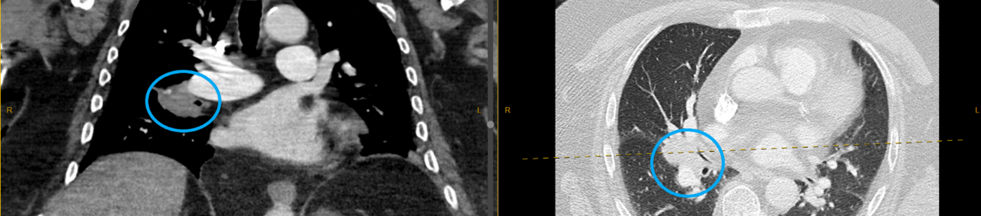
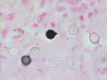
Given his travel history and symptomology, blood was drawn in the emergency department for analysis including a malaria smear. CBC and CMP were significant for elevated bilirubin (Total bilirubin 1.6 mg/dL, Direct bilirubin 0.4 mg/dL), leukopenia (3.60 x 10(9)/L), macrocytosis (92.5 femtoliters), thrombocytopenia (86 x 10(9)/L), and elevated CRP (6.6 mg/dL). His blood differential was significant for neutrophilia (91%), lymphocytopenia (7%), and monocytopenia (1%). The malaria smear was positive, and the patient was given a dose of artemether/lumefantrine in the emergency department. Plasmodium vivax was identified at a parasitemia of 0.5% (Figure 1), and the infectious disease service recommended admission for further workup including testing for G6PD deficiency prior to starting primaquine. He was not G6PD deficient, and an ultrasound of the spleen was unremarkable. The patient was treated with chloroquine for the erythrocytic and primaquine for the exo-erythrocytic stages of P. vivax malaria.

Figure 1. Photomicrograph of Plasmodium vivax ring forms observed in this patient’s Giemsa-stained peripheral blood smear, which are counted to determine the level of parasitemia in a patient’s bloodstream (500x oil immersion).

Discussion

Malaria is an infection caused by protozoan parasites of the genus Plasmodium. These organisms are transmitted by female Anopheles mosquitos upon taking a blood-meal. Human malaria is caused by five defined Plasmodium species: P. falciparum, P. vivax, P. ovale, P. malariae, and P. knowlesi.1 While not endemic to the United States, there is significant disease burden worldwide. In 2019, an estimated 230 million cases of malaria were reported causing approximately 409,000 deaths.2

The two lifecycles of Plasmodium sp. in the human host are classically defined as “erythrocytic” and “exo-erythrocytic”, involving red blood cells and hepatocytes, respectively. Plasmodium sporozoites are inoculated into the human host from the salivary glands of the mosquito upon feeding. From there, the sporozoites travel to the liver where they infect hepatocytes, mature into schizonts and ultimately merozoites. The infected hepatocyte then ruptures, releasing merozoites which enter the circulation and infect erythrocytes, initiating the erythrocytic cycle. This is a unifying trait of all Plasmodium sp. causing human malaria. Importantly, P. vivax and P. ovale form hypnozoites (dormant forms) in the liver, which can reactivate (oftentimes months to years later) following bloodstream clearance, resulting in relapse. It is therefore important that Plasmodium sp. infections be accurately speciated, as management of liver stage parasites differs from that of those in the bloodstream. By contrast, P. malariae and P. falciparum do not form hypnozoites and thus do not chronically infect the liver.

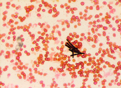
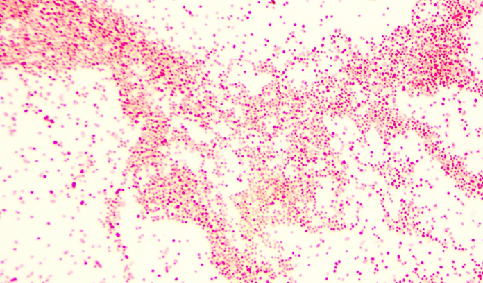
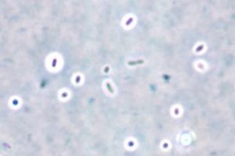
Plasmodium speciationis accomplished by evaluating thin and thick blood spears,4 allowing for assessment of parasite morphology and determination of parasitemia to guide patient management. In cases of P. vivax, the red blood cells are often enlarged (1.5 to 2 times the size of uninfected erythrocytes). Ring forms in all stages of development can be observed in P. vivax infection. These ring forms subsequently mature into trophozoites or gametocytes. P. vivax trophozoites exhibit a large, amoeboid cytoplasm, large chromatin dots, and fine yellow-brown pigment. Trophozoites subsequently develop into schizonts in the infected erythrocytes, subsequently rupturing leading to autoinfection. P. vivax schizonts are large with coalesced pigment and harbor 12 or more merozoites3 (Figure 2). P. vivax gametocytes are large and round to oval shaped and have scattered brown pigment, hemozoin, that may fill the erythrocyte (Figure 3). Gametocytes will migrate to the capillaries which are taken up by a mosquito upon taking a blood-meal, completing the Plasmodium lifecycle.

Figure 2. Photomicrograph of Plasmodium vivax merozoites in a schizont (1000x oil immersion) from this patient.
Figure 3. Photomicrograph of Plasmodium vivax gametocyte with malaria pigment (500x oil immersion) from this patient.

Here we present a case of relapsed P. vivax infection. Blood stage P. vivax parasites are susceptible to chloroquine, but dormant hypnozoites in the liver are resistant to its effects. Hypnozoites can be treated with primaquine, and thus routine management of either P. ovale or P. vivax usually consists of a combination of both antimalarial drugs. It is important to note that primaquine is contraindicated in cases of G6PD deficiency and pregnancy due to hemolytic complications,2 which is why this patient was tested prior to initiating therapy.

P. vivax has a worldwide distribution but has higher prevalence in colder climates as compared to other malaria species. P. vivax is most commonly encountered in Latin America and Southeast Asia. In addition to colder climate adaptation, P. vivax is interesting in that the parasite uses Duffy red cell antigens to enter erythrocytes and in populations with low frequency of Duffy on the surface of RBCs those groups are generally resistant to P. vivax infection. However, there have been rare cases of P. vivax in Africans who are Duffy-null.5

References

  1. Gladwin, M., Mahan, C. S., & Trattler, B. (2021). Malaria. In Clinical microbiology made ridiculously simple (pp. 343–346). essay, MedMaster, Inc.
  2. Menkin-Smith L, Winders WT. Plasmodium Vivax Malaria. [Updated 2021 Jul 23]. In: StatPearls [Internet]. Treasure Island (FL): StatPearls Publishing; 2021 Jan-. Available from: https://www.ncbi.nlm.nih.gov/books/NBK538333/
  3. Procop, G. W., Koneman, E. W., & Winn, W. C. (2017). Malaria. In Koneman’s color Atlas and textbook of diagnostic microbiology (pp. 1467–1470). essay, Lippincott Williams & Wilkins.
  4. Laboratory diagnosis of malaria: Plasmodium vivax. Laboratory Identification of Parasites of Public Health Concern. (n.d.). Retrieved September 14, 2021, from https://www.cdc.gov/dpdx/resources/pdf/benchAids/malaria/Pvivax_benchaidV2.pdf.
  5. Gunalan, K., Niangaly, A., Thera, M. A., Doumbo, O. K., & Miller, L. H. (2018). Plasmodium vivax infections of duffy-negative erythrocytes: Historically undetected or a recent adaptation? Trends in Parasitology, 34(5), 420–429. https://doi.org/10.1016/j.pt.2018.02.006

-Elisa Lin is a fourth-year medical student at UT Southwestern Medical Center in Dallas, Texas. She is interested in AP/CP track residencies.

-Clare McCormick-Baw, MD, PhD is an Assistant Professor of Clinical Microbiology at UT Southwestern in Dallas, Texas. She has a passion for teaching about laboratory medicine in general and the best uses of the microbiology lab in particular.

-Andrew Clark, PhD, D(ABMM) is an Assistant Professor at UT Southwestern Medical Center in the Department of Pathology, and Associate Director of the Clements University Hospital microbiology laboratory. He completed a CPEP-accredited postdoctoral fellowship in Medical and Public Health Microbiology at National Institutes of Health, and is interested in antimicrobial susceptibility and anaerobe pathophysiology.

Microbiology Case Study: A Middle-Aged Man with Generalized Weakness

Case History

A middle age male with a past medical history of liver cirrhosis presented to the emergency department with one day of fever, chills, generalized weakness, and nausea. Complete blood count with differential showed leukopenia and neutropenia. Infectious work up was initiated including collection of 2 sets of blood cultures and imaging studies. A computed tomography (CT) scan of the abdomen revealed an irregularly shaped hypodense lesion in the right hepatic lobe concerning for abscess (Image 1). Ultrasound guided aspiration for the hepatic lesion yielded cloudy yellow bilious fluid, which was sent to the microbiology lab for aerobic and anaerobic cultures.

Image 1. CT scan of abdomen showing irregularly shaped hypodense lesion (yellow circle).

Two sets of blood cultures turned positive and Gram stain showed slender Gram positive rods in chains (Image 2). The aspirated fluid culture also showed 4+ Gram positive rods. Small gray colonies appeared on blood agar, chocolate agar, and Columbia Naladixic Acid (CNA) agar from both specimen types (Image 3). Lactobacillus rhamnosus was identified by matrix-assisted laser desorption/ionization time-of-flight mass spectrometry (MALDI-TOF MS). Minimal inhibitory concentration (MIC) was determined by broth microdilution assay and the organism was susceptible to penicillin while resistant to vancomycin. With appropriate antibiotics and abscess drainage, the patient’s condition improved and he was discharged to home.

Image 2. Gram stain of blood culture demonstrating gram positive rods in chains.
Image 3. Small gray colonies in blood agar from blood culture.

Discussion

Lactobacillus species are facultatively anaerobic, gram positive, non-spore forming rods that can have varying Gram stain morphology including short plump rods or long slender rods in chains or palisides.1 Lactobacillus species are bacterial inhabitant of the human mouth, gastrointestinal tract, and female genital tract. Isolation from clinical specimens could be considered by many to have questionable clinical significance.2 Lactobacillus species are often present in probiotics and fermented dairy products, including yogurt, and have been reported to provide benefits in gastrointestinal health,3–5 which led to increase in consumption of these products by many people, including our patient.

Cases of liver abscess and bacteremia caused by Lactobacillus species have been rarely reported in the literature and risk factors for the infection were immunosuppression, uncontrolled diabetes, hepatopancreaticobiliary disease, bacterial translocation, and use of probiotics or heavy dairy product consumption.6,7 The causative strains included L. rhamnosus, L. acidophilus, and L. paracasei.7

Pathophysiology of liver abscess and bacteremia due to Lactobacillus species is not well understood but it is postulated that several mechanisms may contribute to the pathogenicity of lactobacilli. Some strains are able to bind to intestinal mucosa, which may aid in translocation of the organism into the bloodstream. Also some strains can adhere to extracellular matrix proteins, aggregate platelets, and produce glycosidases and proteases.7 Furthermore, some strains are more resistant to intracellular killing by macrophages and nitric oxide.8

It is worth noting that many species of Lactobacillus are intrinsically resistant to vancomycin. However, they are usually susceptible to penicillin and ampicillin, as it was seen in our patient, and antimicrobial susceptibility testing can be performed by determining MIC of antimicrobials.9

References

  1. Goldstein EJC, Tyrrell KL, Citron DM. Lactobacillus Species: Taxonomic Complexity and Controversial Susceptibilities. Clin Infect Dis. 2015;60(suppl_2):S98-S107. doi:10.1093/CID/CIV072
  2. Chan JFW, Lau SKP, Woo PCY, et al. Lactobacillus rhamnosus hepatic abscess associated with Mirizzi syndrome: a case report and review of the literature. Diagn Microbiol Infect Dis. 2010;66(1):94-97. doi:10.1016/J.DIAGMICROBIO.2009.08.009
  3. Kligler B, Cohrssen A. Probiotics. Am Fam Physician. 2008;78(9):1073-1078. Accessed November 17, 2021. http://www.aafp.org/afp.
  4. Anukam KC, Osazuwa EO, Osadolor HB, Bruce AW, Reid G. Yogurt containing probiotic Lactobacillus rhamnosus GR-1 and L. reuteri RC-14 helps resolve moderate diarrhea and increases CD4 count in HIV/AIDS patients. J Clin Gastroenterol. 2008;42(3):239-243. doi:10.1097/MCG.0B013E31802C7465
  5. Adolfsson O, Meydani SN, Russell RM. Yogurt and gut function. Am J Clin Nutr. 2004;80(2):245-256. doi:10.1093/AJCN/80.2.245
  6. Omar AM, Ahmadi N, Ombada M, et al. Breaking Bad: a case of Lactobacillus bacteremia and liver abscess. J Community Hosp Intern Med Perspect. 2019;9(3):235. doi:10.1080/20009666.2019.1607704
  7. Sherid M, Samo S, Sulaiman S, Husein H, Sifuentes H, Sridhar S. Liver abscess and bacteremia caused by lactobacillus: Role of probiotics? Case report and review of the literature. BMC Gastroenterol. 2016;16(1):1-6. doi:10.1186/S12876-016-0552-Y/TABLES/1
  8. Asahara T, Takahashi M, Nomoto K, et al. Assessment of Safety of Lactobacillus Strains Based on Resistance to Host Innate Defense Mechanisms. Clin Diagn Lab Immunol. 2003;10(1):169. doi:10.1128/CDLI.10.1.169-173.2003
  9. CLSI. M45. Methods for Antimicrobial Dilution and Disk Susceptibility Testing of Infrequently Isolated or Fastidious Bacteria ; Proposed Guideline. Vol 35.; 2015. Accessed November 17, 2021. http://www.clsi.org.

Do Young Kim, MD is a medical microbiology fellow at University of Chicago (NorthShore). His academic interests include clinical microbiology and infectious diseases, epidemiology, and public health.

-Paige M.K. Larkin, PhD, D(ABMM), M(ASCP)CM is the Director of Molecular Microbiology and Associate Director of Clinical Microbiology at NorthShore University HealthSystem in Evanston, IL. Her interests include mycology, mycobacteriology, point-of-care testing, and molecular diagnostics, especially next generation sequencing.

Microbiology Case Study: An Adult Patient with a Tender Mass and Rash

Case History

An adult patient with no significant past medical history presents with a tender right inguinal mass and rash over the right buttock measuring 5×7 cm. A skin punch biopsy was performed on the gluteal rash and sent to histopathology for analysis. Histology (Image 1) revealed an intradermal acantholytic vesicular dermatitis and associated folliculitis. Chronic inflammatory infiltrates surrounded neurovascular bundles as well as adnexal structures. Multinucleated Tzank cells were identified with the characteristic multinucleation, margination, and molding. Scattered eosinophilic Cowdry A inclusions were seen. Stains for bacteria and acid-fast bacilli (AFB) were not performed. A periodic acid-Schiff (PAS) stain (Image 2) demonstrated the absence of fungal elements.

Image 1. A hematoxylin and eosin (H&E) slide reveals a chronic inflammatory infiltrate surrounding (A) neurovascular bundles and (B) adnexal structures. (C) Tzank cells and (D) Cowdry A inclusions are also seen.
Image 2. A PAS stained slide of the same region as Image 1. (A) highlights a chronic inflammatory infiltrate where no fungal hyphae are seen.

Histopathology demonstrated “folliculitis suspicious for herpetic dermatitis.” PCR molecular testing for herpes simplex virus (HSV) and varicella zoster virus (VZV) were ordered on the punch biopsy. HSV was not detected; however, VZV was detected by PCR (Image 3, Image 4).

Image 3. The Simplexa VZV Direct Assay (Diasorin) targets a portion of the VZV DNA polymerase. The PCR amplification curve reveals the presence of VZV DNA (green) as well as that of the internal control (purple).
Image 4. A separate PCR assay targeting TP53 was performed to assess DNA quality of the fixed tissue. The presence of TP53 amplification in both the IC, the patient sample (Sample), as well as other samples on the same run (unlabeled) demonstrates the DNA quality is adequate. The absence of amplification of the NTC demonstrates a lack of nucleic acid contamination.

Discussion

Varicella zoster virus (VZV) is an enveloped double-stranded DNA virus belonging to the herpesviridae family.5 Transmission during primary infection occurs via inhalation of aerosolized respiratory secretions or lesional secretions, and to a lesser extent, via direct contact with lesional secretions. Transmission during secondary infection occurs mainly via physical contact with the secretions of herpetic lesions or the lesions themselves. The window for primary infection of transmissibility is 1-2 days before the onset of the rash lasting until either all lesions have crusted over or 24 hours have passed without the formation of new lesions, whereas secondary infections are only contagious during the presence of active lesions.6 Primary infection causes chicken pox, which is characterized by a vesicular rash, fever, and malaise. After primary infection, VZV resides in the dorsal root ganglia and trigeminal ganglia. VZV may reactivate, possibly as a result of stress or some other immunosuppressive state, as a painful vesicular rash known as shingles or herpes zoster. The rash is limited to the dermatome innervated by the ganglion from which the virus reactivated. Severe cases of shingles may result in meningitis, myelitis, as well as encephalitis, and can be fatal.1 Though the lesions of herpes zoster (secondary VZV infection) are infectious, they are significantly less so than those of varicella (primary VZV infection).6

The histology of VZV infection is characterized by intradermal and sub-epidermal vesicles with associated acantholysis, necrosis, and spongiosis. Tzanck cells demonstrate the characteristic “3 Ms” of multi-nucleation, marginated chromatin, and nuclear molding. The dermis is notable for perivascular, periadnexal, and perineural lymphocytic infiltrates. Folliculitis and syringitis may be present along with small vessel necrotizing vasculitis. Late stage lesions are notable for encrusted ulcers. Though there is significant histologic overlap between VZV infection as those caused by others in the herpes family, VZV histology tends to demonstrate a more substantial follicular involvement.2 Besides other herpes viruses, the differential diagnosis includes erythema multiforme, coxsackievirus, ecthyma contagiosum, pemphigus vulgaris and paravaccinia infection.3

While molecular methodologies are now the gold standard for diagnosis, a number of modalities including immunohistochemistry, immunofluorescence, in-situ hybridization, and serology can be used to aid in diagnosis.3 In the aforementioned case, diagnosis was made using a real time polymerase chain reaction (RT-PCR) assay (Simplexa VZV Direct Assay, Image 3) using previously extracted DNA. Forward and reverse primers target a well conserved portion of the VZV DNA polymerase. In between synthesis cycles, fluorescent probes anneal to the target sequence, separating the fluorophore from the quencher, thus generating a fluorescent signal. Amplification is measure by the cycle threshold (Ct), the number of PCR cycles needed for the fluorescent signal to exceed the background. An internal positive control (IC) is spiked in to assure negative results are not the result the presence of PCR inhibitors. To assess the quality of DNA present, a separate PCR was also performed on TP53, which amplifies if sufficiently high quality DNA is present, irrespective of the presence of VZV DNA (Image 4). A negative control (no template control, NTC) should be run to interrogate the presence of nucleic acid contamination.4

Treatment, if warranted, should be administered as soon as possible. Antiviral options include acyclovir, valacyclovir, or famcyclovir. Central nervous system, ocular, or renal VZV cases are considered emergencies and are typically treated with intravenous acyclovir.6 While resistance is rare, at least three mechanisms of resistance have been shown to endow VZV resistance to the aforementioned drugs: reduced or absent thymidine kinase, altered thymidine kinase activity leading to decreased phosphorylation of the drug, or decreased affinity of VZV DNA polymerase for acyclovir triphosphate.5, 8 If an infection with a resistant strain is identified or suspected, foscarnet is often used in place of acyclovir. Unlike the nucleoside analogs, this pyrophosphate analog does not rely on phosphorylation for the activation of its anti-VZV DNA polymerase activity.7 Historically plaque reduction assays were used, but this method is both labor intensive, low yield, and slow. Thus, molecular testing interrogating mutations in the DNA polymerase or thymidine kinase genes have increased in popularity.8

Two live attenuated vaccines are available, either in isolation or in combination with the measles mumps, and rubella vaccines (MMRV), in a 2 dose series to prevent primary infection. Since the VZV vaccine contains live virus, it should not be administered to pregnant women or the severely immunocompromised. Vaccine administration has been found to be 90% effective in preventing primary infection and 99% effective at preventing severe or complicated disease.7 Additionally, there is a recombinant vaccine consisting of the VZV glycophorin E protein in addition to an adjuvant that is used to prevent shingles. This formulation is recommended for adults over the age of 60 in prevention of secondary infections as well as to immunocompromised individuals at higher risk from exposure to the live attenuated vaccine.9

References

  1. Depledge DP, Sadaoka T, Ouwendijk WJD. Molecular Aspects of Varicella-Zoster Virus Latency. Viruses. 2018;10(7):349. Published 2018 Jun 28. doi:10.3390/v10070349
  2. Busam, K. J. Dermatopathology. 2nd Edition. Published 2014.
  3. Hall, B. Diagnostic pathology: Nonneoplastic Dermatopathology. 3rd Edition. Published 2021.
  4. Simplexa™ VZV Swab Direct REF MOL3655. 2021
  5. Sauerbrei A. Diagnosis, antiviral therapy, and prophylaxis of varicella-zoster virus infections. Eur J Clin Microbiol Infect Dis. 2016;35(5):723-734. doi:10.1007/s10096-016-2605-0
  6. https://www.cdc.gov/chickenpox/about/transmission.html
  7. https://www.cdc.gov/vaccines/vpd/varicella/hcp/index.html
  8. Piret J, Boivin G. Antiviral resistance in herpes simplex virus and varicella-zoster virus infections: diagnosis and management. Curr Opin Infect Dis. 2016;29(6):654-662. doi:10.1097/QCO.0000000000000288
  9. https://www.cdc.gov/vaccines/hcp/vis/vis-statements/shingles-recombinant.html

-Jeremy Adler, MD is a Molecular Genetic Pathology fellow at the University of Chicago Medicine and NorthShore University HealthSystem. He completed his MD at SUNY Stony Brook and his AP/CP residency at the Pennsylvania Hospital of the University of Pennsylvania Health System.

-Paige M.K. Larkin, PhD, D(ABMM), M(ASCP)CM is the Director of Molecular Microbiology and Associate Director of Clinical Microbiology at NorthShore University HealthSystem in Evanston, IL. Her interests include mycology, mycobacteriology, point-of-care testing, and molecular diagnostics, especially next generation sequencing.

Microbiology Case Study: A Female with Diabetes and Renal Disease

Case history

A middle-aged female with a past medical history of diabetes and end stage renal disease resulting in kidney transplant presented for evaluation of right hip and knee pain for the previous two months. An MRI of the hip revealed a large effusion with evidence of septic arthritis, myositis in the surrounding muscle, and osteomyelitis of the hip. Blood cultures remained negative for the duration of her presentation. The patient underwent a joint aspiration, and synovial fluid was sent to the microbiology laboratory for culture. Due to subsequent culture positivity and the extent of the involvement of the surrounding anatomy, the patient was started on ceftriaxone and underwent a total joint replacement. Her symptoms improved post-procedure, and post-operative vertebral MRI and TTE revealed no evidence of osteomyelitis or endocarditis. The patient was discharged on post-operative day six with continued IV ceftriaxone for an additional 5 weeks.

Laboratory identification

The synovial fluid received in the microbiology laboratory was plated onto blood, chocolate, and MacConkey agars. No organisms were visible on direct Gram stain, but the culture revealed scant growth of alpha-hemolytic colonies on blood and chocolate plates. These colonies were comprised of faintly staining gram positive rods (Image 1). The organism was catalase negative. Given the characteristic appearance by Gram stain, the organism was inoculated to a triple sugar iron (TSI) slant where it demonstrated H2S production. A definitive identification of Erysipelothrix rhusiopathiae was achieved by MALDI-TOF MS.

Image 1. Synovial fluid culture sent to the microbiology laboratory. E. rhusiopathiae colonies growing on Sheep’s blood agar are denoted by black arrowheads. Characteristic Gram stain of the E. rhusiopathiae colonies from the plate revealing poorly staining gram positive rods. TSI slant from the colonies demonstrating H2S production.

Discussion

Erysipelothrix rhusiopathiae is a facultatively aerobic, non-spore forming, gram positive pathogen that is a resident of the digestive and respiratory tracts of mammals, bird, fish, and pigs.1 It is the etiological agent of Swine Erysipelas, causing either an acute septicemia, cutaneous disease, endocarditis, or chronic arthritis in pigs. Human infections with E. rhusiopathiae are usually due to exposure to infected animals or contaminated animal products or environments. Certain occupations with frequent animal exposure are at increased risk for infection (including fishermen, veterinarians, farmers, and butchers). Infection requires entry into the skin through cutaneous abrasions, which can be caused by sharp hooks, fish scales, teeth, and other occupational tools or hazards that damage epithelial barriers.1,2

Human E. rhusiopathiae infection can manifest as three distinct forms. An acute, localized cellulitis named eryspieloid (not to be confused with streptococcal erysipelas) is the most common manifestation. This usually impacts the hands, fingers, or other parts of the upper extremities that have contact with animals or animal products.3 A generalized cutaneous form more often associated with systemic symptoms including fever, joint aches, lymphadenitis, lymphadenopathy, and arthritis can also occur. Finally, septicemia frequently associated with endocarditis is a third manifestation. E. rhusiopathiae endocarditis is often subacute, with a tropism for native valves (particularly the aortic valve). Due to its indolent nature, this presentation often requires valve replacement at the time of diagnosis and is associated with increased mortality.1,4 While cases of non-severe eryspieloid may self-resolve, ampicillin or penicillin are the treatments of choice for cutaneous and systemic infections. Cephalosporins and fluoroquinolones are also efficient alternative agents.3 Importantly, the organism is intrinsically resistant to vancomycin, thus accurate and timely identification is critical to ensure appropriate intervention (Image 2). Susceptibility testing is generally not performed but may be useful in the setting of penicillin allergy.

Image 2. E. rhusiopathiae is intrinsically resistant to vancomycin. E. rhusiopathiae exhibits elevated MICs to vancomycin. Penicillin is the treatment of choice.

Laboratory identification of E. rhusiopathiae can be challenging.  Erysiepelothrix can easily decolorize during gram staining and can be mistaken as gram negative due to lack of stain retention. Additionally, the cells can exhibit variable morphologies including pairs, chains, and filaments. Colonies can also exhibit variable morphotypes when grown on routine media, including both rough and smooth forms.2An environmental exposure to animals was investigated in this patient’s case to possibly serve as the source of infection. While a direct link cannot be definitively proven, it was revealed that the patient owned a large fish tank which she regularly cleaned which could have been a potential source of infection. 

References

  1. Wang Q, Chang BJ, Riley TV. 2010. Erysipelothrix rhusiopathiae. Veterinary Microbiology 140:405-417.
  2. Clark AE. 2015. The Occupational Opportunist: an Update on Erysipelothrix rhusiopathiae Infection, Disease Pathogenesis, and Microbiology. Clinical Microbiology Newsletter 37:143-151.
  3. Veraldi S, Girgenti V, Dassoni F, Gianotti R. 2009. Erysipeloid: a review. Clinical and Experimental Dermatology 34:859-862.
  4. Brooke CJ, Riley TV. 1999. Erysipelothrix rhusiopathiae: bacteriology, epidemiology and clinical manifestations of an occupational pathogen. Journal of Medical Microbiology 48:789-799.

-Timothy J. Kirtek, M.D., originally from Grand Blanc, Michigan, graduated from American University of the Caribbean School of Medicine located on the island of Sint Maarten. There, he conducted research on tropical arboviruses including Dengue, Chikungunya, and Zika viruses. He then returned to Michigan to complete his clinical training and, upon graduation from medical school, moved to Dallas, Texas where he is currently an Anatomic and Clinical Pathology resident physician at UT Southwestern.

-Andrew Clark, PhD, D(ABMM) is an Assistant Professor at UT Southwestern Medical Center in the Department of Pathology, and Associate Director of the Clements University Hospital microbiology laboratory. He completed a CPEP-accredited postdoctoral fellowship in Medical and Public Health Microbiology at National Institutes of Health, and is interested in antimicrobial susceptibility and anaerobe pathophysiology.

Microbiology Case Study: A Young Adult in Septic Shock

A 23 year old female with a previous medical history of endocarditis, hepatitis C, IV drug use, and aortic insufficiency status post emergent aortic valve replacement, presented to the ER in septic shock. After one week of hospitalization, she left against medical advice, and did not complete her prescribed course of antimicrobials.

One month later, she returned to the ER with tachypnea, lactic acidosis, and altered mental status, secondary to septic shock and she was admitted to the ICU. She was started on broad spectrum antibiotics based on the cultures from her previous hospitalization. Within one day, blood cultures from her central line were positive for growth of Serratia marcescens. Echocardiogram demonstrated prosthetic valve endocarditis with severe aortic regurgitation. Previous imaging had shown scattered septic emboli throughout her viscera, extremities, and now, MRI/MRA revealed cerebral lesions as well.

Ten and twelve days into her current hospitalization, blood and heart valve tissue cultures (respectively) were both positive for growth of the below-pictured organism. What is this causative organism?

Image 1. Central line blood culture.
Image 2. Heart valve tissue culture.

MALDI-ToF-MS identified the yeast from the blood culture and heart valve as Trichosporon asahii. It is a yeast-like basidiomycete. It is commonly found in soil, but is also a normal colonizer of mucous membranes of the GI and respiratory epithelium, and skin. It may also infect hair shafts and is the causative agent in “white piedra”. It is involved in several opportunistic infections in the immunosuppressed. Of all Trichosporon species, T. asahii is the most common cause of disseminated infection, especially in those with hematologic malignancies (leukemia, multiple myeloma, aplastic anemia, lymphoma), solid tumors, AIDS, and solid tumors. In immunocompetent patients, Trichosporon may cause infections including endophthalmitis following cataract surgery, endocarditis, following prosthetic heart valve replacement (as seen in this patient), and peritonitis in IV drug abusers or those receiving continuous ambulatory peritoneal dialysis (CAPD).

Trichosporon colonies are powdery, cream-colored, and with age, may develop surface wrinkles. On cornmeal Tween 80, yeast can either grow alone or in short chains. True and pseudohyphae may be seen. Barrel-shaped arthroconidia are typically present. Variable growth is seen on media containing cycloheximide. It may also cause Cryptococcal antigen agglutination tests to be falsely positive.

Diagnosis is typically via blood culture.

Combination therapy with amphotericin B and an -azole drugs seems to be the most successful treatment option.

Resources

  1. Brandt, ME, Lockhart, SR. Recent developments with Candida and other opportunistic yeasts. Curr Fungal Infect Rep. 6(3); 170-177. 2012.
  2. Dimorphic Systemic Mycoses | Mycology | University of Adelaide Accessed 10/22/21.
  3. Love, G. Mycology Benchtop Reference Guide. College of American Pathologists. P20. 2013.
  4. Maves, RC. Trichosporon Infections. Emedicine.medscape.com. Updated Feb 12, 2018. Accessed 10/18/21.
  5. Procop, GW, Church, DL, Hall, GS et al. Koneman’s Color Atlas and Textbook of Diagnostic Microbiology. 7th Edition. P 1366-1369. Wolter’s Kluwer Health. 2017.
  6. Ramos, JM, Cuenca-Estrella, M, Gutierrez, F, et al. Clinical case of endocarditis due to Trichosporon inkin and antifungal susceptibility profile of the organism. J Clin Microbiology. 42(5):2341-4. 2004.
  7. Trichosporon | Mycology | University of Adelaide Accessed 10/22/21.

-Jenny Pfeiffer, MD is a 1st year Anatomic and Clinical Pathology resident at the University of Vermont Medical Center.

-Christi Wojewoda, MD, is the Director of Clinical Microbiology at the University of Vermont Medical Center and an Associate Professor at the University of Vermont.

Microbiology Case Study: An Elderly Male Presents with Chest Tightness

Case History

An elderly male with a complex past medical history presented to the Emergency Department with the primary complaint of chest tightness for 2 days. He denied symptoms of diaphoresis, nausea, shortness of breath, palpitations, light-headedness, productive cough, dyspnea, chest pain, fevers, chills, or hemoptysis. He had no known sick contacts or recent travel. A computer tomography (CT) scan of the thorax showed a right hilar mass (Image 1). He underwent a bronchoscopy and right hilar transbronchial needle aspiration (TBNA) and bronchoalveolar lavage (BAL) were collected. The pathology report indicated abnormal lymphocytic proliferation, concerning for a mature small B-cell lymphoproliferative disorder.

Image 1. CT scan of the thorax showing the right hilar mass.

The BAL was submitted for acid-fast bacteria (AFB) culture, Gram stain, aerobic bacterial culture, and fungal culture. The AFB culture, Gram stain, and bacterial culture were all negative. However, 3 tan-yellow creamy colonies of a yeast grew on the sabouraud dextrose agar (SAB) plate in fungal culture after 7 days (Image 2). An India ink stain was performed (Image 3). MALDI-TOF confirmed the identification as Cryptococcus neoformans.

Image 2. Fungal growth on the SAB plate observed after 7 days.
Image 3. India ink staining of the fungus.

Discussion

Cryptococcus neoformans is an encapsulated pathogenic yeast, which is typically associated with bird droppings and contaminated soil.1,2 In immunocompromised patients, it can lead to severe opportunistic infections such as meningitis or disseminated disease. C. neoformans can cause life-threatening fungal infections in these patients, especially those with T-cell mediated immunodeficiency.3,4 The three main virulence factors include the complex capsule, melanin production, and ability to grow at human body temperature.5,6 Signs of pulmonary infection include cough, production of mucoid sputum, pleuritic chest pain, low-grade fever, dyspnea, weight loss, and malaise.

Fungal culture is one of the primary methods of Cryptococcus identification. Upon microscopic examination, Cryptococcus appears as a single bud and a narrow neck between parent and daughter cell and measures 4 – 10 uM.7 It has a fragile cell wall and a polysaccharide capsule that can vary from a wide halo to a nearly undetectable zone around the cells. Colonies can exhibit a wide range of color (i.e. cream, tan, pink, or yellow) and typically grow within one week of inoculation.8,9 India ink smear is a rapid method that allows direct visualization cryptococcal capsule, but is infrequently used now. Certain non-specific histological stains (including Periodic Acid-Schiff and May-Grünwald-Giemsa) can be used to detect fungi directly in fixed specimens. Fontana-Masson is a silver stain used for detecting melanin and has a high sensitivity for cryptococcosis.10  Other useful stains include hematoxylin-eosin, which reveals the clear halo, and mucicarmine and alcian blue, which target the polysaccharide capsule.11 Cryptococcal serology and cryptococcal antigen testing can be used for blood or CSF infections. Radiographic findings (especially in asymptomatic and immunocompetent patients) include patchy pneumonitis, granulomas (typically 2-7 cm), and miliary disease similar to tuberculosis.8 Treatment will vary depending on location of infection and host immune status. In some cases, pulmonary Cryptococcus may not be treated. Some clinical considerations include:

  • CSF chemistry parameters are normal
  • CSF culture, cryptococcal antigen, India ink preparation, and serology results are negative
  • Urine culture results are negative
  • Pulmonary lesion is small and stable/shrinking
  • No predisposing conditions for disseminated disease6

If treatment is required, fluconazole, itraconazole, or amphotericin B with or without flucytosine can be used depending on severity of infection.12

Cryptococcus gattii is another species of Cryptococcus. It differs from C. neoformans in that it typically infects both immunocompromised and immunocompetent patients. Canavine glycine bromothymol blue (CBG) agar can be used to differentiate C. gattii from C. neoformans: C. gattii is able to grow in the presence of canavine, turning the agar blue, while C. neoformans does not, leaving the media color unchanged.13,14

References

  1. Hagen, F., Khayhan, K., Theelen, B., Kolecka, A., Polacheck, I., Sionov, E., Falk, R., Parnmen, S., Lumbsch, H. T., and Boekhout, T. Recognition of seven species in the Cryptococcus gattii/Cryptococcus neoformans species complex. Fungal Genet Biol. 2015; 78: 16-48. 
  2. Lortholary, O., Nunez, H., Brauner, M. W., and Dromer, F. Pulmonary cryptococcosis. Semin Respir Crit Care Med. 2004; 25: 145–57.
  3. Lanternier, F., Cypowyj, S., Picard, C., Bustamante, J., Lortholary, O., Casanova, J. L., and Puel, A.  Primary immunodeficiencies underlying fungal infections.  Curr Opin Pediatr. 2013; 25: 736–47. 
  4. National Organization for Rare Disorders (NORD). Cryptococcosis. Available from: https://rarediseases.org/rare-diseases/cryptococcosis/ Last updated 2007; cited 2021 October 8.
  5. Idnurm, A., Bahn, Y.-S., Nielsen, K., Lin, X., Fraser, J. A., and Heitman, J. Deciphering the model pathogenic fungus Cryptococcus neoformans. Nat Rev Microbiol. 2005; 3(1): 753-64.
  6. Vandeputte, P., Ferrari, S., and Coste, A. T. Antifungal Resistance and New Strategies to Control Fungal Infections. Int J Microbiol. 2012: 713687.
  7. Guarner, J. and Brandt, M. E. Histopathologic Diagnosis of Fungal Infections in the 21st Century. Clin Microbiol Rev. 2011; 24(4): 247-80.
  8. Borman, A. M. and Johnson, E. M. (2020).  Candida, Cryptococcus, and Other Yeasts of Medical Importance. Manual of Clinical Microbiology, 12th Edition. Washington, DC: ASM Press. 2056-86.
  9. Coelho, C., Bocca, A. L., and Casadevall, A. The tools for virulence of Cryptococcus neoformans. Adv Appl Microbiol. 2014; 87: 1-41.
  10. Bishop, J. A., Nelson, A. M., Merz, W. G., Askin, F. B., and Riedel, S. Evaluation of the detection of melanin by the Fontana-Masson silver stain in tissue with a wide range of organisms including Cryptococcus. Hum Pathol. 2012; 43(6): 898-903.
  11. Guery, R., Lanternier, F., Pilmis, B., and Lortholary, O. Cryptococcus neoformans (Cryptococcosis). Antimicrobe. Available at: http://www.antimicrobe.org/new/f04.asp. Last updated: 2014; cited 2021 October 8.
  12. Perfect, J. R., Dismukes, W. E., Dromer, F., Goldman, D. L., Graybill, J. R., Hamill, R. J., Harrison, T. S., Larsen, R. A., Lortholary, O., Nguyen, M.-H., Pappas, P. G., Powderly, W. G., Singh, N., Sobel, J. D., and Sorrell, T. C. Clinical Practice Guidelines for the Management of Cryptococcal Disease: 2010 Update by the Infectious Diseases Society of America. Clinical Infectious Diseases. 2010; 50(3): 291-322.
  13. Larone, D. (2011). Medically Important Fungi. Washington, DC: ASM Press.
  14. Klein, K. R., Hall, L., Demi, S., Rysavy, J. M., Wohlfiel, S. L., and Wengenack, N. L. Identification of Cryptococcus gattii by use of L-canavanine glycine bromothymol blue medium and DNA sequencing. J Clin Microbiol. 2009; 47: 3669-72.

-Marika L. Forsythe, MD is a PGY1 Pathology Resident at University of Chicago (NorthShore). Her academic interests include molecular diagnostics and its growing importance in the field of pathology.

-Paige M.K. Larkin, PhD, D(ABMM), M(ASCP)CM is the Director of Molecular Microbiology and Associate Director of Clinical Microbiology at NorthShore University HealthSystem in Evanston, IL. Her interests include mycology, mycobacteriology, point-of-care testing, and molecular diagnostics, especially next generation sequencing.

Microbiology Case Study: An Adult Male with Left Hand Pain

Case History

An adult male presented to the Emergency Department with edema and pain in his left hand. The patient stated that he was bitten by his cat 24 hours prior to admission. Bloodwork was drawn and patient was found to have mild leukocytosis (11.2 x10³ cells/uL [reference range, 4-10 x103 cells/uL]) with an elevated neutrophil percentage (76.8% [reference range, 40-70%]). Debridement was performed in the operating room and purulent drainage was send to the lab for aerobic and anaerobic bacterial culture. Gram stain of the purulent drainage showed 3+ white blood cells and mixed bacteria. Mixed aerobic flora grew out in culture, but the predominate isolate was Pasteurella multocida, which grew on 5% sheep blood and Chocolate agar plates (Figure 1) as identified using MALDI-TOF MS.

Figure 1. Growth of Pasteurella multocida on Chocolate Agar after 48hrs incubation at 37°C, 5% CO2.
Figure 2. Gram stain of Pasteurella multocida showing gram negative coccobacilli.

Pasteurella multocida

Pasteurella multocida is one of the most common causes of animal bite-related bacterial infections among patients who present to the Emergency Department in the United States.1 Immunocompetent patients with P. multocida infection typically present with cellulitis at the site of physical injury. These infections usually have a rapid course that can develop within 3 – 48 hours following injury. While most commonly associated with animal bites, immunocompromised patients can acquire Pasteurella simply through animal contact. These rare cases can present as more severe infections involving the respiratory tract and invasive infections including meningitis and endocarditis.

P. multocida is a small, non-motile, facultative anaerobe, which contains an outer polysaccharide capsule. The outer polysaccharide capsule is used to further classify serogroup with most human infections caused by serogroups A and D.2 Further classification into sub-serogroups is determined by the composition of the outer lipopolysaccharide (LPS), which further separates isolates into 16 serovars.3 The bacterium stains as a Gram negative coccobacillus (Figure 2). The recommended growth medium to grow Pasteurella multocida is 5% sheep’s blood agar.4 Of note, P. multocida does not grow on MacConkey agar, unlike most other Gram negative organisms, which can aid in identification of the organism. Most strains of the bacterium test positive for oxidase, catalase, and indole.2

While P. multocida is typically susceptible to penicillin,5 due to the polymicrobial nature of animal bites, broad-spectrum anaerobic and oral flora coverage is recommended for animal bite-related infections. Often, amoxicillin-clavulanate (Augmentin) is used, which will cover the most common organisms including Pasteurella species. Antibiotics which should be avoided for therapy include cephalexin, dicloxacillin, and erythromycin as they have been shown to have inadequate activity against the bacterium.6

References

  1. Martin TCS, Abdelmalek J, Yee B, Lavergne S, Ritter M. Pasteurella multocida line infection: a case report and review of literature. BMC Infect Dis. 2018 Aug 23;18(1):420.
  2. Wilson BA, Ho M. Pasteurella multocida: from zoonosis to cellular microbiology. Clin Microbiol Rev. 2013 Jul;26(3):631-55.
  3. Harper M, Boyce JD, Adler B. The key surface components of Pasteurella multocida: capsule and lipopolysaccharide. Curr Top Microbiol Immunol. 2012;361:39-51.
  4. Lariviere S, Leblanc L, Mittal KR, Martineau GP. Comparison of isolation methods for the recovery of Bordetella bronchiseptica and Pasteurella multocida from the nasal cavities of piglets. J Clin Microbiol. 1993 Feb; 31(2):364-7.
  5. Hasan J, Hug M. Pasteurella Multocida. [Updated 2021 May 24]. In: StatPearls [Internet]. Treasure Island (FL): StatPearls Publishing; 2021 Jan-. Available from: https://www.ncbi.nlm.nih.gov/books/NBK557629/
  6. Stevens DL, Bisno AL, Chambers HF, Dellinger EP, Goldstein EJ, Gorbach SL, Hirschmann JV, Kaplan SL, Montoya JG, Wade JC. Practice guidelines for the diagnosis and management of skin and soft tissue infections: 2014 update by the infectious diseases society of America. Clin Infect Dis. 2014 Jul 15;59(2):147-59.

-Tristan R. Grams, is a PhD Candidate at the University of Florida in Gainesville, FL where he studies HSV-1 latency and characterizing SARS-CoV-2 antiviral agents.

-Paige M.K. Larkin, PhD, D(ABMM), M(ASCP)CM is the Director of Molecular Microbiology and Associate Director of Clinical Microbiology at NorthShore University HealthSystem in Evanston, IL. Her interests include mycology, mycobacteriology, point-of-care testing, and molecular diagnostics, especially next generation sequencing.

Microbiology Case Study: Fever and a Blister in a Young Female

A 25 year old female presented to the ED with a mild fever and a blister on her hand, which arose from a rat bite. She stated that she worked with different kinds of animals including rodents. The patient was discharged with clindamycin after two sets of blood cultures were drawn. Anaerobic bottles became positive 48 hours after incubation. Gram stain showed unusual gram negative bacilli (Figure 1). Rapid Multiplex PCR Blood culture identification (FilmArray BCID-2) panel was negative. Poor growth on Blood agar was observed after 48 hours of sub-culture. The organism was identified as Streptobacillus monoliformis by Bruker Biotyper MALDI-ToF.

S. monoliformis appears as pleomorphic gram negative rods, with its classic characteristics of “bulges” or swollen-rods in tangled chains and filaments (Image 1). Sodium polyanethol sulfonate (SPS) inhibits S. monoliformis growth. As aerobic blood culture bottles made by certain manufacturers contain SPS, the growth is mostly seen in anaerobic bottles devoid of SPS. In liquid broth, S. monoliformis grows as “puff balls.”

Since S. moniliformis is a fastidious organism, the Gram stain from direct smears, such as positive blood culture bottles, in the absence of growth on solid agar media provides an adequate preliminary diagnosis. In such circumstances, 16srRNA sequencing provides a definitive identification. When it grows on agar media, it takes from 2-3 days to as long as 7 days and may appear as “fried-egg” colonies.

Wild rodents as well as laboratory rats carry S. moniliformis in their upper respiratory tract. Cats or dogs preying on rodents also carry the organism. Hence, rat bite fever (RBF) typically occurs upon animal bites.

RBF is a systemic disease wherein patients present with fever, vomiting, headache, and muscle pain or joint swelling. While S. monoliformis is the only known cause of RBF reported in the United States, RBF in Asia is caused by Spirillum minus. Signs and symptoms of the RBF caused by these two organisms slightly differ in addition to the fact that S. minus cannot be cultured in vitro. Several cases of endocarditis and septic arthritis due to S. moniliformis have been reported in the United States.

The notion of rat bite fever as a rare infection is likely due to the fact that 1) S. moniliformis is not a reportable disease and 2) the challenges associated with recovery and identification of this organism from culture media. Thus its occurrence maybe underestimated in the past decades. However, the advancement in technology, such as 16srRNA sequencing and MALDI-ToF, has allowed better diagnosis of RBF in recent years. S. moniliformis is generally responsive to penicillin, cephalosporins, carbapenems, aztreonam, clindamycin, clarithromycin, and tetracycline while it may have intermediate susceptibility to aminoglycoside and fluroquinolones. Since there are no definitive minimal inhibitory concentration (MIC) breakpoint guidelines for this organism, clinical laboratories do not generally perform antimicrobial susceptibility testing.

Image 1. Gram stain of Streptobacillus moniliformis from A) positive blood culture bottle and B) subculture growth on blood agar plate (Arrow showing bulging gram negative rods).

References

  1. Elliott SP. 2007. Rat bite fever and Streptobacillus moniliformis. Clin Microbiol Rev 20:13–22.
  2. Manual of Clinical Microbiology. 11th Edition. 2018
  3. https://www.cdc.gov/rat-bite-fever/index.html
  4. Gaastra W, Boot R, Ho HT, Lipman LJ. Rat bite fever. Vet Microbiol. 2009 Jan 13;133(3):211-28. doi: 10.1016/j.vetmic.2008.09.079. Epub 2008 Oct 8. PMID: 19008054.

-Phyu M. Thwe, Ph.D., D(ABMM), MLS(ASCP)CM is Microbiology Technical Director at Allina Health Laboratory in Minneapolis, MN. She completed her CPEP microbiology fellowship at the University of Texas Medical Branch in Galveston, TX. Her interest includes appropriate test utilization and extra-pulmonary tuberculosis.

Microbiology Case Study: A Male Patient with Fever, Chills, and Rigors

Case History

An 80 year old male from central Vermont with a history of coronary artery disease, hypertension, hyperlipidemia, hypothyroidism, polymyalgia rheumatica, and osteoarthritis status post bilateral knee replacements presented to the ED for fevers, chills, rigors, and fatigue complaining of a home temperature reading of 39.4C (103F). He explained that within the past week he visited a fair to enjoy petting farm animals and trying locally grown produce, meat, and dairy. In the ED he continued to complain of fever, chills, and fatigue, but did not endorse headaches, neck pain, chest pain, SOB, cough, abdominal pain, nausea, vomiting, or diarrhea. An open wound on his left arm was noted. Labs revealed mildly elevated lactic acid, hyponatremia, chronic anemia, but no leukocytosis.

Microbiology

Bacteremia was suspected, and blood cultures obtained. The next day, his blood cultures were positive. Before an extensive workup involving additional cultures and MALDI-TOF, a gram stain was performed and is shown in Image 1. Also, a preparation from a young broth culture demonstrated motility. Both findings indicate Listeria monocytogenes.

Gram positive rods seen in Gram stain from blood culture.

Discussion

Listeria monocytogenes is a gram positive rod. Features that aid in its diagnosis include a narrow zone of beta hemolysis, catalase positivity, tumbling motility, and that it is a facultative intracellular organism.  

Listeria monocytogenes has one to five peritrichous flagella and demonstrate a “tumbling motility” after incubation of a broth culture at room temperature from 8-24 hours. Listeria monocytogenes also demonstrate a “Christmas tree” or “umbrella” (shown below) pattern of motility when inoculated into a semi solid agar, or “motility medium” (Image 2). This pattern occurs because it grows best in a zone of reduced oxygen tension about 0.5cm below the surface that is also is not strictly anaerobic.1 It also has intracellular motility via polymerization of actin, referred to as “actin rockets,” and can even use this method to spread from one cell to another.

Image 2. “Christmas tree” pattern when Listeria monocytogenes is inoculated into semi-solid agar.

Listeria monocytogenes prefers colder temperatures and is often acquired through consumption of un-pasteurized refrigerated dairy products, deli meats, or produce. Pregnant individuals are more likely to acquire infection as are the young, elderly, or the immunocompromised. Listeria monocytogenes can cause meningitis, usually in newborns or the elderly. Central nervous system or bloodstream infections are treated with ampicillin and gentamycin.2

References

  1. Allerberger F. Listeria: growth, phenotypic differentiation and molecular microbiology. FEMS Immunol Med Microbiol. 2003 Apr 1;35(3):183-9. doi: 10.1016/S0928-8244(02)00447-9. PMID: 12648835. (n.d.).
  2. Gelfand, M., Thompson, J., Geeta S. (2021). Treatment and prevention of Listeria monocytogenes infection. UpToDate. Retrieved September 2, 2021, from https://www-uptodate-com.ezproxy.uvm.edu/contents/treatment-and-prevention-of-listeria-monocytogenes-infe. (n.d.).

-Joe Teague is a Pathology Student Fellow and Brianna Waller, MD is a 3rd year Anatomic and Clinical Pathology Resident at the University of Vermont Medical Center.

-Christi Wojewoda, MD, is the Director of Clinical Microbiology at the University of Vermont Medical Center and an Associate Professor at the University of Vermont.

Microbiology Case Study: A 67 Year Old Man Develops Severe Leg Pain

Case History

A 67 year old male presented with type II diabetes, hypertension, hyperlipidemia, obstructive sleep apnea, recurrent GI bleeds, and atrial fibrillation (status post ablation and on rivaroxaban). Given the history of recurrent GI bleeds, he was taken off rivaroxaban and underwent a left atrial appendage occluder device implant procedure. Several hours later, he developed severe leg pain and loss of lower extremity pulses. CT angiogram confirmed Watchman device embolization to the abdominal aorta. The patient received emergent surgical removal of the device. In the ICU, the patient developed worsening rhabdomyolysis, anuria, hypotension, ischemic bowel disease, and died within hours. An autopsy was requested by the next of kin, which revealed an unexpected finding of a 6 cm hilar-based lung mass.

Microbiology

Premortem and postmortem cultures were not collected. Fontana-Masson stain of a section from the hilar lung tissue reveals yeast of varying size with a lighter shade in the center and a thick capsule, though the capsule does not stain and can instead be appreciated as a “halo” (Image 1). Fontana Masson stain also reveals narrow based budding (Image 2). Both findings indicate Cryptococcus neoformans.

Image. 1. Fontana-Masson stain revealing yeast of varying size with a lighter shade in the center and a thick capsule.
Image 2. Fontana-Masson stain revealing narrow based budding

Discussion

Cryptococcus neoformans is a saprophytic yeast (5-10 µm) identified best by its thick polysaccharide, antiphagocytic capsule. It can be infectious when inhaled, often from soil or avian droppings.

While most immunocompetent individuals clear the pathogen, in the immunocompromised, it can form a primary focus in the lungs and then disseminate. It is often asymptomatic when localized to the lungs but can present as a cough or dyspnea. Dissemination to the brain presents as meningitis. Cryptococcal neoformans is the most common cause of fungal meningitis.

While C. neoformans most often presents as meningitis in the immunocompromised, a retrospective case analysis found diabetes mellitus II as a newly defined independent factor contributing to morbidity and mortality. This study analyzed cryptococcal infections in patients with DMII from 1997-2015. 57% of the DMII patients did not have any other underlying disease and 69% of patients who presented with pulmonary Cryptococcus neoformans experienced a misdiagnosis and treatment delays.1

Qualities that aid in the diagnosis include urease positivity, positive latex agglutination test due to its thick polysaccharide capsule, and characteristic features on mucicarmine red, methenamine silver, India ink, and Fontana-Masson stains.2

The Fontana-Masson silver (FMS) stain is a histochemical technique that oxidizes melanin and melanin-like pigments as it reduces silver.  FMS can be used to highlight the melanin-like pigment in Cryptococcus spp., including capsule-deficient variants because this pigment is cell-wall (and not capsule) associated. FMS is a very sensitive, but not completely specific stain, for Cryptococcus spp. as other yeasts and fungi can also produce melanin and melanin-like pigments.3 Though two C. neoformans yeast close together can resemble broad-based budding, Image 2 demonstrates the narrow-based budding.

While the India ink stain is often discussed as a popular stain for C. neoformans, it can only be performed on liquid samples (CSF, fluid samples) and cannot be performed on paraffin-embedded tissue samples. Of note, the India ink stain is a “negative stain”, resulting in the classic “halo” effect (image 3) because it is not picked up by the capsule of Cryptococcus spp. Because of this, it will miss capsule-deficient infections.4

Image 3. India Ink Stain showing “halo” effect of capsules.

Prognosis varies by the mechanism of immunosuppression. Acute mortality in in cryptococcal meningitis for HIV patients has improved dramatically with antifungals and ART, ranging from 6-16%. Poor prognostic indicators include abnormal mental status, a high yeast burden defined as CSF antigen titer > 1:1024 by latex agglutination or > 1:4000 by lateral flow assay, or a poor host response defined as CSF WBC count < 20/microL.5

Treatment of cryptococcal infections includes initial therapy with amphotericin B and flucytosine followed by long term fluconazole.6

References

  1. Boulware DR, Rolfes MA, Rajasingham R, von Hohenberg M, Qin Z, Taseera K, Schutz C, Kwizera R, Butler EK, Meintjes G, Muzoora C, Bischof JC, Meya DB. Multisite validation of cryptococcal antigen lateral flow assay and quantification by laser thermal contr. (n.d.).
  2. Li Y, Fang W, Jiang W, Hagen F, Liu J, Zhang L, Hong N, Zhu Y, Xu X, Lei X, Deng D, Xu J, Liao W, Boekhout T, Chen M, Pan W. Cryptococcosis in patients with diabetes mellitus II in mainland China: 1993-2015. Mycoses. 2017 Nov;60(11):706-713. doi: 10.1111/. (n.d.).
  3. McFadden, D., & Casadevall, A. (2001). Capsule and Melanin Synthesis in Cryptococcus neoformans. Medical Mycology, 39, 19-30.
  4. Perfect JR, Dismukes WE, Dromer F, Goldman DL, Graybill JR, Hamill RJ, Harrison TS, Larsen RA, Lortholary O, Nguyen MH, Pappas PG, Powderly WG, Singh N, Sobel JD, Sorrell TC. Clinical practice guidelines for the management of cryptococcal disease: 2010 up. (n.d.).
  5. Saag MS, Powderly WG, Cloud GA, Robinson P, Grieco MH, Sharkey PK, Thompson SE, Sugar AM, Tuazon CU, Fisher JF, et al. Comparison of amphotericin B with fluconazole in the treatment of acute AIDS-associated cryptococcal meningitis. The NIAID Mycoses Study. (n.d.).
  6. Winn, W. C., & Koneman, E. W. (2006). Koneman’s color atlas and textbook of diagnostic microbiology. Philadelphia: Lippincott Williams & Wilkins. (n.d.).

-Joe Teague is a Pathology Student Fellow and Brianna Waller, MD is a 3rd year Anatomic and Clinical Pathology Resident at the University of Vermont Medical Center.

-Christi Wojewoda, MD, is the Director of Clinical Microbiology at the University of Vermont Medical Center and an Associate Professor at the University of Vermont.