Microbiology Case Study: A 60 Year Old Male with Longstanding Skin Lesions

Case History

A 60 year old male from Louisiana presents to his family doctor with a chief complaint of longstanding skin lesions for approximately the last two years. On physical exam, there are several sharply defined reddish-brown plaques on his upper back and extremities. He reports sensory loss involving his chest, back and upper extremities. The lesions have not responded to conventional topical anti-fungal treatments. Punch biopsies along the margin of the most active lesion were obtained and sent to the Microbiology laboratory for bacterial, fungal and mycobacterial cultures and to the Pathology Department for histologic diagnosis.

Tissue sections

mycolep1
Image 1. Section from the right upper extremity skin punch biopsy demonstrates a normal basket-weave stratum corneum and normal epidermis with nodular superficial and deep granulomatous inflammatory infiltrate. A Grenz zone, a narrow layer beneath the epidermis that is not infiltrated or involved in the same way as are the lower layers of the dermis, is noted (H&E, 40x).
mycolep2
Image 2. Inflammation engulfing eccrine glands in the deep portion of the dermis (H&E, 100x).
mycolep3
Image 3. Portion of punch biopsy demonstrating perineural inflammation consisting predominantly of mononuclear cells (H&E, 400x).
mycolep4
Image 4. Fite stain highlighting numerous acid fast bacilli within macrophages surrounding the eccrine glands (1000x oil immersion).


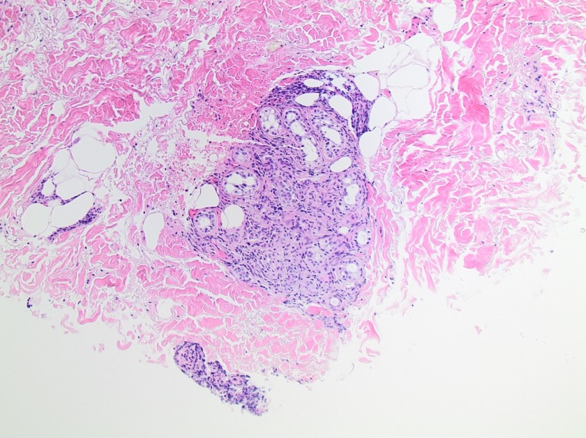
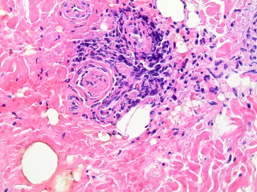
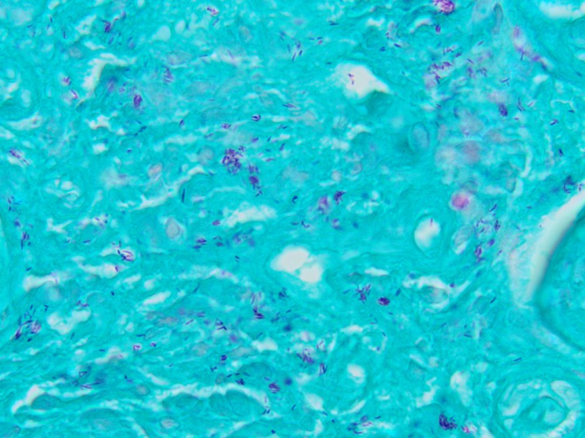
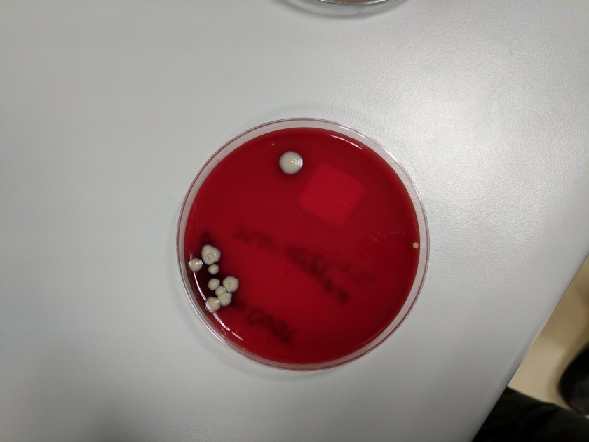
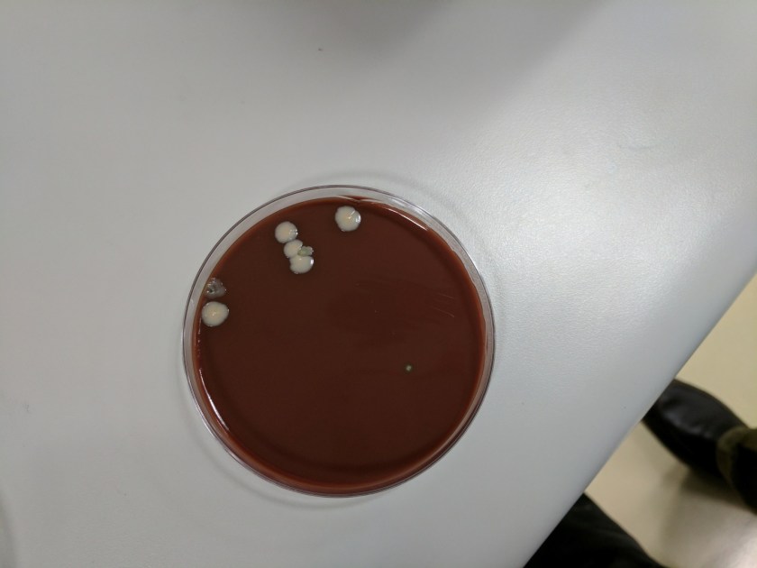
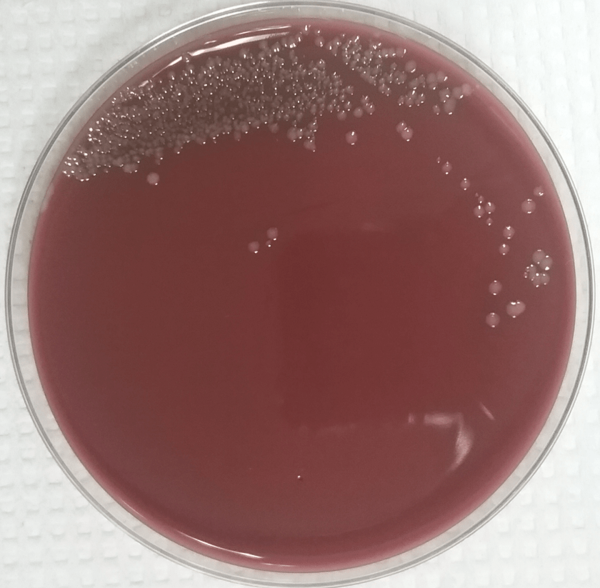
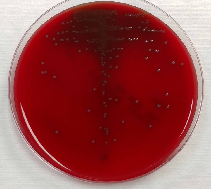
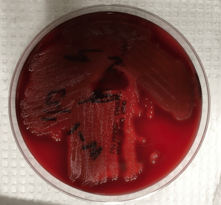
On histologic examination of the skin biopsy, nodular, superficial and deep granulomatous inflammation was noted surrounding eccrine glands and engulfing nerves (Images 1-3). Fite staining illustrated numerous acid fast bacilli (Image 4) and, given the geographic location of the patient and clinical symptoms, was felt to be highly suggestive of Mycobacterium leprae. The case was sent for confirmatory testing by polymerase chain reaction (PCR). All cultures collected were negative.

Discussion

Mycobacterium leprae is a chronic, granulomatous disease which presents as anesthetic skin lesions and peripheral neuropathy with nerve thickening. While rare in the United States (US) today, historically it was one of most prominent pathogens in Mycobacterium genus apart from M. tuberculosis. In the past, leprosy (also known as Hansen’s disease) was prevalent throughout Europe, but due to systematic control programs aimed at underserved and rural locations, the number of cases drastically decreased and countries with the majority of recent cases include India, Brazil and Indonesia. According to National Hansen’s Disease Registry, a total of 178 cases were reported in the US in 2015. Of these, 72% (129) of cases were reported in Arkansas, California, Florida, Hawaii, Louisiana, New York and Texas. Transmission to those who are in prolonged and close contact with an infected person is thought to occur via shedding from the nose. While humans are the only known reservoir of leprosy, infections with organisms indistinguishable from M. leprae have been detected among wild armadillos in parts of the southern US.

The diagnosis of M. leprae is largely a clinical one as the organism is not able to be grown on artificial media, but histology and confirmatory PCR are useful adjuncts. Skin biopsies should be full thickness and include the deep dermis. Ideally, the most active edge of the most active lesion should be biopsied. There is a spectrum of M. leprae which ranges from few lesions and a paucity of bacilli (tuberculoid leprosy) to widespread skin involvement with numerous bacilli (lepromatous leprosy).  Histologically, there are granulomatous aggregates of epithelioid cells, multinucleate giant cells and lymphocytes and inflammation often engulfs sweat glands and nerves. Small lesions that have poorly defined borders and are found on the elbows, knees or ears are where bacilli tend to be located. A Fite stain is useful to highlight the acid fast bacilli located in the macrophages within the inflammatory nodules. M. leprae PCR can also be performed on blood, urine, nasal cavity specimens and skin biopsies as a sensitive diagnostic technique. PCR can also be used to detect certain genes that confer resistance to common treatment drugs such as rifampin, ofloxacin and dapsone.

As with other mycobacterial diseases, the treatment for M. leprae infections consists of a long term multidrug regimen. The six most commonly used medications include rifampin, dapsone, clofazimine, minocycline, ofloxacin, and clarithromycin.


kt

-Katie Tumminello, MD, is a fourth year Anatomic and Clinical Pathology resident at the University of Mississippi Medical Center. 

Stempak

-Lisa Stempak, MD, is an Assistant Professor of Pathology at the University of Mississippi Medical Center in Jackson, MS. She is certified by the American Board of Pathology in Anatomic and Clinical Pathology as well as Medical Microbiology. She is the director of the Microbiology and Serology Laboratories.  Her interests include infectious disease histology, process and quality improvement and resident education. 

Microbiology Case Study: A 51-Year-Old Woman with a Tick Bite

Case History

A 51-year-old female with a past medical history of chronic lower back pain, depression, and anxiety presented to an outpatient dermatology clinic for a lesion on her left cheek that was present for years but has recently grown and become irritated.  Additionally, she reported a “skin taggy thing” that she first noticed on her posterior neck about two days ago.  Upon physical examination, a tick was observed latched onto the right posterior neck.  After being alerted to the presence of a tick, the patient did disclose that she was in the woods three days prior.  The bite site was locally anaesthetized and the tick was removed and sent to the laboratory for arthropod identification.  Furthermore, the patient received a single prophylactic dose of doxycycline 200 mg.

deertick1
Image 1. Ixodes scapularis, adult female, dorsal surface: an anterior capitulum (A) that contains mouth parts (paired palpi (B) and a median (partially intact) hypostome (C)) and the basis capituli (D) with two porose areas (E). The scutum (F) is inornate. 
deertick2
Image 2. Ixodes scapularis, adult female, ventral surface: eight coxa (G) of paired legs I-IV, a genital aperture (H), two spiracular plates (I), and an anal groove (J) that is anterior to the anus (K).


Discussion

Ixodes scapularis (black-legged ticks), also known as deer ticks (their preferred hosts are white-tailed deer), are small arachnids.  As obligate ectoparasites of vertebral hosts, I. scapularis are also willing to feed on humans.  Importantly, infected arthropods can be vectors of multiple pathogens including: the spirochete, Borrelia burgdorferi, that causes Lyme disease; the intracellular gram-negative bacterium, Anaplasma phagocytophilum, that causes human granulocytic anaplasmosis; the Apicomplexan parasites, Babesia spp, that cause babesiosis; and the flavivirus, Powassan virus, that causes encephalitis.

The I. scapularis life cycle, ranging from one to two years in length, is composed of four developmental stages: egg, larva, nymph, and sexually dimorphic adult.  Compared to nymphs and adults that have eight legs, larvae are smaller and have six legs.  The term “three-host cycle” implies that during each of the three motile stages, I. scapularis takes a blood meal from a different host animal, at which time the tick’s saliva is injected and transmission of pathogens can occur.

Hard ticks possess an anterior capitulum (Image 1, A) whereas soft ticks lack a capitulum.  The capitulum is made up of mouth parts that are attached to the basis capituli.  The mouth parts refer to paired appendages called palpi (Image 1, B) that are parallel to a median hypostome (Image 1, C).  The hypostome holds teeth-like structures, called denticles, arranged in a specific formula useful for identification.  The mouth parts (palpi and hypostome) are longer than the width of the basis capituli (Image 1, D) and this ratio is also useful in identification.  The dorsal surface of the basis capituli has two porose areas (Image 1, E) in adult females that secrete wax to waterproof eggs.  The dorsal shield, called a scutum (Image 1, F), is absent in soft ticks and inornate compared to other hard ticks.  In adult males, the scutum covers nearly the entire dorsum.  Other hard ticks have eyes (lateral markings on the scutum) and festoons (grooved bulges on the posterior margin) that are both absent in I. scapularis.  The ventral surface demonstrates coxa (Image 2, G) that are the basal segments of paired legs, numbered I-IV from anterior to posterior.  Posterior to coxa IV are paired spiracular plates (Image 2, I), external openings of the respiratory system.  A median genital aperture (Image 2, H) is present in adults.  The distinct anal groove (Image 2, J) is an inverted U-shaped curve located anterior to the anus (Image 2, K) in all Ixodes species, as opposed to posterior or indistinct anal grooves of other genera of hard ticks.

Following arthropod identification in parasitology, microbiology laboratory reports include: the genus (Ixodes), species (scapularis), developmental stage (adult female), level of engorgement (unengorged), and status of mouth parts (partially intact).  The genus and species are of medical importance because of their characteristic associations with various human pathogens.  Of the developmental stages, nymphs and adults are most frequently associated with human transmission of A. phagocytophilum.  Also, larvae are unable to transmit B. burgdorferi because the spirochete is not transmitted vertically to eggs; as such, a blood meal from a reservoir host is required.  Nymphs, being smaller in size than adults, are more likely to complete feeding undetected and thereby transmit B. burgdorferi.  Feeding is necessary for adult females to achieve fertility; therefore males are less likely to be discovered on hosts.  The level of engorgement, an estimate of feeding time, is relevant because approximately 36 hours are required for B. burgdorferi to multiply in the tick’s midgut and migrate to salivary glands for transmission to a host, by which time ticks are visibly engorged.  Lastly, if the mouth parts (palpi and hypostome) are intact, this suggests that there was only brief host contact and it is less likely that pathogen transmission occurred.

Reference

-Patterson FC and Winn WC. Practical identification of hard ticks in the parasitology laboratory. Pathology Case Reviews 2003; 8(4):187-198.


-Adina Bodolan, MD is a 1st year anatomic and clinical pathology resident at the University of Vermont Medical Center.

Wojewoda-small

-Christi Wojewoda, MD, is the Director of Clinical Microbiology at the University of Vermont Medical Center and an Assistant Professor at the University of Vermont.

Microbiology Case Study: A 65 year Old Male with Decubitus Ulcer

Case History

A 65 year old male with a history of T7-8 paraplegia and lengthy history of a decubitus ulcer presented for surgical debridement of the wound. An ischial bone biopsy for culture was performed.

Lab Identification

The primary gram stain demonstrated mixed gram positive and gram negative organisms, and the tissue sample from the wound grew Corynebacterium striatum and Bacteroides fragilis. The bone sample grew Enterococcus faecalis and anaerobic gram positive cocci. He was initially treated with ertapenem alone but daptomycin was added to cover enterococcus and he continued this dual regimen. Daptomycin was discontinued due to elevated creatinine kinase levels and he was transitioned to intravenous vancomycin. He completed 42 days of ertapenem and vancomycin. He was transitioned to oral amoxicillin/clavulanic acid and he has not developed new fever, chills, sweats, fatigue or increased drainage from his wound.

corstri1

Image 1. Blood agar plate with gray-white, moist, smooth, non-hemolytic bacterial colonies.

corstri2

Image 2. Chocolate agar plate with gray-white, moist, smooth, bacterial colonies.

The blood and chocolate agar plates grew bacterial colonies while the MacConkey agar had no growth. The gram stain was consistent with Corynebacterium and mass spectrometry identified the organism as Corynebacterium striatum.

Discussion

Corynebacterium striatum is a gram positive bacilli that is part of normal skin and mucosal flora. In immunocompromised patients or through direct inoculation of a sterile site, C. striatum can cause infectious endocarditis, bacteremia, pneumonia, lung abscess, arthritis and chorioamnionitis. Studies have shown that C. striatum also can cause wound infections in patients with underlying disease and previous antibiotic use. Foreign medical devices can also be infected by C. striatum, and removal of the device may be necessary. Vancomycin is used to treat C. striatum due to the variable susceptibility to other antibiotics.


-Mustafa Mohammad, MD is a 3rd year anatomic and clinical pathology resident at the University of Vermont Medical Center.

Wojewoda-small

-Christi Wojewoda, MD, is the Director of Clinical Microbiology at the University of Vermont Medical Center and an Assistant Professor at the University of Vermont.

Microbiology Case Study: A 14 Year Old Female with Neck Swelling

Case History

A previously healthy 14-year-old female presents to the emergency department with three days of progressive facial and neck swelling. The swelling started on the left side. Two days ago she visited her primary care physician where she had negative monospot and mumps IgM testing.  She is fully vaccinated, but was exposed to a mumps outbreak at school.

Discussion

Our patient was diagnosed with mumps by positive RT-PCR from a buccal swab. The mumps virus is a member of the Paramyxoviridae family which includes notable human pathogens parainfluenza, Hendra, and Nipah viruses. Members of this family are enveloped, helical viruses with single-stranded, non-segmented RNA genomes with negative polarity. Mumps is an obligate human pathogen that replicates in the epithelial cells of the upper respiratory tract and subsequently moves to regional lymph nodes. It is spread from person to person via direct contact with respiratory secretions or contact with contaminated fomites. Mumps is a highly contagious disease with as high as 85% of naïve individuals becoming infected after contact with a mumps infected individual. It spreads most efficiently in areas where there is close contact among individuals for prolonged periods of time such as college campuses and close-knit religious communities.

Prior to vaccination for mumps in the 1960s, greater than 150,000 cases of mumps occurred each year in the US. The incubation period for infection is 16-18 days, with the majority of infected persons being asymptomatic or having mild respiratory symptoms. Orchitis causing sterility in post-pubescent males is the main concern of mumps infection but other rare but serious complications include mastitis and oophoritis in females, meningoencephalitis, pancreatitis, and deafness.

Due to sporadic outbreaks of measles since the introduction of the vaccine, the vaccine schedule has been revised from one dose of the MMR (measles, mumps, and rubella) vaccine at age 12-15 months to include another MMR booster at age 4-6 years. We are currently in the middle of yet another outbreak with nearly 6,000 cases of mumps reported to the CDC in 2016 and a high rate of infections reported thus far in 2017 (Figures 1 and 2).

mumps1
Figure 1. Number of cases identified by the CDC in 2017 by state. (Figure courtesy of the CDC Mumps website at https://www.cdc.gov/mumps/outbreaks.html. Content source: National Center for Immunization and Respiratory Diseases [NCIRD], Division of Viral Diseases)
mumps2
Figure 2. Number of cases of mumps per year identified by the CDC.
(Figure courtesy of the CDC Mumps website at https://www.cdc.gov/mumps/outbreaks.html. Content source: National Center for Immunization and Respiratory Diseases [NCIRD], Division of Viral Diseases)


Diagnostic Testing for Mumps

Serological testing for IgM and RT-PCR from a buccal swabs are the mainstay of mumps diagnosis. IgM becomes positive in the first 3-4 days after symptom onset and will remain positive for 8-12 weeks. IgG becomes positive 7-10 days following symptom onset and will remain at high levels for many years and detectable for life. In a vaccinated individuals, IgM testing has less utility as it may be non-reactive or weakly positive following a secondary immune response.

RT-PCR from a buccal swab specimen is the most sensitive test for diagnosis of mumps. It should be performed as soon as a patient is symptomatic, as testing by this method is the most sensitive in the first few days following symptom onset and becomes less sensitive as time goes on.

Urine specimens can be used to isolate mumps in viral culture. Urine is not positive for mumps until greater than 4 days post symptom onset and is less sensitive than PCR performed on the bucal swab. For these reasons, viral isolation from urine is no longer a commonly used test for diagnosis of mumps, although viral culture is still considered the gold standard for mumps conformation.

Resolution

The patient and her family were counseled on the infectious nature of mumps. She was instructed to remain in isolation at home for 6 days after resolution of swelling.


References

  1. Manual of Clinical Microbiology, 11th edition
  2. CDC Mumps Website (www.cdc.gov/mumps/index.html)


Erin McElvania TeKippe, PhD, D(ABMM), is the Director of Clinical Microbiology at Children’s Medical Center in Dallas Texas and an Assistant Professor of Pathology and Pediatrics at University of Texas Southwestern Medical Center.

Microbiology Case Study: A 74 Year Old Man’s BAL Specimen

Case History

A laboratory received a bronchioalveolar lavage specimen on a 74 year old male, who is now deceased. The potato flake agar grew white, mucoid colonies, while the Mycosel plate had no growth. The colony was identified as Cryptococcus neoformans by mass spectrometry.

crypto1-1

Image 1. White mucoid colonies on potato flake agar.

Discussion

C. neoformans and C. gattii produce white, mucoid colonies on a variety of agars that usually become visible within 48 hours. Urea and phenoloxidase are positive. L-Canavanine Glycine Bromothymol Blue (CGB) agar helps differentiate C. neoformans colonies from C. gattii, with C. neoformans producing a light green-blue color and C. gattii producing a cobalt blue color. C. neoformans is also described as resembling glass beads on cornmeal agar due the presence of its thick capsule. C. neoformans is generally 5-10 µm in size, however size is variable and they can be increased. Historically, India ink preparation was described to identify the organism due the capsule extruding the ink. Current identification methods include a rapid latex agglutination test for antigen, and mass spectrometry can also be used to identify C. neoformans.

C. neoformans and C. gattii are basidiomycetous, encapsulated yeasts found all over the world. They are commonly found in areas frequented by birds and bats. Patients with recent travel to caves or work in environments that expose them to chickens are at higher risk of infection due to inhalation of C. neoformans. While C. neoformans generally causes pulmonary infections, patients who are immunosuppressed can have disseminated cryptococcosis with CNS involvement. The clinical presentation of cryptococcosis due to the two species is generally indistinguishable. Cryptococcosis can be treated with amphotericin B and flucytosine or fluconazole.


-Mustafa Mohammad, MD is a 3rd year anatomic and clinical pathology resident at the University of Vermont Medical Center.

Wojewoda-small

-Christi Wojewoda, MD, is the Director of Clinical Microbiology at the University of Vermont Medical Center and an Assistant Professor at the University of Vermont.

Microbiology Case Study: A 58 Year Old Female with Fever, Headache, and Vomiting

Case History

A 58 year old female presented to the emergency department with complaints of a fever (reaching 102.9°F) and headache with associated nausea and vomiting for the past 24 hours. Her past medical history was significant for a resection of a recurrent hemangiopericytoma by the neurosurgery service three weeks prior. The patient also noted clear drainage from this surgical site which had begun 5 days ago. Other symptoms noted at presentation included decreased appetite and dehydration. She denied back & neck pain, photophobia or stroke and seizure-like symptoms. Her vital signs were all within normal limits. On physical exam, a healing surgical wound was noted in the posterior auricular area with clear drainage, but no blood or exudates were visualized.  She had no tenderness when her spine was palpated and neurologic exam showed a left sided facial droop and tongue deviation which were noted previously and attributed to her multiple central nervous system surgeries. Complete blood count (CBC) showed a mild increase in white blood cells and anemia. An external ventricular drain was placed and cerebral spinal fluid (CSF) was sent to the microbiology lab for culture. Blood cultures and a swab from the surgical wound were also collected.

Laboratory Identification

sermar1.png

Image 1. Gram stain of the cytospin CSF showed many acute inflammatory cells and numerous Gram negative bacilli (1000x).

sermar2

Image 2. Growth of large, glossy, reddish-orange colonies on sheep blood agar (image taken after 72 hours of incubation).

sermar3.png

Image 3. Growth of large, deep red colonies on MacConkey agar (image taken after 72 hours of incubation).


Gram stain of the CSF showed numerous acute inflammatory cells and many Gram negative bacilli (Image 1). Culture of the CSF and wound swab showed large, glossy red colonies on sheep blood and MacConkey agars (Images 2 & 3). Analysis of the colony by matrix assisted light desorption ionization time of flight mass spectrometry (MALDI-TOF MS) identified the organism as Serratia marscescens.

Discussion:

Serratia marscescens is a facultative Gram negative bacillus that is a member of the Enterobacteriaceae family. S. marscesens is ubiquitous in the environment and the most frequent and clinically important species in the genus. Although S. marscesens usually doesn’t cause infection in healthy individuals, it is notorious for colonizing and causing infections in hospitalized patients, particularly those who are immunocompromised, in intensive care units (especially intubated patients) and those with indwelling catheters.  While respiratory infection are most common, S. marscesens has also been implicated in numerous other opportunistic infections such as urinary tract infections, wound infections and septicemia. Brain abscesses and meningitis are less common. S. marscesens has been implicated as the cause of outbreaks in hospitals and can often be traced back to pieces of medical equipment including nebulizers, bronchoscopes, laryngoscopes and contaminated solutions. Person to person transmission is also recognized and thought to be predominantly transmitted via direct contact.

In the laboratory, S. marscesens can be identified by its characteristic non-diffusible red pigment, prodigiosin. Care should be taken when interpreting the lactose reaction on MacConkey agar, as the red pigment may be confused with a positive reaction, while S. marscesens is known to be lactose negative.  As a member of the Enterobacteriaceae family, S. marscesens is able to ferment glucose, reduce nitrate to nitrite and has a negative oxidase reaction. A unique feature of this genus is that all Serratia spp. produce three proteolytic enzymes: lipase, gelatinase, and DNase. Commercial systems, including MALDI-TOF MS, are helpful in the identification of S. marscesens as well.

Treatment of Serratia marscescens infections can be difficult due to various antimicrobial resistance mechanisms, such as expression of extended spectrum beta lactamases (ESBLs), AmpC cephalosporinases and carbapenemases, exhibited by the organism. In the case of our patient, she was empirically started on vancomycin and piperacillin-tazobactam and taken to surgery for wound wash out, removal of hardware and repair of CSF leak. Her antibiotics were changed to meropenem and gentamicin. She was discharged to a rehabilitation facility and received meropenem for a total of 6 weeks.


DM

-David Marbury, MD, is a 3rd year Anatomic and Clinical Pathology resident at the University of Mississippi Medical Center.

Stempak

-Lisa Stempak, MD, is an Assistant Professor of Pathology at the University of Mississippi Medical Center in Jackson, MS. She is certified by the American Board of Pathology in Anatomic and Clinical Pathology as well as Medical Microbiology. Currently, she oversees testing performed in both the Chemistry and Microbiology Laboratories. Her interests include infectious disease histology, process and quality improvement and resident education.

Microbiology Case Study: A 64 Year Old with Coronary Artery Disease

Case History

The patient is a 64 year old man with a past medical history significant for coronary artery disease, chronic systolic heart failure, poorly controlled type 2 diabetes mellitus, atrial fibrillation, chronic respiratory failure on home oxygen, squamous cell carcinoma of the larynx status post chemotherapy, and radiation and lung adenocarcinoma status post microwave ablation. On the morning of presentation, the patient’s wife was unable to wake him and found him to have low oxygen saturation with a home monitor. Three days prior to presentation, the patient had been evaluated in the emergency department for leg and back pain for which he was prescribed hydromorphone. He had also been experiencing nausea for several days, with fever (101° F) and chills. His wife endorses several sick contacts at home. He has been admitted to the hospital numerous times within the past year for respiratory failure, most recently 6 months ago. The patient also was given a course of antibiotics one month prior for a “cold.”

Enroute to the emergency department, EMS administered naloxone without significant response. Upon arrival to the hospital, his vital signs were: temperature 97.9° F, heart rate 69, respiratory rate 23, blood pressure 104/57, 95% SpO2. He was found to have a white blood cell count of 15,690/cm2 and a chest x-ray showed diffuse patchy airspace opacities concerning for multifocal pneumonia. A viral swab was collected and blood cultures were drawn. He was started on levofloxacin for suspected community acquired pneumonia.

His blood cultures were found to be growing gram negative coccobacilli.

pastmult1

Figure 1. Gram stain from a positive blood culture illustrating small Gram negative coccobacilli (100x, oil immersion).

pastmult2.png

Figure 2. Chocolate agar illustrating the convex, smooth and gray colonies.

MALDI-TOF identified the organism as Pasteurella multocida. Further investigation revealed that the patient recently acquired a puppy (4-month-old) that bit him while playing. Antibiotics were switched to IV ceftriaxone and the patient recovered. He was later discharged on IV antibiotics and home healthcare.

Discussion

Pasteurella multocida is a non-motile gram negative coccobacillus that is oxidase-positive and glucose fermenting. Most isolates don’t on MacConkey agar. It is a known cause of disease amongst humans and animals, but is commonly a commensal organism found in domesticated and wild animals. It is most notably found in the oropharynx of cats, dogs, pigs, and birds. The majority of P. multocida infections are due to animal exposure, either from traumatic inoculation or proximity. Soft tissue infections are most common, resulting from animal bite or scratch. It has also been known to cause pulmonary disease in the form of multifocal pneumonia in those with pre-existing chronic lung disease. It is also a rare cause of septic arthritis, osteomyelitis, endocarditis and meningitis in those with disseminated disease.

P. multocida virulence factors include an endotoxin and an antiphagocytic capsule. It grows well on routine laboratory media but not on MacConkey. Colonies appear convex, smooth, gray and nonhemolytic. Mucoid variants can also occur. It has been described as having a musty or mushroom odor, although sniffing plates is not recommended. Susceptibility testing is usually not required as it is universally susceptible to β-lactams. However, CLSI does provide breakpoints for suggested drugs should the need for testing arise.

References

Tille, P. M., & Forbes, B. A. (2014). Bailey & Scott’s Diagnostic Microbiology (Thirteenth edition.). St. Louis, Missouri: Elsevier.

Kuhnert P; Christensen H, eds. (2008). Pasteurellaceae: Biology, Genomics and Molecular Aspects. Caister Academic Press.


-Clayton LaValley, MD is a 1st year anatomic and clinical pathology resident at the University of Vermont Medical Center.

Wojewoda-small

-Christi Wojewoda, MD, is the Director of Clinical Microbiology at the University of Vermont Medical Center and an Assistant Professor at the University of Vermont.


Help Researchers Tackle Antimicrobial Resistance in Tuberculosis

Researchers at the University of Oxford are researching antibiotic resistance in Tuberculosis, and they want help reading MIC plates. You don’t have to fly to England, though–you can do it online! Visit the project Bash the Bug on Zooniverse to learn more, view a short tutorial, and get started.

You can read more about the Bash the Bug project here.


Microbiology Case Study: An 11 Year Old with Abdominal Pain

Case History

An 11 year-old patient with a history of a relapsed lymphoma presented to the hematology/oncology clinic with worsening abdominal pain. The patient was recently started on metronidazole to treat a C. difficile infection. In the clinic, the patient was found to be hypotensive, hypoxemic and pancytopenic. Blood cultures were drawn and the patient was admitted directly to the pediatric ICU and started on empiric antibiotics.

The blood cultures turned positive with Gram-positive cocci, which went on to produce small, gray, alpha-hemolytic colonies on the blood agar plate (Image 1). The colonies were catalase negative and PYR negative. The isolate was analyzed by a Bruker MALDI-TOF mass spectrometer and was identified as Streptococcus lutetiensis (score 2.19). Susceptibility testing revealed the isolate susceptible to ceftriaxone, penicillin, and vancomycin.

streplut

Image 1. 5% sheep blood agar growing small, grey, alpha-hemolytic colonies


Discussion

S. lutetiensis is part of the complex of organisms previously identified as the Streptococcus bovis group. This group of organisms, which possess the Lancefield Group D antigen, has undergone considerable reclassification schemes as phenotypes and genotypes have been investigated. The original biochemical classification schemes were based on their ability to ferment mannitol as well as the presence or absence of beta-glucuronidase activity. Early observations of the DNA properties from these organisms, such as %-GC base content and DNA-DNA hybridizations, identified six unique clusters of Group D streptococci [1]. One cluster group (cluster group #4) had heterogeneous biochemical phenotypes. A subcluster of this cluster group #4 was separated from the other members of the cluster based upon esculin hydrolysis. This subcluster would later go on to be named S. infantarius, so named as several isolates originated from the feces of human infants [2].

Further DNA-DNA hybridizations and ribotyping analysis led to the declaration of two S. infantarius subspecies: subsp. infantarius and subsp. coli [3]. The 16S rRNA ribotyping was problematic, however, as several species in this genus are 97-99% sequence identical.

In an attempt to address some of the limitations of relying on the 16S rRNA gene, one group analyzed the features of the conserved gene encoding the manganese-dependent superoxide dismutase gene (sodA). They observed substantial differences between S. infantarius subsp. infantarius and S. infantarius subsp. coli [4]. Thus, for the latter organism, a new species of streptococci was proposed: S. lutetiensis. It was named for Lutetia, a historical name for the city of Paris [4].

The species designation S. lutetiensis was not widely accepted, however. Based on further DNA-DNA hybridization experiments and the prior studies of the 16S rRNA, others have rejected the species name “S. lutetiensis” and maintain that it is a subspecies of S. infantarius as previously described [5].

So which name is correct? There appears to be no clear consensus about the designation of these streptococci, whether it is S. infantarius subsp. coli or whether it is another species altogether as S. lutetiensis. The Judicial Commission of the International Committee on Systematic Bacteriology reportedly met to discuss the name changes, however no resolution appears to have been determined [6]. Both names are seen in the literature as well as the names for reference organisms.

The important clinical aspect to recognize is that this organism, as well as the S. bovis group in general, can be a cause of bacteremia, endocarditis, and meningitis in children. Treatment with beta-lactam antibiotics is generally sufficient to cover these organisms.

References

  1. Farrow, J., et al., Taxonomic Studies on Streptococcus bovis and Streptococcus equinus: Description of Streptococcus alactolyticus sp. nov. and Streptococcus saccharolyticus sp nov. System. Appl. Microbiol, 1984. 5: p. 467-482.
  2. Bouvet, A., et al., Streptococcus infantarius sp. nov. related to Streptococcus bovis and Streptococcus equinus. Adv Exp Med Biol, 1997. 418: p. 393-5.
  3. Schlegel, L., et al., Streptococcus infantarius sp. nov., Streptococcus infantarius subsp. infantarius subsp. nov. and Streptococcus infantarius subsp. coli subsp. nov., isolated from humans and food. Int J Syst Evol Microbiol, 2000. 50 Pt 4: p. 1425-34.
  4. Poyart, C., G. Quesne, and P. Trieu-Cuot, Taxonomic dissection of the Streptococcus bovis group by analysis of manganese-dependent superoxide dismutase gene (sodA) sequences: reclassification of ‘Streptococcus infantarius subsp. coli’ as Streptococcus lutetiensis sp. nov. and of Streptococcus bovis biotype 11.2 as Streptococcus pasteurianus sp. nov. Int J Syst Evol Microbiol, 2002. 52(Pt 4): p. 1247-55.
  5. Schlegel, L., et al., Reappraisal of the taxonomy of the Streptococcus bovis/Streptococcus equinus complex and related species: description of Streptococcus gallolyticus subsp. gallolyticus subsp. nov., S. gallolyticus subsp. macedonicus subsp. nov. and S. gallolyticus subsp. pasteurianus subsp. nov. Int J Syst Evol Microbiol, 2003. 53(Pt 3): p. 631-45.
  6. Beck, M., R. Frodl, and G. Funke, Comprehensive study of strains previously designated Streptococcus bovis consecutively isolated from human blood cultures and emended description of Streptococcus gallolyticus and Streptococcus infantarius subsp. coli. J Clin Microbiol, 2008. 46(9): p. 2966-72.


IJF

-I.J. Frame MD, PhD, is a 1st year Clinical Pathology Resident at UT Southwestern Medical Center.

Erin McElvania TeKippe, PhD, D(ABMM), is the Director of Clinical Microbiology at Children’s Medical Center in Dallas Texas and an Assistant Professor of Pathology and Pediatrics at University of Texas Southwestern Medical Center.

Microbiology Case Study: A 28 Year Old Woman with Acute Onset Fever Post Delivery

Case History

A 28 year old woman at 37 weeks and 2 days presented in labor to our ED. After 22 hours, she delivered a healthy baby boy and sustained a second degree perineal laceration requiring repair. On hospital day 2, she reported feeling lightheaded, nauseous and “shaky.” She attempted to walk around the unit but became tremulous and unsteady, requiring assistance to get back into bed. Her vital signs were as follows: febrile at 38.8 C, BP 108/54, HR 104 and normal respiration rate at 12 breaths/min. On exam, she appeared pale and lethargic, and was noted to have a tender uterus on palpation. Based on her presentation and status post SVD, the diagnosis of endometritis was established. Blood cultures were obtained and within 16 hours, blood culture bottles were positive for gram-positive cocci. The patient was started on antibiotic therapy with ampicillin, gentamycin and clindamycin, and clinically improved within 36 hours.

streppyo1
Image 1. Blood culture on blood agar.
streppyo2
Image 2. Blood culture on chocolate agar.

Discussion

Streptococcus pyogenes is one of the most aggressive pathogens encountered in clinical microbiology. It is a beta hemolytic streptococcus and is notoriously associated with Streptococcal Toxic Shock Syndrome (STSS), necrotizing fasciitis, as well as more benign (yet still problematic) conditions, like Scarlet Fever, Impetigo, Rheumatic heart disease and Acute Post-streptococcal Glomerulonephritis. A gram-positive cocci, it possesses several virulence factors, including protein F, M protein (involved in antigen mimicry leading to valvular heart disease) hemolysins and exotoxins. These factors allow S. pyogenes to attach to and invade epithelial tissue, and in the case of hyalurondiase, potentially use hyaluron as a carbon food source. S. pyogenes agglutinates with Lancefield group A antisera and is pyrrolidonyl arylamidase (PYR) positive and VP, hippurate and CAMP test negative. Penicillin (PCN) remains the drug of choice in treating most S. pyogenes infections. Alternative antibiotic therapy includes macrolides and certain cephalosporins (e.g. cefixime, cefpodoxime). Vancomycin should be used in more severe infections such as sepsis or for patients with a PCN allergy.


-Christina Litsakos is a Pathology Student Fellow at University of Vermont Medical Center.

Wojewoda-small

-Christi Wojewoda, MD, is the Director of Clinical Microbiology at the University of Vermont Medical Center and an Assistant Professor at the University of Vermont.