Undetermined, Undetermined

I like to think most people who go into healthcare professions do so with the hope of helping others. For those of us who do autopsies, the greatest sense of reward comes when we can explain to someone how and why their loved one died. Inevitably, though, there will be situations where we need to accept what we don’t know – despite how disappointing it may be. In these situations, the most intellectually honest course of action is to issue a manner of “undetermined.”

Let’s recap briefly what we know about manners of death (see https://labmedicineblog.com/2022/11/25/please-dont-tell-me-i-died-of-cardiac-arrest/ for a more in-depth discussion). Manner of death describes the circumstances in which the cause of death is sustained, and there are five choices in most jurisdictions – natural, accident, homicide, suicide, or undetermined. Natural deaths are those due entirely to non-traumatic diseases (like cancer or coronary artery disease). Accidental deaths involve trauma or toxicity without an intention to harm. Homicide is death at the hands of another person, whereas suicide is death at one’s own hands. The final option is undetermined.

There are two main pathways by which we can arrive at an “undetermined” manner. There can either be 1) reasonable competing evidence between two manners of death, or 2) we may be unable to identify a cause of death due to loss or destruction of bodily tissue. Let’s look at some examples.

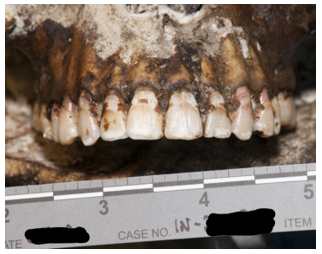
In the first pathway, consider an autopsy of a person with a single gunshot wound to the head. In a readily accessible region like the temple or beneath the chin, this wound could easily be self-inflicted. While this would be a “typical” location for a suicidal injury, such a wound could also be inflicted by another person. There are indicators we look for at autopsy which favor one scenario over the other. For example, most suicidal gunshot wounds (broadly speaking, of course) are contact wounds or intra-oral. A self-inflicted gunshot wound to the back of the head would be unusual, but (contrary to popular conception), not impossible depending on the firearm used. However, the same type of pattern could be elicited with another person holding a firearm to that individual’s head. We may examine the length of the firearm to determine if it’s possible for the decedent to have pulled the trigger themselves (keeping in mind that other items like a cane, coat hanger, or even the decedent’s toe, may have been used to depress the trigger). Similar questions can arise in autopsies of people who have fallen from height. There is no way an autopsy can tell with certainty whether an individual was pushed, fell accidentally, or left the edge of an elevated structure intentionally. The cause of death in both situations is undisputed – a gunshot wound in the first, and blunt force injuries in the second. This is why contextual information, like scene photographs and investigative records, is indispensable for forensic pathologists. Without context, we have no way to discern homicides, suicides, and accidents. Occasionally even with context, there can be competing narratives (one witness claims a gunshot wound was self-inflicted, while another claims it was inflicted by the first) or suspicious circumstances to cast doubt. Without clear cut evidence to support one story, the manner of undetermined is appropriate.

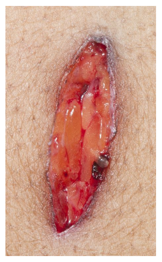
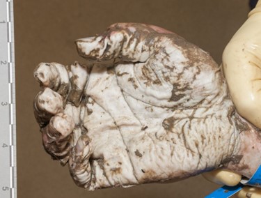
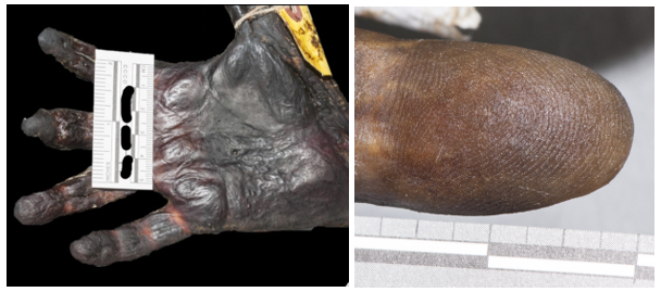
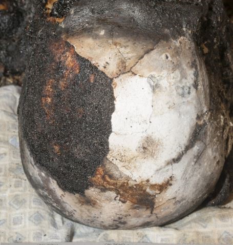
The second pathway by which we reach an undetermined manner is when extensive decomposition or other soft tissue loss (such as fire damage) interferes with our ability to determine a cause of death. Think of completely skeletal remains discovered in an abandoned building. Sometimes, indicators of potentially lethal injuries can still be identified – for example, a gunshot wound of the skull or knife marks on a rib. But, as the aphorism goes, “an absence of evidence isn’t necessarily evidence of absence” – a bullet or blade could be lethal while only striking soft tissue (especially in regions like the abdomen or neck). If we cannot rule out non-natural causes of death, the best choice for manner is “undetermined.”

An undetermined manner of death can understandably frustrate family members or law enforcement. I always try to explain that manner determinations are, as one of my mentors says, “written on paper and not in stone.” We reserve the right to change the ruling in the future if additional evidence comes to light. As forensics pathologists our primary responsibility is to speak honestly and truthfully, and sometimes that means admitting the limitations of our science.

-Alison Krywanczyk, MD, FASCP, is currently a Deputy Medical Examiner at the Cuyahoga County Medical Examiner’s Office.

It’s What’s on the Inside That Counts: Uses of Radiography at Autopsy

In several previous blogs, I’ve mentioned the topic of post-mortem radiography (or “x-rays”). While postmortem CT scanning is a hot topic in the field, plain films are a tool which has been in widespread use for decades. Autopsy standards of the National Association of Medical Examiners require, at a minimum, radiographs be performed on all infants, gunshot wound victims, explosion victims, and charred or decomposed remains. Let’s examine the reasons for these requirements and look at a few specific examples.

All infants must get full body x-rays to check for acute or healing rib and long bone (extremity) fractures, which could be indicative of physical abuse. The extremities are not usually dissected in the course of a typical post-mortem examination, but fractures that can’t be attributed to birth trauma (especially in a pre-mobile child) are a concerning finding that needs further investigation and dissection.

Radiographs taken of gunshot wound victims document the presence and location of retained projectiles, all of which need to be recovered as evidence.  

This x-ray of a gunshot wound victim shows two separate types of ammunition, seen as radio-opaque (white) in the image. The smaller, circular pieces are “birdshot” shotgun ammunition (blue arrows) while the larger pieces are traditional handgun ammunition (red arrow).

Radiographs are commonly performed in any type of penetrating trauma, including sharp force injury and explosive injuries, to identify retained foreign bodies (especially broken fragments of the blade, which may pose a risk to the pathologist). Similar to projectiles, these fragments need to be recovered as evidence. Radiographs can also document the presence of an air embolism, which can be missed at autopsy if special dissection techniques aren’t performed.  

This individual had sharp force injuries to their neck, which injured large veins. The x-ray in this case was performed to see if any fragments of the weapon were still in the body, but also showed a large air embolism in the right atrium and ventricle (blue arrows), seen as radiolucency (gray/black). Open injuries to veins in the head and neck can cause air emboli as breathing creates negative intra-thoracic pressure, drawing air inward though any open channels.

In pedestrians who have been struck by motor vehicles, radiography is the first step in examining trauma. As mentioned earlier, the extremities aren’t typically dissected during a traditional autopsy – but in pedestrians, lower extremity fractures can document the site of initial impact, and the distance of the fracture from the foot may indicate the bumper height of the car.

This x-ray of a pedestrian struck by a motor vehicle shows displaced fractures of the right femur, tibia, and fibula. Note the body bag zipper in the lower right corner.

Fire victims may have extensive thermal injuries and charring which can hide evidence of other injuries, and radiographs can help identify them. Radiographs are also one step toward identifying the victim. Decomposed bodies must be x-rayed for similar reasons – external and internal soft tissue alterations make assessment for trauma more difficult, and radiographs may be needed to confidently identify the body.

In this x-ray of a decomposed person, dental fillings are easily visible (red arrow), which are typically unique for an individual and can be used for identification. Note the irregular shapes of the fillings, which are just as important as their location. Also note the air-fluid level in the cranial cavity (blue arrow), indicating complete liquefication of the brain.

On occasion, radiographs can be performed on individual organs to help define anatomy or previous surgical alterations. Especially in the heart, radiographs can demonstrate coronary artery or valve calcifications, surgical clips from bypass grafts, or other radiopaque prostheses. Knowing the location of devices before dissection gives the pathologist a better chance at preserving and evaluating important structures.

This heart was x-rayed after removal, as the decedent had a history of heart disease with prior interventions. Coronary stents (red arrows) are easily visible; in practice, they can be difficult to find on gross dissection in patients with heavily calcified arteries.

Not all offices can afford the installation (or maintenance) of CT scanners, but access to x-ray machines is more widespread. As we’ve seen here, x-rays are a versatile tool which can document injuries, help identify decedents, and direct the pathologist to perform special autopsy procedures which aren’t part of the daily routine.

-Alison Krywanczyk, MD, FASCP, is currently a Deputy Medical Examiner at the Cuyahoga County Medical Examiner’s Office.

The Ins and Outs of Gunshot Wounds

In the United States, victims of gunshot wounds represent a significant majority of all homicides (and a high proportion of suicides). There’s a propensity among other medical specialties to think of forensic pathologists as “bullet pullers,” just collecting the used projectiles and moving on to the next case. However, autopsies of multiple gunshot wound victims can be some of the most detailed examinations we perform. Even though the cause of death isn’t a mystery, thorough observation and documentation is crucial for other questions which may arise – was the victim immediately incapacitated? Did the shooter reload during the assault? How close to the victim were the shots fired? To address these questions, it’s first helpful to understand the basics of gunshot wounds.

Gunshot wounds can be penetrating (entering the body without exiting) or perforating (entering and exiting the body). Full body x-rays are taken in all gunshot wounds to identify retained projectiles, all of which must be recovered as evidence. It’s important to not use metal tools (like forceps or scissors) when removing a projectile, which may scratch metal and interfere with ballistics comparison.

Under typical circumstances, distinguishing entrance from exit wounds is straightforward. Classic entrance wounds are circular, as the bullet hasn’t yet been deformed, and there is a surrounding rim of abrasion where the edges of the bullet scrape against the skin. In contrast, exit wounds have a stellate or slit-like appearance, but the wound edges can typically be reassembled and are not abraded. As with any area of medicine, though, real life doesn’t always follow the rules. An entrance wound can be atypical if the bullet has passed through an ‘intermediary target’ before striking the victim (say a piece of furniture or a car door). If the area of exit is pressed against a firm object (even a tight pants waistband), the skin edges will be abraded (or “shored”). Sometimes, the entrance and exit truly cannot be distinguished – this is more likely with superimposed decomposition, insect activity, or superficial wounds.

Range of fire is another important feature to note and is the reason we always take photographs of a wound before cleaning the body. When a gun is fired, smoke, soot, and unburnt particles of gunpowder exit the barrel as well as the bullet. The smoke creates “fouling,” dark discoloration which easily wipes away and can be seen if the end of the barrel is within approximately one foot of the victim. Stippling, caused by unburnt grains of gunpowder, are actual abrasions and can be seen when the projectile fired within 18” of the victim. These are gross generalizations, though, and in each individual circumstance the weapon itself must be tested to identify the distances. If soot or gunpowder particles aren’t visible on the skin surface, they may have been deposited on the victim’s clothing. ‘‘Bullet wipe’ is slightly different from fouling, in that it is dirt and residue on the actual bullet which gets ‘wiped’ off around the entrance wound on clothing – so it doesn’t tell you range of fire, but it can be helpful to identify tricky entrance wounds.

Now, for some of the common misconceptions around gunshot wounds …

Can you tell the caliber of the gun from the size of the wound? The answer is an emphatic “no.” Much of the injury caused by a bullet comes from the temporary wound cavity created by dissipation of kinetic energy; so no matter the size of the bullet, tissue will stretch and distort around it.

Can you tell the order in which gunshot wounds were sustained? Most often, no. In some autopsies, the first wounds cause so much blood loss that the later wounds lack hemorrhage, but this is the exception rather than the rule.

Can I tell what position the victim was in when they were shot? Again, usually not. In isolation, an autopsy can only tell you the trajectory of the bullet through the body; to determine the position of the victim when they were shot requires knowledge of either where the gun was when it was fired or where the bullet landed–two factors which are often not available.

These are just a few of the complexities faced by a pathologist when working with gunshot wounds, and we haven’t even covered different types of ammunition or firearms. Stay tuned for more in the future!

This is an example of a classic entrance gunshot wound – nearly circular, with a thin rim of abraded (or scraped) skin.
In contrast, this exit wound is slit shaped, and the wound edges can be neatly reapproximated.
This entrance wound has stippling on the surrounding skin; occasionally gunpowder particles are still visible embedded in the abrasions.

-Alison Krywanczyk, MD, FASCP, is currently a Deputy Medical Examiner at the Cuyahoga County Medical Examiner’s Office.

Autopsy Examination of Sharp Force Injuries

In television and movies, you’ll sometimes see a scene where the pathologist describes to a detective the specific dimensions of a knife used in an attack, all based on the autopsy findings. While this is no doubt extremely helpful to an investigation (and the limits of a 60-minute run time) it’s also impossible to provide this level of detail from an autopsy. Unfortunately the pervasiveness of such creative liberties in pop culture creates unrealistic expectations from juries, lawyers, and law enforcement, which we often need to gently correct. There are certain characteristics we can identify which may clarify what kind of blade was used, but conversely may not match the dimensions of the weapon at all.

While any object with a pointed or edged tip can be used to inflict sharp force injuries, we will focus on knives as the stereotypical ‘sharp force’ weapon. As a reminder, sharp force injuries have clean, neat edges and no tissue bridging in the wound depths. These are the features that are used to distinguish sharp force from lacerations, which are skin tears due to blunt force injury (see last month’s blog post for a refresher!).  The most important first step for any instance of sharp force injury is to fully x-ray the body; this allows for identification of potential evidence to discover (such as a broken knife tip), and identifies potential sharp hazards for the prosector.

The first issue when interpreting sharp force injuries at autopsy is distortion of wounds by skin elasticity. Depending on the location and orientation of the wound, the edges may be pulled by tension along Langer’s lines. If this is the case, re- approximating the wound edges is needed before measuring for better accuracy. The ends of a stab wound may reflect whether a blade has one or two edges, depending on whether both ends are tapered (two-edged) or if one end is blunt and the other tapered (single-edged). The morphology isn’t always clear, though. Occasionally a double-edged blade will have a blunt-edged segment known as the “ricasso” just before the crossguard or handle. If the blade is fully inserted, the impression of the ricasso may make the wound appear to have two blunt edges.

The second difficulty faced is that knife wounds are rarely inflicted on a stationary body – there is typically (at least initially) a struggle involving motion of both the victim and the blade. This can result in wounds which are distorted from the dimensions of the weapon. These kinds of wounds also don’t necessarily imply that the person holding the knife was ‘torturing’ the victim – it just means that there was motion between the body and the blade. For example, a ‘dovetail’ or “V”-shaped wound can be created if the knife is inserted and removed at different angles – this could happen if either the victim is moving, if the assailant moves the angle of the knife, or both. 

Another relatively frequent source of confusion is depth of wound relative to the length of the blade. All soft tissues are compressible, but certain parts of the body have a little more give. For example, the relatively soft abdominal pannus and viscera are easily compressed, whereas the chest is held in a rigid position by the rib cage. Therefore it’s very possible for a 5” blade to leave a wound 8” in depth if inflicted in a soft location like the abdomen. Additionally, if a blade isn’t inserted to the full length, the wound depth could also be shorter than the weapon.

One concerning trope on television is the immediate reconstruction of a crime based on autopsy findings. As one example, much emphasis is placed in the media on identifying ‘defensive wounds’. However, there is no way to tell at autopsy if a wound was sustained defensively or aggressively (and to be fair, even if the incident was caught on camera there could be differing interpretations). There are characteristics which are more commonly seen in ‘defensive’ wounds, such as locations on the hands and forearms as the victim attempts to block or grab the weapon. Hand wounds can also be seen on the assailant, though, as blood can make their hand slip onto the cutting blade during thrusts. Similarly, it is never possible to ascertain the “handedness” of an assailant from the wound pattern.

Serrated knives may leave a characteristic pattern of abrasions on the body but may leave no mark depending on the angle of the wound. So, if such marks are present, it’s distinctive of a serrated weapon – but if they’re absent, the weapon could be either a serrated or straight edged blade.

Contrary to what is shown on TV, there is no way to definitively ‘match’ one specific knife to a particular wound. The closest we could get would be finding a broken segment of the blade within the body – and even then, one must imagine there is more than one knife in the world that has a broken tip. While the world would be a much simpler place if such answers were possible, it’s our duty as scientists and physicians to be honest about the limitations of science and avoid speculation.

This is a stab wound inflicted by a knife with a single-edged blade, characterized by a squared-off, blunt end at the top of the photo and a tapered, “sharp” end at the bottom of the photo.
This stab wound initially has very unclear morphology (on the left) due to tension placed on the wound by skin elasticity and nearby sutures. Once the wound is reapproximated (on the right), there are clearly two tapered edges which may be consistent with a double-edged blade.

-Alison Krywanczyk, MD, FASCP, is currently a Deputy Medical Examiner at the Cuyahoga County Medical Examiner’s Office.

Drowning: A Diagnosis of Exclusion

With warmer weather approaching (or already arrived, depending on your location), it’s a good opportunity to review investigation of drowning deaths in forensic pathology. Drowning is the leading cause of deaths for children between the ages of 1 and 4 in the United States, but it can affect any age group.

Drowning is a diagnosis of exclusion – there are no pathognomonic signs of drowning, and a complete autopsy is required to rule out competing causes of death. Keeping an open mind during the investigation is the first step – discovering a body in water doesn’t automatically mean the cause of death is drowning. The body of a homicide victim may be disposed of in water as an attempt to destroy evidence, or someone may die of a cardiac arrythmia while they happen to be swimming.

The questions to answer in possible drowning deaths are much like those when faced with a body found after a fire (see “The Basics of Deaths by Fire” from February 23, 2023). Was the person alive when their body entered the water? And if they were, did they die from drowning – or did they die of another cause, and then become submerged?

Autopsies of drowned individuals commonly reveal findings supportive of, but not specific for, drowning. The lungs are typically over-expanded and edematous, and foamy fluid may be in the airways. A “foam cone” may protrude from the nostrils and/or mouth. Pulmonary effusions can be present, and the petrous ridges at the base of the skull may show red-purple discoloration due to vascular congestion and hemorrhage. The stomach and sphenoid sinus may contain large amounts of watery fluid. Wrinkling and pallor of the skin of the hands and feet (formerly called “washerwoman’s hands”) is often identified but doesn’t necessarily indicate drowning as it can occur with pre-mortem or post-mortem submersion.

A posterior neck dissection is necessary in most drownings to rule out high cervical spine injuries, which are often overlooked without this special autopsy technique. This type of trauma can be seen in divers or jumpers who strike head-first in shallow bodies of water and may cause death by itself or contribute to the decedent’s inability to self-extricate from the water. Pathologists also need to be aware that post-mortem injuries can happen as the body is passively carried by currents and bumps into rocks or other debris, known as “travel abrasions”.

The body of water is another consideration. It would be highly unlikely for a neurologically alert teenager or adult to drown in a bathtub, whereas an infant could easily drown if left unsupervised. In contrast, it may take a river or ocean with strong currents to overpower experienced swimmers. Personal medical history is important, as well – an adult with epilepsy could drown even in shallow water if they experience a seizure. The temperature of the water can also play a role. Cold water can trigger cardiac arrhythmias, contribute to fatigue of skeletal muscles, or incite hypothermia leading to a loss of consciousness. Toxicology testing is an important ancillary test in drowning deaths to provide context and may reveal intoxications that help explain someone’s inability to remove themselves from the water. Alcohol can contribute to the impairment of physical coordination and/or increase risk-taking behaviors, and has been associated with up to 70% of water recreation-associated deaths.

If the autopsy doesn’t show any indicators of drowning but reveals potentially lethal natural disease (such as severe coronary artery stenosis), then it’s likely the person died while they happened to be in the water and not because they were in the water. In every situation, though, the autopsy findings must be correlated with the decedent’s history, the results of scene investigation, and toxicology testing before a final diagnosis can be rendered. In this way, autopsies of water-associated deaths highlight the importance of context and investigation in forensic pathology.

Figure 1. A classic example of the “foam cone” seen at autopsy in instances of drowning. This finding results from marked pulmonary edema, and isn’t specific for drowning – it can be seen in many other conditions including opiate overdoses and heart disease.
Figure 2. Foamy fluid in the trachea and mainstem bronchi can be seen in any condition that causes pulmonary edema, and also is not specific for drowning.
Figure3. Wrinkling and paleness of the hands and feet is often seen in bodies recovered from water, whether or not the cause of death was actually drowning.

References

  • Armstrong EJ, Erskine KL. Investigation of drowning deaths: a practical review. Academic Forensic Pathology, Jan 2018.
  • Centers for Disease Control and Prevention, National Center for Injury Prevention and Control. Drowning Prevention. <https://www.cdc.gov/drowning/facts/index.html&gt; Accessed 6/21/2023.

-Alison Krywanczyk, MD, FASCP, is currently a Deputy Medical Examiner at the Cuyahoga County Medical Examiner’s Office.

Toxicology and Forensic Pathology: More Than a Numbers Game

I was recently reviewing new toxicology reports from my pending autopsies, and came across a report with the following results: 

Looking at this in isolation, it would be easy to assume this person died from an overdose. Even low levels of fentanyl can be dangerous to an opioid-naive individual – a level this high is rare. Then there’s the added presence of fluoro fentanyl, a fentanyl analog, which would seem to support the notion of an overdose. The problem with this assumption? This person died from blunt force trauma as a pedestrian struck by a car. He was, according to witness accounts, walking and talking right until the moment of impact. Autopsy had shown multiple blunt force injuries incompatible with life.

This situation illustrates some of the complexity of postmortem forensic toxicology. Despite methodology being nearly the same, toxicology in a forensic setting differs in many important ways from that performed in a clinical setting.

The first major difference occurs in the pre-analytical phase. The results of clinical testing may be used to alter therapy or make a diagnosis. However, forensic toxicology results are meant to be used in a court of law, meaning the chain of custody needs to be maintained. If there is no documentation of who touched the sample and when, the integrity of the specimen can be called into question and results may be impermissible.

Not all forensic toxicology is performed on deceased patients. Specimens may be taken from the living during evaluation of an alleged assault, driving under the influence, or for workplace monitoring. In autopsy specimens though, postmortem redistribution (PMR) is another pre-analytical factor to consider. After death the stomach, intestines, and liver can serve as a drug reservoir and passively transfer the drug to surrounding vasculature. Other organs can also act as reservoirs, depending on where the drug is concentrated in life. Drugs which are highly lipid-soluble and/or have a high volume of distribution will diffuse down their gradient from adipose tissue into the bloodstream – antidepressants are notorious for this, and elevated postmortem levels need to be interpreted with caution.

Autopsy specimens are also more varied in type and quality than typical clinical specimens. Vitreous fluid, bile, and liver tissue are commonly collected at autopsy, in addition to central (heart) and peripheral (femoral or subclavian) blood. Femoral blood vessels, being relatively isolated from PMR-causing drug reservoirs, are a preferred source of specimens. Decomposition or trauma can limit the types or quantity of specimens and may even alter results. After death, bacteria from the GI tract proliferate and can produce measurable levels of ethanol in the blood. Decomposition also produces beta-phenethylamine, which can trigger a ‘positive’ result for methamphetamine on ELISA-based tests.

The post-analytical phase of autopsy toxicology also poses unique challenges. Lawyers and law enforcement will sometimes ask what the ‘lethal level’ of a drug is, and they’re invariably disappointed by my response. While there are published ranges of toxicity and lethality for most drugs, these are only general guidelines. There is no absolute lethal blood level for prescription or illicit drugs. Opioid users develop tolerance, making them relatively immune to a dose which would kill an opioid-naive person. In the example of the pedestrian described above, he had a long history of heroin abuse and could therefore tolerate much higher levels than most. For stimulants like cocaine and methamphetamine, there are no documented ‘safe’ levels as any amount could act as an arrhythmic agent. To add to the complexity, most overdose deaths involve multiple substances which may have synergistic effects and interactions that are difficult to parse.

Because of the reasons given above, the National Association of Medical Examiners still recommends full autopsy for possible overdoses. Deciding if a death was due to overdose is more complex than just reading a toxicology report – it requires interpretation and correlation with the autopsy findings and overall investigation.   

References

D’Anna T, et al. The chain of custody in the era of modern forensics: from the classic procedures for gathering evidence for the new challenges related to digital data. Healthcare. 2023 Mar;11(5):634.

Davis GG, et al. National Association of Medical Examiners Position Paper: Recommendations for the Investigation, Diagnosis, and Certification of Deaths Related to Opioid Drugs. Acad Forensic Pathol 2013 3(1):77-81.

Pelissier-Alicot AL, et al. Mechanisms underlying postmortem redistribution of drugs: A review. J Anal Toxicol. 2003 Nov-Dec;27(8):533-44.

Thoracic Aortic Disease at Autopsy: An Opportunity for Intervention

When I perform an autopsy, I know that anything I find will be discovered too late to save my patient’s life. I generally hope the autopsy report helps explain why and how someone died to family members, law enforcement, lawyers, and potential jurors.  It’s less common that an autopsy finding can have immediate impact on the healthcare of the decedent’s family, but this is the case with thoracic aortic disease.

Thoracic aortic aneurysms and/or dissections (TAAD) are relatively common mechanisms of sudden, unexpected, and natural death we encounter in forensic pathology. The classic teaching is that TAAD are caused by hypertension, bicuspid aortic valves, pregnancy, and cocaine use. Genetic disease1,2,3 is included late in the list, almost as an afterthought, with Marfan syndrome given as the prototype. However, the evidence for a genetic underpinning of TAAD has been steadily expanding. Family pedigrees in the late 1990s revealed up to 20% of patients with TAAD had an affected first-degree relative.4,5 If extended to 2nd degree relatives, up to 43% of patients with TAAD had at least one affected family member.6 There are also now more than 15 types of defined connective tissue diseases, and more than 29 genes identified which are mutated in heritable TAAD (H-TAAD). Clearly, the etiologies of hereditary TAAD (H-TAAD) expand far beyond Marfan syndrome.

H-TAAD can be categorized as “syndromic” or “non-syndromic” and has wide variability in clinical presentation. Syndromic forms show multisystem involvement, and often have externally apparent phenotypes. The most common forms of syndromic H-TAAD are Marfan syndrome, vascular Ehlers-Danlos, and Loeys-Dietz syndrome. However, the physical appearances associated with these syndromes can be subtle and aren’t always present.7 Non-syndromic H-TAAD affects only the aorta and aortic valve and includes both bicuspid aortic valve-associated TAAD and “familial” H-TAAD.

Thoracic aortic disease often remains subclinical until serious, life-threatening complications occur. Forensic pathologists can therefore play an important role in preventative health, proactively identifying patients at high risk for TAAD. Because a relatively large percentage of thoracic aortic disease is hereditary, it is prudent to ask whether forensic pathologists should initiate genetic testing in these situations.  Unfortunately, postmortem genetic testing is still out of reach for most Medical Examiner and Coroner systems. Insurance companies don’t reimburse for postmortem genetic tests, even when requested by healthcare providers of the surviving family. Selective testing of decedents with high-risk features may be more affordable, but there is no consensus on what those “high-risk” features are – and because of the variable penetrance and expressivity of H-TAAD, patients can present at nearly any age (even in their 80s).5,6

Despite the limitations of access to postmortem genetic testing, the most practical resource forensic pathologists have at our disposal is the telephone. According to 2022 guidelines from the American College of Cardiology and American Heart Association, aortic imaging is recommended in all first-degree relatives of patients with TAAD to screen for occult disease.8 Family members therefore need to be notified of this recommendation, regardless of the pathologist’s choice to pursue genetic testing. Developing a collaborative relationship with a local hospital or academic center may be beneficial; medical examiner offices then have a place to refer families for screening, and genetic counselors in these locations are better suited to evaluate the entire family and potentially order targeted genetic tests.9

Forensic pathologists are in a crucial position to recognize potential H-TAAD. Surgical outcomes for patients with TAAD are much better when performed prophylactically rather than emergently, so identifying aortic disease prior to rupture or dissection is crucial. Premature attribution to hypertension, or overreliance on the presence of Marfan-like features to identify hereditary disease, will result in missed opportunities to save lives. Forensic pathologists can therefore have a significant impact on public health, by recognizing the heritability of TAAD and contributing to improved screening of families at risk.

References

  1. Prahlow JA, Barnard JJ, Milewicz DM. Familial thoracic aortic aneurysms and dissections. J Forensic Sci. 1998 Nov;43(6):1244-1249.
  2. Gleason T. Heritable disorders predisposing to aortic dissection. Semin Thorac Cardiovasc Surg. 2005 17:274-281.
  3. Gago-Diaz M, Ramos-Luis E, Zoppis S. Postmortem genetic testing should be recommended in sudden cardiac death cases due to thoracic aortic dissection. Int J Legal Med. 2017 Sep;131(5):1211-1219.
  4. Biddinger A, Rocklin M, Coselli J, et al. Familial thoracic aortic dilatations and dissections: a case control study. J Vasc Surg. 1997;69:506-511.
  5. Coady MA, Davies RR, Roberts M, et al. Familial patterns of thoracic aortic aneurysms. Arch Surg. 1999;134:361-367.
  6. Chou AS, Ma WG, Mok SCM, et al. Do familial aortic dissections tend to occur at the same age? Ann Thorac Surg. 2017 Feb;103(2):546-550.
  7. Isselbacher EM, Cardenas CLL, Lindsay ME. Hereditary influence in thoracic aortic aneurysm and dissection. 2016. Circulation 133(24):2516-2528.
  8. Isselbacher EM, Preventza O, Hamilton Black 3rd J, et al. 2022 ACC/AHA Guideline for the diagnosis and management of aortic disease: A report of the American Heart Association/American College of Cardiology Joint Committee on Clinical Practice Guidelines. Circulation. 2022 Nov 2. Online ahead of print.
  9. Krywanczyk A, Rodriguez ER, Tan CD, Gilson T. Thoracic aortic aneurysm and dissection: Review and recommendations for evaluation. Am J Forensic Med Pathol. 2023 Mar 6.

-Alison Krywanczyk, MD, FASCP, is currently a Deputy Medical Examiner at the Cuyahoga County Medical Examiner’s Office.

The Importance of Patient Identification in Forensics

Body identification is one of the core responsibilities of a forensic pathologist yet is also probably the most common one to be overlooked. Most of the deceased people who come to our office are visually identifiable, and the identity may already have been confirmed if they were transported to the hospital. In some situations, though, the body may be disfigured by fire, decomposition, injury, or there may only be partial remains recovered. In incidents with multiple fatalities, we need to be sure the correct remains are returned to the correct family. Particularly when foul play is involved, there may be intentional attempts to conceal the decedent’s identity or disguise them as another person.

There are two different levels of identification: “positive” identification (the gold standard) and “presumptive” identification.

The three generally accepted forms of “positive” identification are DNA comparison, fingerprint comparison, and radiograph comparison. While television shows have DNA “matches” coming back in the time it takes for a commercial break, DNA identification can pose challenges. A pre-existing specimen from the decedent or close family members is needed for comparison, which means you need to already have some suspicion of who they are. If they’ve been previously arrested or charged with a felony (the laws vary slightly by state), their DNA may have been uploaded to the Combined DNA Index System (CODIS), and a match may potentially be obtained blindly by uploading the decedent’s sample. By comparison, one can collect fingerprints from a decedent and submit them to the Automated Fingerprint Identification System (AFIS) for relatively rapid identification. Many people have been fingerprinted in their lifetime, whether for relatively minor arrests, employment, or background checks. However, there are still limitations. The hands (or at least fingertips) need to be intact, with printable skin. For mummified remains, the tissue can be rehydrated by soaking in sodium carbonate or sodium hydroxide to obtain legible prints. “Degloved” remains, where the skin has sloughed from the hands due to decomposition, can be fingerprinted by inserting one’s gloved hand into the sloughed skin.

Radiographs, like DNA, are limited by the need to have a pre-existing sample from the decedent (meaning you need to know who they might be). Radiographs are invaluable when trying to identify someone with no usable fingerprints, or no fingerprints on file. A variety of locations can be used for comparison, including the dentition, frontal sinuses, vertebral processes, healed fractures, or orthopedic implants. Serial numbers on implanted devices can also be traced back to the decedent, although not all implantable devices have such markings.

“Presumptive” identifications are based on many other common sense factors including context, visual identification, tattoos, belongings, and clothing. Depending on the context of the case, a presumptive identification may suffice. For a decomposed body in a secure apartment occupied by a single, elderly person the neighbors haven’t seen in days, monogrammed dentures within the mouth may be sufficient. But in a fire with three charred female victims, aged 20-25, it’s much more important to confirm the identifications by a positive method. As I mentioned earlier any situation involving foul play may provide motivation for to conceal a victim’s identity, and so all homicide victims must be positively identified. It’s often taken for granted that the tag on our patient’s toe is accurate, but we need to approach our autopsies with the same level of diligence a laboratorian has when evaluating the label on a blood tube. Knowing who your patient is, and who your sample comes from, is the first critical step for any pathologist.

The mummified remains of a young adult were found in an abandoned house; while the fingertips were initially too dessicated to yield fingerprints, rehydration revealed excellent ridge details. Fingerprints were then uploaded to AFIS, and the decedent was identified within an hour.
For skeletal remains with intact teeth, dental radiographs of the remains can be used for identification; however, for edentulous patients, a different strategy must be used.

-Alison Krywanczyk, MD, FASCP, is currently a Deputy Medical Examiner at the Cuyahoga County Medical Examiner’s Office.

The Basics of Deaths by Fire: Answering Your Burning Questions

Emergency services were called to a fire in a small apartment building, in which the structure was completely engulfed. Most of the occupants had been evacuated – however, once the fire was extinguished, the charred remains of an adult woman were found in the debris.

At the autopsy of severely fire-damaged human remains, two key questions must be answered: 1) who is the decedent?, and 2) were they alive when the fire started?

Question #1 is particularly relevant in this case, as many people lived in the building. Presumptive identification based on the tenant list may seem reasonable at first, but this victim could represent a visitor, contractor, or subletter. When facial identification isn’t possible, radiographic identification can be done with dental x-rays or x-rays of other bones which may have unique features from healed trauma or degeneration. Additional methods of positive identification could include fingerprints (if still intact), or DNA comparison to first degree relatives.

Question #2 is of importance because fire can be used in an attempt to disguise the identity of a victim of violent crime and destroy evidence. Cutaneous evidence of trauma may be disguised by burns, so full body x-rays are taken of every fire-damaged body. X-rays can also reveal retained bullets, knife tips, or fractures unlikely to have been caused by the fire.

When deciding if a fire victim was alive when the fire started, we first examine the upper and lower airways for soot.  Most fire victims do not die from cutaneous burns, but from smoke inhalation – including carbon monoxide (CO) toxicity, which is often apparent by cherry red discoloration of the blood and viscera. Postmortem carboxyhemoglobin measurements in house fire victims are typically greater than 50%. There are exceptions to this rule, of course. Rarely, someone who was clearly alive when the fire began will have minimal or no soot in their airways and a negligible carbon monoxide concentration. This can happen in a “flash fire”, such as one ignited by gasoline or oxygen tanks, in which thermal injury to the upper airway may cause rapid occlusion by laryngospasm or edema. People with underlying heart or lung conditions will be more susceptible to the effects of carboxyhemoglobin, and may not survive long enough to obtain a level above 50%. Fires also produce other toxic products of combustion such as cyanide, and can lower ambient oxygen saturations to result in asphyxiation by lack of ambient oxygen (even without CO).

Forensic pathologists need to be aware of the artifacts that fires can create. Pugilistic posturing of fire victims (limb flexion) is due to heat-related contraction of muscle fibers. Epidural hematomas can result from boiling blood and bone marrow within the calvarium extravasating into the epidural space. The heat can induce fractures in exposed bone once the surrounding soft tissue is consumed or fully charred. Finally, the heat can split apart skin and soft tissue, resulting in sharp-force-like defects which occur parallel to the orientation of muscle fibers (rather than across them, which is more suspicious for penetrating trauma).

Of utmost importance in fire-related deaths, however, is scene investigation. The manner of death in fire fatalities is related to the origin of the fire. Most fire deaths are accidental, as the fire is unintentionally sparked by some electrical malfunction or unattended flame. However if the fire started intentionally, the manner of death can be homicide (if started by another) or suicide (started by the victim). It is therefore crucial to review the final fire investigation report before finalizing the autopsy report and death certificate. 

This image shows dark black soot lining the main and lobar bronchi; this indicates the victim was breathing during the fire.
Heat-related epidural hematomas have a brown, amorphous appearance rather than the bright red color of traumatic epidural hematomas.
The scalp has been consumed by fire, and the exposed bone is calcined and brittle with fractures of the outer table.

-Alison Krywanczyk, MD, FASCP, is currently a Deputy Medical Examiner at the Cuyahoga County Medical Examiner’s Office.

Determining Time of Death: Separating Science from Pseudoscience

One of the most common questions I’m asked by family members is “do you know when they died?” If death occurs in the hospital, or is witnessed, the time of death isn’t controversial. It’s common though in forensics that people may not be found for hours, days, weeks, or more. Forensics television shows usually depict an investigator measuring body temperature at the scene, and then confidently declaring they’ve been dead for 44 hours. Unfortunately, there aren’t any existing methods that actually give that level of precision – but there is a way we can systematically approach the question.

When determining time of death (TOD), it’s most important to keep in mind that it will be an estimate. The estimate starts with the “window of death” – the time between when the decedent was last known alive and when their body was found. The smaller this window, the greater accuracy is possible.

Once the window is known, one can assess postmortem changes of the body. Livor mortis is the gravity-dependent settling of blood within vessels, which can appear as soon as twenty minutes after death. Sparing of lividity will be present in areas of pressure, such as parts of the body pressed against the floor or with tight clothing. Livor is initially blanchable, but after 8 to 12 hours blood extravasates from vessels and it becomes “fixed”. Clearly though, this only allows one to differentiate between ‘less than’ or ‘greater than’ 8 to 12 hours.

Rigor mortis (stiffening of the body after death) occurs because of postmortem ATP depletion. Muscle fibers require a supply of ATP to both contract and relax – once ATP levels are sufficiently low, muscle will remain contracted until the fibers are broken down by decompositional changes. Generally speaking, rigor starts to develop within an hour of death, peaks from 12 to 24 hours, and dissipates by 36 hours. However, these are average intervals. The onset of rigor is hastened by vigorous physical activity, seizures, electrocution, or increased body temperature, which preemptively deplete ATP. Rigor is also harder to detect in people with low muscle mass (e.g. infants), and can’t be assessed in frozen bodies with those with extensive thermal damage.

Cooling of the body after death, known as algor mortis, is similarly prone to interfering elements. One can find many formulas for estimating the time of death based on the temperature of the body – unfortunately, none of them are particularly useful because of the assumptions that must be made. Change in temperature after death is affected by numerous variables, including body habitus, clothing, wind, actual body temperature at the time of death (not many people are constantly at 98.6℉), sepsis, terminal seizures, and many others. If the environment is warmer than the body, the temperature can even increase after death.

I’ll briefly mention vitreous potassium measurement, which is probably the most recently discovered (and debunked) “holy grail” of time of death. Similar to algor and rigor mortis, vitreous potassium does a reasonably decent job predicting time of death in a controlled experiment – but in this line of work, people don’t tend to die in controlled environments.

At the end of the day, time of death is best estimated by thorough scene investigation, correlated with the evidence the body provides. Newspapers or mail not retrieved from the mailbox, expiration dates on perishable groceries, last refills of prescriptions, and unreturned text messages or phone calls can all narrow down the window of death.

As stated earlier, the longer the interval between death and discovery of the body, the more difficult time of death determination becomes. In advanced decomposition, there is no rigor, livor, or algor remaining to assess (there may even be scant residual soft tissue). In one such situation, despite months of a potential “window of death”, dates on unopened bills and crossed-off calendar dates helped us place the time of death within one or two days. It’s not as flashy as multivariate equations for temperature or potassium levels, but it’s far more accurate and scientifically defensible.

Image 1. The quilting pattern of this decedent’s mattress is visible in the livor mortis on his back.
Image 2. This decedent’s right arm is defying gravity due to rigor – he was initially face down, and his arm musculature became temporarily fixed in this position. Rigor can be forcibly broken if needed, but will also break down as decomposition proceeds.

-Alison Krywanczyk, MD, FASCP, is currently a Deputy Medical Examiner at the Cuyahoga County Medical Examiner’s Office.