Dying in a Winter Wonderland: Staying Safe as the Temperature Drops

A 40 year old man was found deceased in a parking garage in a Midwest city. It was late October and had rained the previous evening. He was identified by his sister who was a tenant in the adjacent apartment building. Unknown to her, he had recently been discharged from the hospital after a one-week psychiatric admission. His sister stated he was homeless and would occasionally sleep in the parking garage for shelter.

At the scene the decedent was prone on the ground, clad only in a pair of boxers. His water-soaked shoes, socks, sweatpants, and shirt were strewn about him. Autopsy revealed an atraumatic, thin adult man. Prominent pink discoloration was noted over the hips and knees. Internal examination showed only patchy black-brown discoloration of the gastric mucosa and pale kidneys. Histology was remarkable for subnuclear vacuolization of the renal tubular epithelium. The cause of death was certified as environmental hypothermia, and the manner of death accidental.

Hypothermia is defined as a core body temperature below 95℉ (35℃) and can result from endogenous illnesses like hypothyroidism or sepsis. The most common cause, though, is exposure to cold environments. On exposure, the hypothalamus initiates shivering and increases cellular metabolism to produce heat. Another crucial survival response is vasoconstriction, particularly of vessels in skin and skeletal muscle. If the overall loss of heat overtakes the body’s ability to produce or retain heat, hypothermia will result.

Developing hypothermia doesn’t require frigid weather – in dry air, temperatures of 50℉ can still result in hypothermia. Wind removes warmed air surrounding the body, and water conducts heat three times faster than air; therefore, with either of these factors present, people can develop hypothermia at even warmer temperatures,

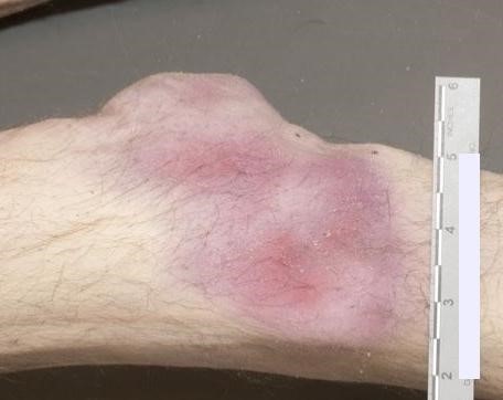
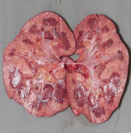
The autopsy findings of hypothermia are not specific. External examination may show bright pink discoloration of the skin over joints (“frost erythema”). There may be black-brown spots on the gastric mucosa, (“Wischnewsky spots”), thought to result from terminal vasodilation of submucosal vessels. The kidneys may be pale with microscopic subnuclear vacuolization of the tubular epithelium (the “Armanni-Ebstein” lesion). Acute hemorrhagic pancreatitis has also been described. However, these findings require a period of survival to develop—many cases, particularly if the decedent succumbs quickly, show no findings at all. The diagnosis of hypothermia therefore relies heavily on scene investigation. “Paradoxical undressing” (demonstrated in this case), refers to the phenomenon of a terminally hypothermic person taking off their clothes. This is caused by a feeling of warmth resulting from failure of vasoconstriction in the skin, and contributed by altered mentation.

Those at greatest risk are people spending extended time outdoors, including the homeless and outdoor recreationalists. The elderly and very young have a lower ability to centrally regulate body temperature. Children’s increased body surface area also leads to more rapid heat loss. People who are intoxicated with alcohol or drugs may not sense the cold or lack judgment to seek shelter. Alcohol also acts as a vasodilator, impairing vasoconstrictive adaptation to cold.

As the weather cools down, be mindful of how easily hypothermia can develop. Temperatures can be above freezing, yet those who are vulnerable are still at risk of hypothermia. Prepare yourself well for any snowy excursions, and keep an eye on those in your community who may not be able to seek shelter.

Stomach mucosa showing spots of black or dark brown discoloration
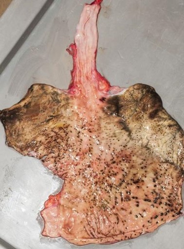
known as Wischnewsky spots. These are not specific to hypothermia and may just be an indicator of physiologic stress.
Bright pink discoloration over the knees, or “frost erythema”.
Pallor of the renal cortices corresponds to the microscopic “Armanni-Ebstein” lesion. This isn’t specific to hypothermia and can be seen in ketoacidosis from any cause.

-Alison Krywanczyk, MD, FASCP, is currently a Deputy Medical Examiner at the Cuyahoga County Medical Examiner’s Office.

Microbiology Case Study: Bacteremia with Long, Gram Negative Rod in a 34 Year Old Patient

Case History

A 34 year old male presented to the emergency department (ED) with acute onset abdominal pain, nausea, vomiting, persistent fever, and chills. His physical examination at that time was consistent with appendicitis. Patient was treated with Zosyn for broad coverage. Imaging showed a normal appendix. Three days later after blood was drawn, his blood cultures flagged positive for gram negative, elongated, thin rods. Growth was determined to be Fusobacterium mortiferum by MALDI-TOF. Ampicillin/sulbactam was started and patient was given Amoxicillin/clavulanic acid for outpatient treatment. Further follow-up of the patient showed normal white blood count and normal urinalysis. Repeat blood cultures were negative.

Images of Gram stain demonstrating long, slender, gram negative rods (top) and bacterial growth on anaerobic plate (bottom) from positive blood culture bottle.

Discussion

Fusobacteria are anaerobic, gram negative, spindle-shaped rods with pointed ends. They are part of the upper respiratory and gastrointestinal flora in humans but can cause diseases ranging from tonsillitis to septic shock.1 Fusobacterium nucleatum and necrophorum are commonly isolated in human diseases, although other species such as Fusobacterium mortiferum, as described in our case, have occasionally been documented as a secondary cause of septicemia 2 or bacteremia 1 and in rare instances implicated in the development of thyroid abscess.3

F. nucleatum is a member of oropharyngeal flora and unsurprisingly involved in gingival and periodontal diseases.4 It has been also described as the most likely cause of extra-oral infections among oral anaerobes.5 F. nucleatum has been detected in various fetal and placental tissues associated with adverse pregnancy outcomes, such as preeclampsia, chorioamnionitis and preterm rupture of membranes.6 Recent studies have reported this species to be abundant in colon, esophageal carcinoma, pancreatic and breast cancers. It is associated with poor prognosis in colon, rectal, pancreatic and esophageal cancers by promoting pro-tumorigenic immune microenvironment and reduction in the number of tumor-infiltrating lymphocytes.7, 10 One of the proposed theories is the involvement of the Fap2 virulence factor that has been described to inhibit tumor cell clearance in colorectal cancer cells.8 The other commonly isolated species is F. necrophorum, which is associated with oropharyngeal infection followed by septic thrombophlebitis of the internal jugular vein with sepsis and metastatic diseases typically involving the lungs. This syndrome is known as Lemiere’s disease first described in 1936 by Andre Lemierre. F. necrophorum usually causes infection in young, otherwise healthy adults in contrast to F. nucleatum1 which is associated more with the elderly population. According to Afra et al most of the mortality cases were due to F. nucleatum as opposed to F. necrophrum. This could be attributed to co-morbidities in elderly patients with positive F. nucleatum cultures.

Fusobacterium species can be identified using mass spectrometry MALDI-TOF. Typically, Fusobacterium species are resistant to vancomycin, but susceptible to colistin and kanamycin disk identification tests; however, F. nucleatum is susceptible to all three drugs. F. mortiferum and F. varium grow in the presence of bile. F. necrophorum shows positive indole and negative nitrate testing. Sequencing of the 16S RNA gene and 16S-23S rRNA gene spacer region can be used to determine the different species3,9

Fusobacterium species are usually susceptible to penicillin, clindamycin, metronidazole, and chloramphenicol and resistant to macrolides. F. nucleatum and F. necrophorum may produce beta-lactamases.3 In rare cases, surgical intervention is warranted for abscess formation.

References

  1. Afra K, Laupland K, Leal J, Lloyd T, Gregson D. Incidence, risk factors, and outcomes of fusobacterium species bacteremia. BMC Infect Dis. 2013;13(1). doi: 10.1186/1471-2334-13-264.
  2. Prout J, Glymph R. Fusobacterium mortiferum septicemia. Clinical Microbiology Newsletter. 1985;7(4):29. doi: 10.1016/s0196-4399(85)80052-0.
  3. Stavreas NP, Amanatidou CD, Hatzimanolis EG, et al. Thyroid abscess due to a mixed anaerobic infection with fusobacterium mortiferum. J Clin Microbiol. 2005;43(12):6202. doi: 10.1128/jcm.43.12.6202-6204.2005.
  4. Moore WE, Moore LV. The bacteria of periodontal diseases. Periodontol 2000. 1994 Jun;5:66-77. doi: 10.1111/j.1600-0757.1994.tb00019.x. PMID: 9673163.
  5. Bolstad AI, Jensen HB, Bakken V. Taxonomy, biology, and periodontal aspects of Fusobacterium nucleatum. Clin Microbiol Rev. 1996 Jan;9(1):55-71. doi: 10.1128/CMR.9.1.55. PMID: 8665477; PMCID: PMC172882.
  6. Han YW. Fusobacterium nucleatum: A commensal-turned pathogen. Current Opinion in Microbiology. 2015;23:141. doi: 10.1016/j.mib.2014.11.013.
  7. Alon‐maimon T, Mandelboim OO, Bachrach G. Fusobacterium nucleatum and cancer. Periodontology 2000. 2000;89(1):166. doi: 10.1111/prd.12426.
  8. Umaña A, Sanders BE, Yoo CC, Casasanta MA, Udayasuryan B, Verbridge SS, Slade DJ. Utilizing Whole Fusobacterium Genomes To Identify, Correct, and Characterize Potential Virulence Protein Families. J Bacteriol. 2019 Nov 5;201(23):e00273-19. doi: 10.1128/JB.00273-19. PMID: 31501282; PMCID: PMC6832068.
  9. Garcia-Carretero R, Lopez-Lomba M, Carrasco-Fernandez B, Duran-Valle MT. Clinical features and outcomes of fusobacterium species infections in a ten-year follow-up. The Journal of Critical Care Medicine. 2017;3(4):141. doi: 10.1515/jccm-2017-0029.
  10. Brennan CA, Garrett WS. Fusobacterium nucleatum — symbiont, opportunist and oncobacterium. Nat Rev Microbiol. 2018;17(3):156. doi: 10.1038/s41579-018-0129-6.

-Dr. Hayk Simonyan was born and raised in Yerevan, Armenia. He attended Yerevan State Medical University after Mkhitar Heratsi where he received his doctorate degree. He did his research at The George Washington University. His studies were focused on transcription factor activation in the SFO-PVN axis that leads to cardio-metabolic changes mediated by obesity, oxidative stress, and angiotensin-II. One of his other projects included collaboration with the National Cancer Institute, working on alternative treatment for glioblastoma multiforme. His academic interests include surgical pathology and molecular. In his spare time, Hayk enjoys spending time with family, playing soccer, tennis, and skiing. Hayk is pursuing AP/CP training. 

-Rebecca Yee, PhD, D(ABMM), M(ASCP)CM is the Chief of Microbiology, Director of Clinical Microbiology and Molecular Microbiology Laboratory at the George Washington University Hospital. Her interests include bacteriology, antimicrobial resistance, and development of infectious disease diagnostics.

Histio Makes History

An 81 year old female presented to the head and neck clinic after being diagnosed with cutaneous T cell lymphoma of the posterior mid-parietal scalp at an outside institution. She was initially treated with Brentuximab every three weeks but developed significant toxicities. The patient’s previous “T cell lymphoma” material was reviewed at our institution and the immunophenotypic report described the neoplastic cells as being positive for CD45, CD2, CD4, BCL6+, CD3 (subset), and CD123 (scattered), while negative for CD7, CD8, CD20, CD30, CD56, EBER ISH, PAX5, and lysozyme. Immunohistochemical slides were not provided for review. Flow cytometric analysis determined that there was no immunophenotypic evidence of a clonal T cell population in the patient’s peripheral blood.

A second scalp biopsy was performed at another outside institution, and the findings were similar to the parietal scalp; however, there were atypical pleomorphic cells which displayed irregular contours, hyperchromasia, and multiple nucleoli. The atypical cells were predominantly positive for CD4 and diffuse positivity for CD1a. These same pleomorphic cells were negative for CD3, CD8, CD20, CD30, ALK1, BCL6, CD56, EBER, AE1/AE3, SOX10, Desmin, PAX5, MUM1, CD5, and Cam 5.2.

The smears contained large, highly pleomorphic cells with irregular, elongated, and multilobated nuclei, frequent nuclear grooves and folds, fine chromatin, prominent nucleoli, and variable amounts of pale, eosinophilic cytoplasm, alt.

The outside tissue block on the original scalp biopsy was requested, and our pathology department performed additional immunostains. The neoplastic cells of interest were positive for CD1a, S100, CD68 (a small subset), and negative for lysozyme, CD21, CD30, and CD3. Ki67 proliferation index was interpreted at approximately 60%. An unstained FFPE tissue section was sent to a reference laboratory, and the neoplastic cells were strongly positive for Langerin.

While the Brentuximab treatment initially appeared to have a positive impact on the overall disease burden, the PET CT following 3 cycles showed a mixed response, including resolution of cervical lymphadenopathy and identification of multiple new lung nodules and bulky mediastinal lymphadenopathy. Between that and numerous reported toxicities, the treatment protocol was discontinued. The patient was then referred to radiology for a CT-scan guided right lower lobe lung biopsy measuring 2.2 x 1.3 centimeters with an SUV or 29.6.

In the CT Scan suite, we received multiple FNA passes from the interventional radiologist and made air-dried and alcohol-fixed smears, rinsing the residual needle material into a tube of balanced salt solution for a cell block preparation. We determined our specimen was adequate for scant tumor cells, as depicted on the Diff-Quik smears below.

Images 1-2. Lung, right lower lob, CT-guided FNA. Diff-Quik stained smears.

In comparison to the material from the second scalp biopsy, the cells from the lung biopsy appeared identical. Our Pap-stained smears and H&E cell block sections also demonstrated the highly pleomorphic cells described above.

Images 3-6. Lung, Right Lower Lobe, CT-guided FNA. 3-4: Pap-stained smears, 5-6: H&E sections (5: 100x, 6: 400x).

Immunostains performed on the cell block slides with adequate controls show that the tumor cells are positive for CD1a, CD4, partially positive for CD45 and S100, negative for AE1/3, TTF-1, and p40.

Images 7-8. Lung, Right Lower Lobe, CT-guided FNA. Cell block section immunohistochemistry. 7: CD1a-positive; 8: partially S-100-positive.

Our pathologists felt the cells from the second scalp biopsy and the lung biopsy were representative of a Langerhans cell sarcoma, a form of malignant histiocytosis, rather than a T-cell lymphoma. It is possible that the first scalp biopsy’s diagnosis of T-cell lymphoma was due to sampling error and the pleomorphic cells of interest were missed. The Ki-67 proliferative index of 60% helped to distinguish between Langerhans cell histiocytosis and Langerhans cell sarcoma.

Molecular testing performed on the core biopsy was negative for a BRAF mutation and positive for an NF1 inactivating mutation. The tumor may then be sensitive to mTOR inhibitors and MAPK pathway inhibitors, such as MEK inhibitors. Appeals for a MEK inhibitor were denied by insurance, but fortunately, the tumor also demonstrated high PD-L1 expression at 90%, making this specific patient a candidate for pembrolizumab, which was fully covered by insurance.

____________________________________________________________________________________________

I can’t help but think about the disparities associated with cancer and the inaccessibility of potentially lifesaving or life-prolonging treatments. Sure, there may be viable alternatives, such as this case, but what if we had equal access to cutting edge, personalized therapies? What if the only therapy available was too costly to bear? Just because a cancer might be rare, such as Langerhans cell sarcoma, it doesn’t mean access to a proven effective therapy should also be rare. Even with drug assistance programs, so many patients face the harsh reality of tapping into their life savings to just to save their own life. When we became medical laboratory professionals, we promised to provide timely and accurate for all of our patients. Now, it’s time that pharmaceutical companies and our healthcare system as a whole work together to provide high quality, low-cost, readily accessible and personalized treatment options to every patient. They deserve that chance to overcome or at least manage their cancer.

-Taryn Waraksa-Deutsch, MS, SCT(ASCP)CM, CT(IAC), has worked as a cytotechnologist at Fox Chase Cancer Center, in Philadelphia, Pennsylvania, since earning her master’s degree from Thomas Jefferson University in 2014. She is an ASCP board-certified Specialist in Cytotechnology with an additional certification by the International Academy of Cytology (IAC). She is also a 2020 ASCP 40 Under Forty Honoree.

Microbiology Case Study: A 70 Year Old with Fevers, Rigors, and Dizziness

Case Description

A 70 year old female arrived in the hospital with chief complaints of 6 days of fever, rigors, weakness, headache, and dizziness; she has a history of asthma, type 2 diabetes, supraventricular tachycardia and exercise-induced ventricular tachycardia. The patient was also seen 5 days before the current visit for abdominal pain, nausea, and fever. The abdominal pain has gone, but she has had a loss of appetite. She admitted that she sleeps with her dog in bed during that visit. No scleral icterus, rash, cough, urinary tract burning, or neck stiffness was reported on any visits.

CT scan, CBC with differential, BMP, liver function panel, Coag, blood culture, and blood parasite tests were ordered. On the CBC, the cells below were flagged for review (Figure 1).

Figure 1. A Cellavision capture of morulae inside a neutrophil.

Discussion

The round light purple dots pointed by the arrow in Figure 1 are morula indicative of Anaplasma phagocytophilum, formally named “human granulocytic anaplasmosis (HGA)”. Historically, Ehrlichia phagocytophila and Ehrlichia equi were recognized separately (Sexton & McClain, 2022). HGA is a tick-borne illness more commonly found in the northeast U.S., and the case number has continuously increased in recent years (Centers of, 2022). The tick bite is not painful, and the first symptom usually shows after about a week from the bite. Early diagnosis can be hard at the initial stage since laboratory serology tests often give negative results for the antibodies. It is essential to carefully review the clinical signs and symptoms, travel history, outdoor activity, and animal contacts (Centers of, 2022). PCR is the most sensitive and specific method of diagnosis. Blood smears can be made to confirm the parasite morphology, although patients can have leukopenia leading to decreased sensitivity.

Lab results showed critical hyponatremia (121 mmol/L) and thrombocytopenia (33 K/uL) in this case. The patient was admitted to the floor and prescribed 10 days of doxycycline.

Extreme hyponatremia related to anaplasmosis is not common, and the causing mechanism is unclear; however, all the reported cases fit the description of SIADH – syndrome of inappropriate secretion of antidiuretic hormone (Ladzinski et al., 2021).

References

  1. Centers for Disease Control and Prevention. (2022, August 15). Epidemiology and statistics. Centers for Disease Control and Prevention. Retrieved 2022, from https://www.cdc.gov/anaplasmosis/stats/index.html
  2. Ladzinski, A. T., Baker, M., Dunning, K., & Patel, P. P. (2021). Human granulocytic anaplasmosis presenting as subacute abdominal pain and hyponatremia. IDCases, 25. https://doi.org/10.1016/j.idcr.2021.e01183
  3. Sexton, D. J., & McClain, M. T. (2022, March 21). Human ehrlichiosis and anaplasmosis. UpToDate. Retrieved 2022, from https://www.uptodate.com/contents/human-ehrlichiosis-and-anaplasmosis

-Sherry Xu is a Masters Student in the Department of Pathology and Laboratory Medicine at the University of Vermont Larner College of Medicine.

-Christi Wojewoda, MD, is the Director of Clinical Microbiology at the University of Vermont Medical Center and an Associate Professor at the University of Vermont.

Microbiology Case Study: An Elderly Adult Presenting with Foodborne Illness Related to Shellfish Consumption

Case History

An adult consumed shellfish at a restaurant. Approximately 12 hours after this dinner, the patient experienced the first signs of loose stools, fever, and abdominal cramping. The patient had watery diarrhea for the next three days with 8 bouts a day. The patient did not have a fever after the first day. The patient denied blood in stool or nausea or vomiting. The patient did not have a recent travel history and denied recent antibiotic use. On the 4th day of symptoms, the patient was seen by their primary care provider. The physical exam was unremarkable except for dehydration. A stool and blood sample were obtained and aggressive hydration was recommended. Blood smear, complete blood panel, and basic metabolic panel resulted in normal. Shigella, Salmonella, Campylobacter, and Shiga-toxin-producing gene were not detected by PCR. The stool sample was set up for culture. Mucoid colonies were noticed after 12 hours on the blood agar plate. MALDI revealed Grimontia hollisae.

Discussion

The genera of Grimontia is one of the new members of the Vibrionaceae family. Grimontia hollisae, previously known as Vibrio hollisae, is currently the only known pathogenic species in the Grimontia genera. Vibrio hollisae was first described and named by Hickman et al. in 1982.1 However, based on phylogenetic and phenotypical differences V. hollisae was placed into a novel genus, named Grimontia.2 It is named after French microbiologist Patrick P. A. Grimont.

G. hollisae are halophilic, gram negative, oxidase-positive, indole-positive, ornithine-negative, and motile by a single polar flagellum.2 One of the most important features of G. hollisae is its failure to grow on thiosulfate-citrate-bile salts-sucrose (TCBS) agar, the main phenotypical difference from vibrios.2 However, it does grow well on sheep blood agar and marine agar.3 G. hollisae is generally transmitted via shellfish (mostly oysters, mussels, and prawns etc.).2 However, it can also be transmitted through infected ocean water, and other foods that are cross-contaminated with the organism.4 To date, the person-to-person spread has not been documented.4

Diagnosis of G. hollisae can be challenging since it does not grow on Vibrio-selective media (TCBS agar) or on MacConkey.5 However, the organism grows well on blood agar plate. Spot oxidase and indole tests may be helpful to rule-in a possible Vibrio or Grimontia species in suspicious cases.5 It is important that the stool sample should be collected as soon as possible in patients suspicious for vibrio gastroenteritis.5 Cary-Blair medium should be used as transport medium.5

The incubation period of G. hollisae is usually 12-24 hours (ranging between 4-96 hours).4 It primarily causes moderate to severe gastroenteritis.3 Signs and symptoms of G. hollisae gastroenteritis include fever, abdominal cramping, watery diarrhea, nausea, and vomiting. Although it is mostly self-limited, it may also cause serious conditions such as hypovolemic shock, sepsis, hepatitis, and ileus.3, 6-8 Rarely, grossly bloody stool can be seen in severe cases.9 Treatment is mostly supportive, oral hydration is preferred over intravenous in tolerating patients.

G. hollisae disease, clinically, is still considered Vibriosis.4 Janda et al. showed that among the all other causes of Vibriosis, G. hollisae comprises only 1.2% of the cases.5 In 83% of these cases, the organism was isolated from the gastrointestinal system.5 Skin and soft tissue specimens were other resources where G. hollisae was isolated.5 In the same study, it has been shown that unlike V. cholerea, V. mimicus, and V parahaemolyticus, G. hollisae has never caused an epidemic, a pandemic, or an outbreak.5 However, unfortunately, the numbers of vibriosis are in increasing trend due to rising sea surface temperature.10 Considering the record high temperatures and heat waves in recent years, it is more than a lucky guess that we may see more and more Vibriosis cases in the next years, especially in the summer seasons. As microbiologists and healthcare workers we should be aware of these organisms, their capabilities, their limits, and how to prevent the spread of them.

References

  1. Hickman FW, Farmer JJ 3rd, Hollis DG, Fanning GR, Steigerwalt AG, Weaver RE, Brenner DJ. Identification of Vibrio hollisae sp. nov. from patients with diarrhea. J Clin Microbiol. 1982 Mar;15(3):395-401. doi: 10.1128/jcm.15.3.395-401.1982. PMID: 7076812; PMCID: PMC272106.
  2. Thompson FL, Hoste B, Vandemeulebroecke K, Swings J. Reclassification of Vibrio hollisae as Grimontia hollisae gen. nov., comb. nov. Int J Syst Evol Microbiol. 2003 Sep;53(Pt 5):1615-1617. doi: 10.1099/ijs.0.02660-0. PMID: 13130058.
  3. Hinestrosa F, Madeira RG, Bourbeau PP. Severe gastroenteritis and hypovolemic shock caused by Grimontia (Vibrio) hollisae infection. J Clin Microbiol. 2007 Oct;45(10):3462-3. doi: 10.1128/JCM.01205-07. Epub 2007 Aug 17. PMID: 17704283; PMCID: PMC2045321.
  4. https://www.oregon.gov/oha/PH/DiseasesConditions/CommunicableDisease/ReportingCommunicableDisease/ReportingGuidelines/Documents/vibrio.pdf
  5. Janda JM, Newton AE, Bopp CA. Vibriosis. Clin Lab Med. 2015 Jun;35(2):273-88. doi: 10.1016/j.cll.2015.02.007. Epub 2015 Apr 9. PMID: 26004642.
  6. Edouard S, Daumas A, Branger S, Durand JM, Raoult D, Fournier PE. Grimontia hollisae, a potential agent of gastroenteritis and bacteraemia in the Mediterranean area. Eur J Clin Microbiol Infect Dis. 2009 Jun;28(6):705-7. doi: 10.1007/s10096-008-0678-0. Epub 2008 Dec 17. PMID: 19089475.
  7. Gromski MA, Relich RF, Siwiec RM. Grimontia hollisae: A Cause of Severe Ileus in a Seafood-Loving Traveler: 968. American Journal of Gastroenterology: October 2015 – Volume 110 – Issue – p S415-S416
  8. Edouard S, Daumas A, Branger S, Durand JM, Raoult D, Fournier PE. Grimontia hollisae, a potential agent of gastroenteritis and bacteraemia in the Mediterranean area. Eur J Clin Microbiol Infect Dis. 2009 Jun;28(6):705-7. doi: 10.1007/s10096-008-0678-0. Epub 2008 Dec 17. PMID: 19089475.
  9. Abbott SL, Janda JM. Severe gastroenteritis associated with Vibrio hollisae infection: report of two cases and review. Clin Infect Dis. 1994 Mar;18(3):310-2. doi: 10.1093/clinids/18.3.310. PMID: 8011809.
  10. Baker-Austin C, Trinanes J, Gonzalez-Escalona N, Martinez-Urtaza J. Non-Cholera Vibrios: The Microbial Barometer of Climate Change. Trends Microbiol. 2017 Jan;25(1):76-84. doi: 10.1016/j.tim.2016.09.008. Epub 2016 Nov 12. PMID: 27843109.

-Kadir Isidan, MS, MD is a pathology resident at University of Chicago (NorthShore). His academic interests include gastrointestinal pathology and cytopathology.

-Paige M.K. Larkin, PhD, D(ABMM), M(ASCP)CM is the Director of Molecular Microbiology and Associate Director of Clinical Microbiology at NorthShore University HealthSystem in Evanston, IL. Her interests include mycology, mycobacteriology, point-of-care testing, and molecular diagnostics, especially next generation sequencing.

Feed the Safety Need

Ben was excited to bring the new analyzer into the laboratory until he discovered the manufacturer’s newest security feature. Anytime a user was to log into the analyzer’s computer to diagnose issues or to perform maintenance, a unique numeric passcode would have to be entered, and that code would be sent via text to the app that staff could download on their cellphones. John knew that the use of cell phones in the lab violated the personal electronic device policy.

Emily was proud of the work she had done to design the new outpatient collection draw area. It included a row of collection rooms each with their own computer for order entry. The central area outside the rooms had a phone and printer set up for an efficient workflow. However, every time she performed a site visit she noticed her staff were using cell phones in the patient collection rooms. When she asked why, they told her they often had to make calls to clarify orders, and that talking on the central phone meant discussing patient information in front of people seated in the waiting area.

When a basic need of a human being is not met, conflict is automatically set up in the mind, and humans will deal with that conflict with a workaround or possibly with aggression. Often laboratories and their procedures are designed without considering all of the potential needs of the staff who will work there. Conflict will arise, and policies will not be followed, and you may also wind up with unhappy employees.

When it comes to safety policies and procedures, it is important to educate why they must be followed. It is vital to discuss the possible outcomes of not using safe practices. That may mean exposures to chemicals and biohazards, and it may also mean injuries. It can take time to explain that the use of a smart watch with contaminated gloves can lead to infection and potentially severe illness at work and in the home.

While this understanding is important, it must be coupled with a system of practices that allows staff to easily follow the prescribed safe practices. It must be easy for staff to perform safe acts, there should be no hindrances in their way for that to happen. Otherwise, conflict will occur, and the set policies will not be followed. Staff may know the regulations, they may even understand the potential consequences of not following them, but they will not conform to the policies because of some software glitch or because some vital tool is missing in their environment.

When you notice a lab safety violation, or if a safety incident has occurred, the first thing to look for in the investigation is something in the system that may have caused it. Unless the incident occurred because of a blatant act by the employee, blame should never first be focused on the person. What departmental design flaw exists? What engineering control could have been in place? What PPE should have been readily available? What was the temperature and humidity in the department, etc.?

Upon further discussion with the vender, Ben learned that the manufacturer’s security code system could not be bypassed, but that the app could be downloaded onto an electronic tablet rather than a cell phone. Ben purchased a tablet that could be used in the lab and remain there so as not to create any infection control issues. The tablet was also used for lab safety and quality audits so that pictures of issues could be taken and that results of audits could be entered directly. It became a real time saver, and no cell phones were needed in the laboratory.

Upon review, Emily realized that access to phones in the new outpatient collection area needed to be better. There was no way to even call or help from a collection room should there be an adverse reaction to phlebotomy. Emily was able to acquire portable phones in the short term until she could get permanently-mounted telephones into each of the three blood collection rooms. Staff no longer needed to use cell phones in the biohazardous areas.

Humans have basic needs like food, shelter, and clothing. When those needs are not met, some may act in surprising ways to obtain them. The same holds true in the laboratory. There is a need to be safe, there is a need to follow safety regulations and policies, and unsafe behaviors will arise if it cannot be achieved. Feed the safety needs of your employees. Provide a safe working environment with good engineering controls, PPE, and polices that allow for workdays that have no safety conflict.

Dan Scungio, MT(ASCP), SLS, CQA (ASQ) has over 25 years experience as a certified medical technologist. Today he is the Laboratory Safety Officer for Sentara Healthcare, a system of seven hospitals and over 20 laboratories and draw sites in the Tidewater area of Virginia. He is also known as Dan the Lab Safety Man, a lab safety consultant, educator, and trainer.