Triaging Times

As a clinical instructor and lead cytologist at my institution, I like to remind our newer cytologists and cytology students of the importance of being prepared for FNA biopsies so they develop good habits or best practices as they become more experienced. This level of preparation helps to create a culture of ongoing learning and improvement, which is necessary for the laboratory. In my experience, I’ve met some cytologists who prefer to go into a case blind, with the mindset that knowing the patient’s clinical history in advance muddies their knowledge, skills, and abilities, limiting their mindset by excluding the possibility of other diagnoses. While diving into the unknown might seem exciting, it is also a hindrance and could result in errors, especially when the clinical history helps us triage the patient’s sample. For example, knowing that the patient has a history of lymphoma or that the presentation state includes bulky lymphadenopathy prompts us to collect additional needle passes to send for flow cytometry analysis. Another concern is not knowing whether the patient has a history of breast, gastric, or esophageal cancer, and consequently processing the specimen routinely, which may result in an extended cold ischemic time. This delay in fixation along with insufficient formalin fixation can yield false negatives on ER/PR IHC in breast cancers and HER2 FISH in breast, gastric, and esophageal cancers, which could restrict the use of hormone therapies, such as tamoxifen and aromatase inhibitors for hormone receptor-positive (HR+) cancers, or trastuzumab for HER2+ cancers. I cannot overemphasize the importance of familiarizing yourself with clinical history and communicating case specifics while you act as a mediator between clinician and pathologist.

Whether the clinical history impacts the pre-analytical phase, such as specimen collection (limiting cold ischemic time or collecting additional needle passes for ancillary studies) or the analytical phase, as such processing (formalin fixation) and diagnosis (selecting an appropriate immunoprofile), we must remain vigilant and proactive in laboratory medicine. In this case, knowing the patient’s clinical history was of the utmost significance as it helped to reduce the number of immunostains and ancillary studies necessary to make the diagnosis. Using morphologic criteria in tandem with the patient’s clinical history narrowed the differential diagnoses to just two possible types of cancer, presented below.

A 59 year old male patient presented to the emergency room after an automobile accident. On imaging, the X-ray and CT scan identified a left humerus mass and fracture, and bloodwork was performed. His medical record was sparse and uneventful with no recent visits or encounters. To build a more comprehensive wellness profile and prepare for surgery, he was also offered a one-time screening for Hepatitis C, as an adult who was born between 1945 and 1965.

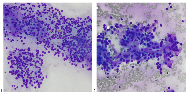
The left humerus mass was biopsied via CT-scan guidance and two passes were obtained. The Diff-Quik stained smears demonstrate large polygonal cells, some with abundant, granular cytoplasm and some isolated cells with naked nuclei. Vessels also appear to traverse some of the cell groups.

Images 1-2: Bone, Humerus, Left, CT-guided FNA. Diff-Quik-stained smears.

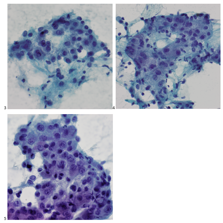
The Pap-stained smears also demonstrate polygonal cells with granular cytoplasm, nuclei with coarse chromatin, and prominent nucleoli. An interesting feature frequently identified in this case is the intranuclear inclusions, and in hindsight, a focus on these may have further reduced the number of immunostains performed.

Images 3-5: Bone, Humerus, Left, CT-guided FNA. Pap-stained smears.

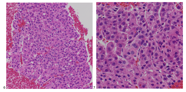
The H&E-stained cell block sections show trabeculae with endothelial wrapping around the cell cords. While renal cell carcinoma was listed as a differential diagnosis due to its telltale oncocytic cytoplasm and vascularity, hepatocellular carcinoma was favored.

Images 6-7: Bone, Humerus, Left, CT-guided FNA. H&E sections (6: 100x, 7: 400x).

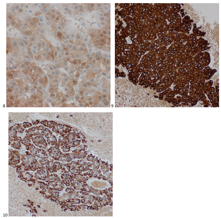
Immunostains were performed using proper positive and negative controls on the cell block sections, and the tumor cells show positive staining for Arginase, cam5.2, and Hepar1, while negative staining for CK7 and PAX8 (not shown).

Images 8-10: Bone, Humerus, Left, CT-guided FNA. Cell block section immunohistochemistry. 8: Arginase-positive; 9: cam5.2-positive; 10: Hepar1-positive.

Fortunately, before ordering immunostains, both our cytologist and pathologist working on the case peered into the patient’s medical record and noticed that he had recent bloodwork which demonstrated a positive Hepatitis C screening. This diagnosis was as recent as the identification of his humerus mass. Had it not been for his car accident, I can’t imagine how long he would have gone undiagnosed for both hepatitis and metastatic hepatocellular carcinoma. Incidental findings save lives, folks.

Granted, in settings of unknown primaries with widespread metastatic disease, such as carcinomatosis, an extensive workup is almost always inevitable. Narrowing down possible etiology based on information such as gender, age, and environmental or occupational exposure can help, but that doesn’t always yield a definitive answer as time- or cost-effectively as possible. In this case, that one clue of untreated Hepatitis C was all the cytopathology team needed. A rarity, sure, but as we are asked to do more personalized tests with less material, think of the patient’s specimen as a puzzle and keep your eye out for a clue both under the microscope and behind the computer. You never know what you might find that reduces errors and unnecessary testing while efficiently leading to a definitive diagnosis.

-Taryn Waraksa-Deutsch, MS, SCT(ASCP)CM, CT(IAC), has worked as a cytotechnologist at Fox Chase Cancer Center, in Philadelphia, Pennsylvania, since earning her master’s degree from Thomas Jefferson University in 2014. She is an ASCP board-certified Specialist in Cytotechnology with an additional certification by the International Academy of Cytology (IAC). She is also a 2020 ASCP 40 Under Forty Honoree.

Microbiology Case Study: Severely Immunocompromised Female with Respiratory Failure

Case History

A 50 year old female with a complex medical history consisting of lymphoma, diabetes mellitus (type II), sarcoidosis, congestive heart failure, chronic renal failure (stage 3), and pancytopenia  presented to the emergency department with shortness of breath, cough, fever. She was found to be positive for SARS-CoV-2 and was transferred to the ICU due to hypoxic respiratory failure. She was treated for sepsis and respiratory failure, but her status continued to decline. The patient had multiple admissions due to COVID-19 in the past, received remdesivir and was on corticosteroid therapy due to the interstitial lung disease from last year. Initial evaluation included complete blood count which revealed anemia (hemoglobin=8.7 mg/dl), leukocytosis (WBC = 21,900/mcl), lymphopenia (910/mcl) and thrombocytopenia (Plt = 27000/mcl). The patient was treated with broad antibiotics and additional steroids. Additional tests revealed hyperproteinemia and hypoalbuminemia. Chest x-ray showed worsening infiltrates in lungs and chest CT scan revealed left apical hydropneumothorax, loculated left pleural effusion, pneumomediastinum, and chest wall subcutaneous emphysema. Lung biopsy revealed necrosis. Histopathology examination revealed broad, branching hyphae with sporulation in lung tissue biopsy and bronchoalveolar lavage. Respiratory cultures of lung biopsy and BAL grew rapidly and lactophenol cotton blue tape preps showed broad hyphae with round sporangium and rhizoids between the stolons. The patient was diagnosed with mucormycosis, infection with Rhizomucor, and was treated with Amphotericin B. Surgical debridement of the tissue was not possible due to her declining condition. She passed away after 5 days.

Figure 1. H&E stain of the lung biopsy (top, left) and Papanicolaou stain of bronchoalveolar lavage (top, right) revealed broad, ribbon-like, right-angle branching hyphae (visible in lung biopsy) with sporulation (credits to Dr. Elham Arbzadeh, George Washington University School of Medicine and Health Sciences). Rapid growth was observed from the respiratory cultures of the tissue biopsy by day 2 (bottom, left) where lactophenol cotton blue tape preps showed broad hyphae with sporangium (bottom, right) and intermodal rhizoids (not shown in this image).

Discussion

The term mucormycoses refers to infections caused by the Zygomycetes which is further separated into Mucorales and Entomophthorales. Some of the members of Mucorales are Rhizopus spp., Mucor spp., Lichtheimia (Absidia) spp., Syncephalastrum spp., and Rhizomucor spp.1,2 These organisms live in soil, dung, and vegetative matter. Infection is usually acquired by inhalation/ingestion of their spores or direct inoculation and contamination of wounds. The mold can invade the walls of the blood vessels causing angioinvasion and often results in dissemination of mycotic thrombi and development of systemic infection. Zygomycetes are most commonly known for causing rhinocerebral, pulmonary, cutaneous, and disseminated disease. Infections with Zygomycetes most commonly occur as opportunistic infections in immunocompromised hosts. Risk factors include diabetes, those with acidosis, neutropenia, and sustained immunosuppression such as after transplantation.

Zygomycetes grow very fast (within 48 to 72 hrs.) and is often called a “lid lifter”. The colonies have a wooly mycelium and can be described as cotton candy-like. Lactophenol tape preps of the mold would reveal broad hyphae, aseptate or pauciseptate, ribbon-like hyphae with irregular width. At the tip of the sporangiophore, there is a sack-like structure called a sporangia with contains all the spores. Fungal elements and hyphae seen on tissue biopsies from patients with mucormycosis typically have near right angle branching (usually >40o) broad, non-septate hyphae. In contrary, those with aspergillosis show acute angle branching (usually <45o) with narrow, septate hyphae.3  

Genus-level identification can be achieved by microscopic morphology. Rhizomucor is an intermediate between Rhizopus and Mucor. Rhizoids found in Rhizomucor are few in number and are located on stolons, between the sporangiophores, as opposed to Rhizopus where the rhizoids are often seen directly at the nodes and Mucor which does not produce rhizoids. Sporangia (40-80 µm in diameter) are brown in color and round in shape. Apophysis is absent, which allows for differentiation from Lichtheimia (Absidia) where apophysis can be seen.4 The genus Rhizomucor includes three species: Rhizomucor pusillusRhizomucor miehei, and Rhizomucor tauricus.5

Treatment of mucormycosis consists of antifungal and surgical therapy. Amphotericin B is the most commonly used antifungal agent. Liposomal amphotericin B has also been successfully used in some cases with zygomycosis due to Rhizomucor.6  Early diagnosis and treatment are crucial and mortality rate is high.7  Of note, Zygomycetes are intrinsically resistant to voriconazole.

References

  1. Rippon J W. Medical mycology. The pathogenic fungi and the pathogenic actinomycetes. Philadelphia, Pa: Saunders; 1974. Mucormycosis; pp. 430–447. 
  2. Scholer H J, Müller E. Beziehungen zwischen biochemischer Leistung und Morphologie bei Pilzen aus der Familie der Mucoraceen. Pathol Microbiol. 1966;29:730–741.
  3. Mohindra S., Mohindra S., Gupta, R., Bakshi, J., Gupta, S. K. Rhinocerebral mucormycosis: the disease spectrum in 27 patients. Mycoses. doi: 10.1111/j.1439-0507.2007.01364.x.
  4. de Hoog, G. S., J. Guarro, J. Gene, and M. J. Figueras. 2000. Atlas of Clinical Fungi, 2nd ed, vol. 1. Centraalbureau voor Schimmelcultures, Utrecht, The Netherlands)
  5. Schipper M A A. On the genera Rhizomucor and Parasitella. Stud Mycol. 1978;17:53–71. 
  6. Bjorkholm, M., G. Runarsson, F. Celsing, M. Kalin, B. Petrini, and P. Engervall. 2001. Liposomal amphotericin B and surgery in the successful treatment of invasive pulmonary mucormycosis in a patient with acute T- lymphoblastic leukemia. Scand J Infec Dis. 33:316-319.
  7. Ribes, J. A., C. L. Vanover-Sams, and D. J. Baker. 2000. Zygomycetes in human disease. Clin Microbiol Rev. 13:236-301.

-Maryam Mehdipour Dalivand, MD is a Pathology Resident (PGY-1) at The George Washington University Hospital. She is pursuing AP/CP training.

-Rebecca Yee, PhD, D(ABMM), M(ASCP)CM is the Chief of Microbiology, Director of Clinical Microbiology and Molecular Microbiology Laboratory at the George Washington University Hospital. Her interests include bacteriology, antimicrobial resistance, and development of infectious disease diagnostics.

Answering Your Questions about Monkeypox

I have been hearing many concerned questions about Monkeypox lately, and I wanted to add onto the great job already done by Dan Scungio in his previous post on how laboratorians should be safe around Monkey pox suspected samples. As a part of the queer community, I’ve heard from several people who are very concerned as this is predominately spread among men who have sex with men. I’ll be focusing on what is new about Monkeypox, how it is different, where it is spreading, and what can be done about it so far. I’ll address questions like should we be sequencing Monkeypox like COVID-19 and does your smallpox vaccination will protect you.

What is Monkey Pox?

This is an orthopox virus that is from the same family as smallpox, which was so effectively cleared from human circulation that vaccines were discontinued in the U.S. in the early 1980’s. It causes a systemic disease characterized by lesions that start as a red, flat rash (macula) then form vesicles that break open, crust and resolve in 2-4 weeks. If you ever had chicken pox, you may recall how painful it was, and this is the major symptom that requires medical management.

Figure 1. Transmission electron microscope image of Monkey pox (purple). https://www.cdc.gov/poxvirus/monkeypox/index.html

Is it really that big of a concern?

Initially case increases were attributed to undiagnosed disease just as happened with COVID-19 initially. However, now that commercial labs are testing for it and access to testing is not an issue, we still see case counts increasing. This indicates that the rapid spread is real and concerning. That rapid spread is one reason that it has now been declared a national public health emergency.

How is it tested for?

Initially testing was sent out to one of the CDC regional testing centers. However, there were only 60-70 of these sites and they had limited capacity for high throughput testing. Then Labcorp and Quest they can each perform PCR testing, which has expanded access greatly. However they have different specimen types they accept:

  • Labcorp: Lesion swab in VTM sent frozen or refrigerated (room temp not acceptable)
  • Quest: Lesion swab in VTM

What is new?

  • It has been in Sub-Saharan Africa for a long time.
  • Early summer it began to spread into other continents like Europe.
  • U.S. now has the highest levels of Monkeypox cases.

This all reinforces the impact of communicable diseases in a global society.

Figure 2. Global distribution of cases Jan 1, 2022-Aug 9, 2022. https://www.cdc.gov/poxvirus/monkeypox/response/2022/world-map.html

How are symptoms different?

  • The rash may begin without the typical prodrome symptoms of fever, malaise, etc.
  • Spread occurs by skin to skin contact
Figure 3. Examples of Monkey pox lesions. https://www.cdc.gov/poxvirus/monkeypox/symptoms.html

Does my smallpox vaccine protect me?

  • Smallpox vaccines are 85% effective for 3-5 years
  • Unknown how well they work after many years
  • They likely decrease severity of disease even if it was given many years ago.

What can be done to prevent it?

  • Monkeypox vaccination: requires 2 shots. Space out 4 weeks apart.
  • FDA recently approved 1) decreasing the dose and 2) performing subcutaneous injection.
  • This would increase the effective doses by 5x and still produce a robust immune response.

Should we be sequencing it?

  • There are 3 major clades of the virus with the Congo clade being more severe (10% death rate) than the one we are seeing (West African: 3%)
  • These differences can be found by PCR based tests
  • There is no treatment difference based on the clade.
  • If this continues to spread and mutate, then there could be a reason to sequence the virus.
  • Some evidence suggests the mutation rate is 2x higher than would be expected.
    • However, the last known samples were sequenced >5 years ago and not many were sampled to get a very accurate measure of the mutation rate.
    • So this news about mutation rate should be taken with a grain of salt.

References

Jeff SoRelle, MD is Assistant Instructor of Pathology at the University of Texas Southwestern Medical Center in Dallas, TX working in the Next Generation Sequencing lab. His clinical research interests include understanding how lab medicine impacts transgender healthcare and improving genetic variant interpretation. Follow him on Twitter @Jeff_SoRelle.

Tick Identification: Why Do We Do It and What Does It Tell Us?

During the warmer months here in the Midwest, ticks are abundant and our microbiology lab receives several tick submissions per day for identification. When possible, we provide species level identification as well as sex for any tick submitted. While this is common practice in most microbiology laboratories, our molecular laboratory accidently received a tick specimen and, in the process of routing it to the microbiology lab, was curious as to why the tick identification matters—what does that tell us clinically? This led to an impromptu plate rounds with both labs and prompted me to write this post.


How do we determine tick identity?

A tick is submitted in a cup and sent to the laboratory. Ideally the tick would be submitted whole without missing appendages or damaged in any way. The tick is placed in ethanol to kill the organism and to allow for examination under a microscope. The mouth parts, scutum, and festoons are examined for defining features. Thorough examination is challenging when the tick arrives damaged or only partially intact.

Why do we provide tick identification?

Certain ticks carry specific pathogens. For instance, Amblyomma americanum (lone star tick) can transmit ehrlichiosis, Francisella tularensis, Heartland virus, Bourbon virus, and Southern tick-associated rash illness, while Ixodes scapularis can transmit Borrelia burgdorferi & Borrelia mayonii (both are causative agents of Lyme disease), Anaplasma phagocytophilum, and Erhlicia muris as well as Powassan virus. Knowing which tick that the patient was bitten by can allow providers to understand what potential pathogens they may or may not have been exposed to. If Amblyomma americanum is submitted, for example, that tick does not carry Borrelia burgdorferi. However, it is important to note that the majority of patients who develop tick-borne illness have no recollection of a tick bite! So while one tick may be discovered and sent to the lab, the patient could still have been unknowingly bitten by a different tick, which could carry other pathogens. When a patient exhibits clinical symptoms that are consistent with a tick-borne disease, such as Lyme Disease, the patient should be tested for that disease regardless of their tick history.

The patient has an Ixodes tick! They are worried about Lyme Disease. Should we send the tick out for molecular testing?

We discourage the use of molecular testing on the ticks themselves because ticks carry a variety of pathogens and there is a high likelihood of carrying a particular pathogen in a high prevalence area. For Ixodes ticks in Lyme Disease endemic areas, 15-70% of ticks may carry the causative agent, Borrelia burgdorferi. However, just because a tick carries a particular pathogen, it does not mean that the patient is now infected. This can lead to unnecessary treatment and misdiagnosis. Moreover, ticks must feed for a certain amount of time before pathogens can be transmitted. For example, Ixodes ticks must typically feed for more than 24 hours before it can transmit Lyme Disease or other pathogens.

Image 1. A male Dermacentor variabilis (also known as the American dog tick) submitted by one of our patients.

In summary, tick identification can provide a glimpse into what the patient was potentially exposed to and if symptoms do arise days to weeks later, the tick identification may offer additional clues. However, just because a person was bitten by a tick does not mean that they are infected. Identification is just a piece of the puzzle!

References

-Paige M.K. Larkin, PhD, D(ABMM), M(ASCP)CM is the Director of Molecular Microbiology and Associate Director of Clinical Microbiology at NorthShore University HealthSystem in Evanston, IL. Her interests include mycology, mycobacteriology, point-of-care testing, and molecular diagnostics, especially next generation sequencing.