Microbiology Case Study: A 67 Year Old Man Develops Severe Leg Pain

Case History

A 67 year old male presented with type II diabetes, hypertension, hyperlipidemia, obstructive sleep apnea, recurrent GI bleeds, and atrial fibrillation (status post ablation and on rivaroxaban). Given the history of recurrent GI bleeds, he was taken off rivaroxaban and underwent a left atrial appendage occluder device implant procedure. Several hours later, he developed severe leg pain and loss of lower extremity pulses. CT angiogram confirmed Watchman device embolization to the abdominal aorta. The patient received emergent surgical removal of the device. In the ICU, the patient developed worsening rhabdomyolysis, anuria, hypotension, ischemic bowel disease, and died within hours. An autopsy was requested by the next of kin, which revealed an unexpected finding of a 6 cm hilar-based lung mass.

Microbiology

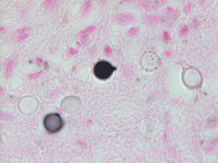
Premortem and postmortem cultures were not collected. Fontana-Masson stain of a section from the hilar lung tissue reveals yeast of varying size with a lighter shade in the center and a thick capsule, though the capsule does not stain and can instead be appreciated as a “halo” (Image 1). Fontana Masson stain also reveals narrow based budding (Image 2). Both findings indicate Cryptococcus neoformans.

Image. 1. Fontana-Masson stain revealing yeast of varying size with a lighter shade in the center and a thick capsule.
Image 2. Fontana-Masson stain revealing narrow based budding

Discussion

Cryptococcus neoformans is a saprophytic yeast (5-10 µm) identified best by its thick polysaccharide, antiphagocytic capsule. It can be infectious when inhaled, often from soil or avian droppings.

While most immunocompetent individuals clear the pathogen, in the immunocompromised, it can form a primary focus in the lungs and then disseminate. It is often asymptomatic when localized to the lungs but can present as a cough or dyspnea. Dissemination to the brain presents as meningitis. Cryptococcal neoformans is the most common cause of fungal meningitis.

While C. neoformans most often presents as meningitis in the immunocompromised, a retrospective case analysis found diabetes mellitus II as a newly defined independent factor contributing to morbidity and mortality. This study analyzed cryptococcal infections in patients with DMII from 1997-2015. 57% of the DMII patients did not have any other underlying disease and 69% of patients who presented with pulmonary Cryptococcus neoformans experienced a misdiagnosis and treatment delays.1

Qualities that aid in the diagnosis include urease positivity, positive latex agglutination test due to its thick polysaccharide capsule, and characteristic features on mucicarmine red, methenamine silver, India ink, and Fontana-Masson stains.2

The Fontana-Masson silver (FMS) stain is a histochemical technique that oxidizes melanin and melanin-like pigments as it reduces silver.  FMS can be used to highlight the melanin-like pigment in Cryptococcus spp., including capsule-deficient variants because this pigment is cell-wall (and not capsule) associated. FMS is a very sensitive, but not completely specific stain, for Cryptococcus spp. as other yeasts and fungi can also produce melanin and melanin-like pigments.3 Though two C. neoformans yeast close together can resemble broad-based budding, Image 2 demonstrates the narrow-based budding.

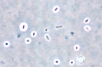
While the India ink stain is often discussed as a popular stain for C. neoformans, it can only be performed on liquid samples (CSF, fluid samples) and cannot be performed on paraffin-embedded tissue samples. Of note, the India ink stain is a “negative stain”, resulting in the classic “halo” effect (image 3) because it is not picked up by the capsule of Cryptococcus spp. Because of this, it will miss capsule-deficient infections.4

Image 3. India Ink Stain showing “halo” effect of capsules.

Prognosis varies by the mechanism of immunosuppression. Acute mortality in in cryptococcal meningitis for HIV patients has improved dramatically with antifungals and ART, ranging from 6-16%. Poor prognostic indicators include abnormal mental status, a high yeast burden defined as CSF antigen titer > 1:1024 by latex agglutination or > 1:4000 by lateral flow assay, or a poor host response defined as CSF WBC count < 20/microL.5

Treatment of cryptococcal infections includes initial therapy with amphotericin B and flucytosine followed by long term fluconazole.6

References

  1. Boulware DR, Rolfes MA, Rajasingham R, von Hohenberg M, Qin Z, Taseera K, Schutz C, Kwizera R, Butler EK, Meintjes G, Muzoora C, Bischof JC, Meya DB. Multisite validation of cryptococcal antigen lateral flow assay and quantification by laser thermal contr. (n.d.).
  2. Li Y, Fang W, Jiang W, Hagen F, Liu J, Zhang L, Hong N, Zhu Y, Xu X, Lei X, Deng D, Xu J, Liao W, Boekhout T, Chen M, Pan W. Cryptococcosis in patients with diabetes mellitus II in mainland China: 1993-2015. Mycoses. 2017 Nov;60(11):706-713. doi: 10.1111/. (n.d.).
  3. McFadden, D., & Casadevall, A. (2001). Capsule and Melanin Synthesis in Cryptococcus neoformans. Medical Mycology, 39, 19-30.
  4. Perfect JR, Dismukes WE, Dromer F, Goldman DL, Graybill JR, Hamill RJ, Harrison TS, Larsen RA, Lortholary O, Nguyen MH, Pappas PG, Powderly WG, Singh N, Sobel JD, Sorrell TC. Clinical practice guidelines for the management of cryptococcal disease: 2010 up. (n.d.).
  5. Saag MS, Powderly WG, Cloud GA, Robinson P, Grieco MH, Sharkey PK, Thompson SE, Sugar AM, Tuazon CU, Fisher JF, et al. Comparison of amphotericin B with fluconazole in the treatment of acute AIDS-associated cryptococcal meningitis. The NIAID Mycoses Study. (n.d.).
  6. Winn, W. C., & Koneman, E. W. (2006). Koneman’s color atlas and textbook of diagnostic microbiology. Philadelphia: Lippincott Williams & Wilkins. (n.d.).

-Joe Teague is a Pathology Student Fellow and Brianna Waller, MD is a 3rd year Anatomic and Clinical Pathology Resident at the University of Vermont Medical Center.

-Christi Wojewoda, MD, is the Director of Clinical Microbiology at the University of Vermont Medical Center and an Associate Professor at the University of Vermont.

Looking into the Pathology Mirror

Conversing with people early in their career has always been an exciting experience for me and, I hope, for those with whom I have spoken. I tend to get enthusiastic in discussing all the possibilities that lie ahead and try to keep the conversation focused on the individual in question. I try to avoid talking about my own career path unless someone specifically asks—but I keep it brief. One-on-one conversations tend to be very productive for the individual because we can delve deep into their questions, fears, concerns, hopes, and goals. Group discussions often end up being more informative for me, and I have learned a ton from listening to dynamic young people. I was recently gifted with the opportunity to lead 9 focus groups as part of a grant-funded project which included several groups with medical students and pathology residents. Although our focus was on forensic pathology, the groups were quite diverse. I would call the experience overall very positive and enlightening for all of us, but I was struck by a few observations that I felt the need to explore further on my own—so, you get to read a blog about it.

Pathology is a fascinating specialty after medical school that covers a large range of diseases and patient types, an even larger range of scopes of practice, and includes some of the lowest and highest paid jobs in the field. At the same time, the practices of pathology and medicine are evolving at an extremely rapid rate while medical knowledge is expanding exponentially. There is an entire industry based around paraphrasing the current literature for a given specialty because, even within a specialty, you can’t read every new study or follow every new development. It is this expansion that has created the demand by pathologists in the last 2 decades to be sub-specialists so that a focus on one particular area of practice will keep their expertise sharp, their diagnoses hyper-accurate, and their risk profile minimal. This expansive phenomenon in medicine in general but specifically in pathology is an excellent indicator that the field of knowledge is ripe for a disruptive innovation. It is common knowledge that the practice of anatomic pathology, for example, is based on a technique that is more than 100 years old—histology; however, what is not common knowledge is that the amount of data generated by reviewing a histology slide from, for example, a tumor, is 1/1000th or less than the data generated by performing genetic sequencing of that same tumor. Add to the mix the ability to perform transcriptional analysis, mass spectroscopy, metabolomics, lipidomics, phospholipidomics, glycobiological analysis, etc. and it becomes clear that what is contributed by an H&E pales in comparison to what we can know about a piece of tissue. There are barriers, you say? Cost, integration of information, usable outputs, or process:volume ratios? All true. But the technological ability to characterize a tumor across all these different attributes and mathematically reduce that to a multiplex assay which can perfectly classify and predict therapeutic responsiveness exists. Still don’t believe me? A collection of companies is focused on testing that has been variably called, “Universal Cancer Screening”, “Multi-cancer Screening”, and “Multi-cancer Early Detection”. These systems currently use sequencing across multiple loci to detect from 20 to 50 different cancer types. One such company can do so with stool to look for gastrointestinal cancer and is on the market today. Why am I going down this path of which many of you are already aware? Because when I was talking to a trainee recently, they told me that they originally wanted to go into forensic pathology but were talked out of it and were now considering doing GI pathology. Let’s break this down so you can understand my frustration.

GI pathology as a career is largely generating revenue through colonoscopy from screening. Yes, the field is diverse and the most complicated parts like liver, pancreas, IBD, etc. are part and parcel to the practice. But, from a C-suite perspective, the fiscal bulk of the value of the service is in biopsy reads from screening. Because of the interest in the field in the last two decades (increase in pathologists in GI) juxtaposed to the much-needed control and reduction of 88305 reimbursement (due to rampant misuse and overuse), there are a lot of GI pathologists in the United States. So many, in fact, that jobs for GI pathology are sort of hard to find. Add to the mix a product, already on the market, that can detect colon cancer in stool without screening colonoscopy and its risks, which is only the harbinger of a group of products that will arrive on the market which can do the same for many other cancers from stool, blood, etc., and one gets nervous about where GI pathology’s current revenue volume is headed. But then there is the recent recommendation that the screening age for colonoscopy be reduced to 45 (from 50). The increase in volume of biopsies from screening (if everyone was 100% compliant) would overwhelm some practices. Where is GI pathology as a specialty going? Do we have too many and should we be concerned about disruptive innovations to screening decimating revenue generating volumes? Or are we facing an overwhelming number of biopsies with the new screening guidelines? I wouldn’t dare try to predict where this is headed but there is clearly some “uncertainty” in the practice of GI pathology. And a practicing pathologist talked a resident out of forensics and into GI??

Let’s contrast this with forensic pathology so my point is clear. There are currently only about 500 FPs in the United States and there is a need—to meet minimum requirements for coverage—of 1200 FPS. That’s a difference of 700 FPs, all of which must be board certified pathologists. There are more than 50 current open full-time positions for FPs that are funded (i.e., actively recruiting to hire today) that were identified on the most common sites for these listings. Seven of these programs offer tuition repayment for FPs from $100,000 to $250,000. Outside of those seven programs, there are three federal programs that specifically offer loan repayment for FPs and a fourth for which they are also eligible. Doing the math, basically, anyone wanting to practice forensic pathology likely qualifies for a loan repayment program (hint: that’s not true for the majority of pathology jobs). Although the average salary for an FP is often reported as ~$110,000 (about half of the average salary for a pathologist according to publicly available data), the current open positions I mentioned have an average of $240,612 (with a range of $175,000 to $350,000). The work of forensic pathologists includes death scene investigation, varying levels of postmortem examination (e.g., chart review, external examination, complete autopsy, etc.), medicolegal reporting including court appearances, participation in public health investigations, participation with local government, etc. This role is vital to the functioning of society and is required by law to be performed. Stated another way, we will always need FP (and we desperately need them now!). It is very difficult to imagine a disruptive innovation or even a transformative innovation that will replace this role in the next several decades. That same can’t be said for other parts of pathology (see my GI example above). And yet, we struggle to find FPs. Why?

Certainly not the only reason but a valid and real reason that we struggle is the presence of microaggressions in the medical community. These are common for pathology in general but can be extremely harsh and rampant for forensics (even coming from other pathologists!). The real example I have given you of the resident selecting GI after being talked out of forensics is a true story. And, more importantly, it was reiterated by nearly every medical student and resident (and fellow) with whom we talked about their experiences. Considers these statements (which are direct quotes):

“You’re too smart to do pathology.”

“Why would you waste your brain on forensics?”

“You’re too good with people and patients to be a pathologist.”

“Forensics is a dead specialty (pardon the pun)”.

Excuse me?? Are you kidding? It’s not that these microaggressions are inappropriate because they are damaging to a young person’s passions and interests. It is that these microaggressions, which are heard repeatedly, are simply wrong. Pathology, if nothing else, is a data and knowledge heavy specialty where we spend most of our time thinking, solving problems, and receiving, processing, interpreting, and synthesizing data into a useful answer on which a clinician can act. And we don’t do it one patient at a time. We produce literally thousands if not tens of thousands of tests results per day in an average laboratory. Forensics requires highly intelligent, detail-oriented individuals who can not only synthesize an entire patient’s life and death into a succinct story—but they have to defend their opinion in court. Every day! I’d like you to ask your primary care doctor if every decision he/she made for each of their patients in one day they would be comfortable defending in court. Every decision! It requires a special person who is not only amazing with data and knowledge but extremely talented at interacting with people—many of which are trying to prove you wrong. Moreover, few medical specialties call upon the physician to routinely deal with families at the lowest point in their lives in every single encounter. A person that is good with people and patients is exactly the person that can become a successful forensic pathologist—one that provides meaningful care when care is most needed. And lastly, forensics is thriving as a job market (as I described). And yet, our “mentors” who train our medical students and pathology residents continue to provide microaggressions (or outright rebuke) for those brave, brilliant individuals who would choose forensics as a career. Considering the state of the field and the perks of the practice at the moment, forensics seems like a pretty smart choice today. But stepping back from this rhetoric to a 10,000 foot view—because, remember, this is me thinking through a problem and forcing you to read about it—the overall observation I have is that the field of pathology (internally) needs to understand where it is going, what its scope of practice will look like tomorrow, 5 years, and 10 years from now, and, more importantly, what the needs of our patient community are (alive or dead). Without a global view of the total need in pathology, how can we possibly have meaningful conversations with individuals early in their career that both enhance their passion and meet the needs of the community of practice?

milner-small


-Dan Milner, MD, MSc, spent 10 years at Harvard where he taught pathology, microbiology, and infectious disease. He began working in Africa in 1997 as a medical student and has built an international reputation as an expert in cerebral malaria. In his current role as Chief Medical officer of ASCP, he leads all PEPFAR activities as well as the Partners for Cancer Diagnosis and Treatment in Africa Initiative.

Microbiology Case Study: A 28 Year Old Woman with Fever and Rash

Case History

A 28 year old female presented to the emergency department (ED) with fevers, chills, and rash. A maculopapular rash began two days prior to her presentation starting at her shins, then spreading to her abdomen, chest, and arms. On presentation she had left knee and right elbow pain and had pain with walking. The patient denied neck pain, headache, blurry vision. Her past medical history is significant for bacterial meningitis between the ages of 10-12. She does not recall the causative pathogen but does recall that her mother had to quarantine for a period of time. On physical exam, the patient was in no distress, had a low-grade fever (38.2 °C) with normal heart rate and blood pressure. Her left knee and right knee were swollen, warm, and painful to the touch with limited range of motion. She was found to have round, erythematous papules and plaques, some with central purpura primarily involving both legs as well as the back and arms (Figure 1).

Initial labs showed a WBC of 11.24/L, Hgb 10.2 g/dL, platelets of 172/L. Her acute inflammatory marker was elevated (CRP 24.7 mg/dL) and her sed rate was 30 mm/hr. One of two blood cultures on admission grew Neisseria meningitides. She was initially started on vancomycin and piperacillin-tazobactam and later transitioned to ceftriaxone to complete a 7 day course. Further blood cultures remained sterile. Dermatology was consulted for her skin lesions and a biopsy of a thigh lesion revealed leukocytoclastic vasculitis. Orthopedic surgery was consulted given her left knee and right elbow swelling. X-ray revealed trace effusions in both joints. A tap of both joints was unsuccessful. The patient developed acute renal insufficiency secondary to acute tubular necrosis during her hospitalization, which improved prior to discharge. Her fevers resolved and the joint swelling and pain improved prior to discharge. She had off and on headaches during her hospitalization but denied neck pain/stiffness and never required a lumbar puncture. She was discharged on prophylactic amoxicillin.

Two months after discharge she was seen in immunology clinic at which time she was tested for humoral and complement immune deficiencies. Her total complement (CH50) was found to be low at 15 U/mL while C3 and C4 were normal, suggesting a terminal complement deficiency. Her humoral immunity panel was normal.

Discussion

Meningococcemia without meningitis occurs in 20-30% of patients with invasive meningococcal disease.1 This patient showed evidence of invasive disease with a maculopapular rash, joint involvement, and renal injury. However, her disease never reached the level of septicemia or disseminated intravascular coagulation (DIC) likely because she presented early enough in the course of her infection. A prior history of bacterial meningitidis raised suspicion for a terminal complement deficiency that was later confirmed by a low CH50. Patients with terminal compliment (C5-C9) deficiency are 1,400-10,000x more likely to develop meningococcal disease and 40-50% of these individuals can have recurrent infection.2 Deficiencies in other components of the complement pathway such as C3 and C4, especially in association with systemic lupus erythematous (SLE), and properdin (a promoter of the alternative complement pathway) can lead to invasive meningococcal disease.3 Patients with anatomic or functional asplenia and patients on eculizumab therapy are also at increased risk.4

Neisseria meningitidis is a gram negative diplococcus and is an obligate human pathogen that is also a common human commensal found in the nasopharynx of 3-25% of the population. Meningococcal disease most commonly manifests as meningitis (40-65%) and meningococcemia (20-30%) followed by pneumonia (10%), septic arthritis (2%), and chronic meningococcemia.1 The major virulence factor associated with meningococcal disease is a capsular polysaccharide, which are classified into 13 serogroups (A-L, W-135, X, Y, Z).5 Serogroups A, B, C, W-135, X, and Y are associated with invasive disease and prevalence of each serogroup is geographically varied. Diagnosis is by visualizing gram-negative diplococci on gram-stain (see Figure) from CSF or other sterile body fluid and by culture of these sterile body fluids. It is worth noting that intravenous antibiotics can sterilize meningococci in the CSF within 3-4 hours after administration.6 Transmission is by large respiratory droplets and direct contact from those with carriage or infection. Disease, especially among high-risk patients with complement deficiency, is preventable with the meningococcal conjugate vaccine. Chemoprophylaxis with rifampin, ceftriaxone, ciprofloxacin, or azithromycin is recommended for close contacts regardless of vaccination status.

Figure 1: Maculopapular rash with central purpura of both legs.

References

  1. Stephens, D., Neisseria meningitidis. 9th ed. Principles and Practices of Infectious Diseases, ed. J. Bennett. Vol. 2. 2015.
  2. Ram, S., L.A. Lewis, and P.A. Rice, Infections of people with complement deficiencies and patients who have undergone splenectomy. Clin Microbiol Rev, 2010. 23(4): p. 740-80.
  3. Fijen CA, K.E., te Bulte MT, Daha MR, Dankert J, Assessment of complement deficiency in patients with meningococcal disease in The Netherlands. Clin Infect Dis, 1999. 28(1): p. 98-105.
  4. Lebel, E., et al., Post-eculizumab meningococcaemia in vaccinated patients. Clin Microbiol Infect, 2018. 24(1): p. 89-90.
  5. Chang, Q., Y.L. Tzeng, and D.S. Stephens, Meningococcal disease: changes in epidemiology and prevention. Clin Epidemiol, 2012. 4: p. 237-45.
  6. Crosswell, J.M., W.R. Nicholson, and D.R. Lennon, Rapid sterilisation of cerebrospinal fluid in meningococcal meningitis: Implications for treatment duration. J Paediatr Child Health, 2006. 42(4): p. 170-3.

-Denver Niles is the Medical Microbiology fellow at UT Southwestern Medical Center. Prior to his Medical Microbiology fellowship, he completed pediatric infectious disease training at Baylor College of Medicine/Texas Children’s Hospital.

-Dominick Cavuoti is a professor of Pathology at UT Southwestern Medical Center who specializes in Medical Microbiology, ID Pathology and Cytology.

-Clare McCormick-Baw, MD, PhD is an Assistant Professor of Clinical Microbiology at UT Southwestern in Dallas, Texas. She has a passion for teaching about laboratory medicine in general and the best uses of the microbiology lab in particular.

Microbiology Case Study: A Female in her 60s with Retro-orbital Headaches

Case History

The patient was a previously healthy female who presented with a five day history of retro-orbital headaches, lightheadedness, and intermittent falls. Her presentation was consistent with meningitis and further studies were pursued. Head computed tomography (CT), CT angiogram of the head and neck, brain magnetic resonance imaging (MRI), and electroencephalogram (EEG) were unremarkable. Analysis of the cerebrospinal fluid (CSF) demonstrated an elevated white blood cell count (605 white blood cells/µL) of which 88% were lymphocytes, 9% were monocytes, and 3% were neutrophils. CSF glucose was slightly decreased at 33 mg/dL and protein was elevated at 81 mg/dL. Additional tests requested on the CSF included herpes simplex virus (HSV), varicella zoster virus (VZV), West Nile virus (WNV), and Epstein-Barr virus (EBV). The CSF was positive for HSV-2 and negative for HSV-1, VZV, and EBV by PCR. WNV IgG and IgM were negative. Of note, the patient had two episodes of viral meningitis in the past of unknown etiology. The patient received a one week course of valacyclovir and was discharged. Per the patient, she continues to have fluctuating headaches and occasional lightheadedness. Follow-up imaging has been unremarkable.

Figure1. Results of the HSV-1 and HSV-2 PCR. HSV-2 (green) and internal control (purple) amplified. HSV-1 (red) was not detected.

Discussion

Herpes simplex virus 1 and 2 (HSV-1 and HSV-2) are enveloped, double stranded DNA viruses that are members of the Herpesviridae family. They are common viruses that cause cold sores or fever blisters. HSV is a lifelong infection, and latent infection can cause reactivation. While both HSV-1 and HSV-2 can affect any area, HSV-1 is typically associated with non-genital sites whereas HSV-2 typically causes genital infections. In addition to herpetic gingivostomatitis, herpes labialis, and herpes genitalis, other associated clinical conditions include encephalitis, meningitis, keratitis, esophagitis, neonatal herpes, and disseminated primary infection. Most cases of HSV encephalitis have been linked to HSV-1 while HSV meningitis is typically caused by HSV-2. As seen in our patient, HSV-2 has been implicated in recurrent, aseptic, and self-limiting meningitis, also known as Mollaret meningitis.1 There are no specific treatment guidelines for HSV-2 meningitis with the main therapeutic strategy being symptom management. The utilization of acyclovir to manage uncomplicated HSV-2 management is controversial and there is no current consensus.2

Clinically, patients with meningitis typically present with acute onset of fever, headache, and neck stiffness. Other associated symptoms include malaise, rash, nausea, vomiting, sore throat, lymphadenopathy, and genitourinary symptoms. In order to differentiate between the infectious etiologies (i.e. viral, bacterial, tuberculous, or fungal) that cause meningitis, a lumbar puncture may be performed. For viral meningitis, CSF will usually show an elevated white count with predominantly mononuclear cells. The CSF:serum glucose ratio and protein levels are often elevated. The most common CSF viral pathogens in the non-immunosuppressed population are enteroviruses, HSV-1, HSV-2, and VZV, which can all be detected by real time polymerase chain reaction (RT-PCR) technology Molecular methods are faster, more sensitive, and more widely available that viral culture.3 Antibody tests are not recommended for HSV as ~70% of adults will be positive for HSV-1 and ~20-50% of adults will be positive for HSV-2.4

Given the broad range of infectious etiologies that can cause meningitis, there has been interest in the development of a multiplex molecular test. Currently, the FilmArray meningitis/encephalitis panel is the only one that has received FDA clearance. This panel includes 14 bacterial, fungal, and viral targets, including HSV-1 and HSV-2. However, this panel should be used cautiously as several studies have shown a high proportion of false negatives in the detection of HSV-1, HSV-2, and Crytococcus neoformans/gattii. It has been suggested that for HSV-1 and HSV-2, the multiplex panel does not work as well if the viral load is near the limit of detection of the assay or if the patient is having a reactivation of HSV. If there is a high clinical suspicion, particularly in neonates and immunosuppressed patients, an assay for detection of only HSV-1 and HSV-2 should be performed.5

References

  1. Koelle DM and Corey L. (2008) Herpes simplex: insights on pathogenesis and possible vaccines. Annual Review of Medicine, 59: 381-395.
  2. Bamberger DM. (2010) Diagnosis, initial management, and prevention of meningitis. American Family Physician, 82: 1491-1498.
  3. Logan SAE and MacMahon E. (2008) Viral meningitis. The BMJ, 336: 36-40.
  4. Schiffer JT, Corey L. (2020) Herpes simplex virus. Bennett’s Principles and Practice of Infectious Diseases, 9th edition.
  5. Tansarli GS and Chapin KC. (2020) Diagnostic test accuracy of BioFire FilmArray meningitis/encephalitis panel: a systematic review and meta-analysis. Clinical Microbiology and Infection, 26: 281-290.

-Melissa Tjota, MD, PhD is a Molecular Genetic Pathology fellow at the University of Chicago Medicine and NorthShore University HealthSystem. She completed her MD/PhD (Immunology) and AP/CP residency at the University of Chicago.

-Paige M.K. Larkin, PhD, D(ABMM), M(ASCP)CM is the Director of Molecular Microbiology and Associate Director of Clinical Microbiology at NorthShore University HealthSystem in Evanston, IL. Her interests include mycology, mycobacteriology, point-of-care testing, and molecular diagnostics, especially next generation sequencing.

Microbiology Case Study: A Middle Aged Man with a Non-Healing Ulcer

Case History

A middle-aged man with osteogenesis imperfecta, poorly controlled HIV, and hepatitis C presented for outpatient management of an infected non-pressure ulcer on his ankle. The patient had no history of recent travel and was current on vaccinations. He was afebrile but reported increasing difficulty with ambulation due to pain from the lesion. Per the patient, the wound began as an itchy “bug bite” two weeks prior, which he had scratched, causing skin breakage. The ulcer became progressively larger with corresponding increases in pain, warmth and swelling. Specimens from the ulcer were collected for culture, and the patient started empiric doxycycline in addition to prophylactic amoxicillin/clavulanate he was already taking for management of a pre-existing ulcer on the opposite foot. The non-healing wound progressed to the size of a nickel with worsening pain. Due to these symptoms and the associated microbiological data, the patient was instructed to present to the emergency department where he was admitted for additional evaluation. Upon admission, the ulcer exhibited no surrounding erythema, but a slight exudate and pitting edema was noted (Image 1). An X-ray of the ankle was obtained which revealed soft tissue swelling, but no fracture or crepitus.

Image 1. Photograph of the ulcerated lesion when seen at hospital admission.

Microbiology

Specimens of the ulcerated lesion were submitted to the microbiology laboratory for routine bacterial culture. No growth was observed on MacConkey agar plates, while two beta-hemolytic morphotypes and one non-hemolytic morphotype were observed on blood agar. The two beta-hemolytic species were identified as Streptococcus dysgalactiae and Arcanobacterium haemolyticum by MALDI-TOF MS. The third, non-hemolytic organism was a catalase-positive, gram positive coryneform rod (Image 2A and B), and was identified by MALDI-TOF MS as Corynebacterium diphtheriae. This identification was subsequently confirmed by both a commercial reference laboratory and the US Centers for Disease Control and Prevention.

Image 2. Growth of the isolate of C. diphtheriae on a blood agar plate and associated Gram stain.  A) Non-hemolytic colonies observed after 24 hours incubation.  B) gram positive coryneform rods of C. diphtheriae.

Discussion

Corynebacterium diphtheriae is the etiological agent of diphtheria, a toxin-mediated disease classically associated with respiratory and cutaneous infections. C. diphtheriae is infrequently encountered in the United States due to a robust national vaccination program but remains endemic in other parts of the world. Respiratory manifestations include pharyngitis with dysphagia, lymphadenitis (often described as a “bull-neck” appearance), and the development of a characteristic white/grey pharyngeal pseudomembrane which can cause airway obstruction. The pathogenesis of C. diphtheriae is mediated by diphtheria toxin which inhibits host cell protein synthesis leading to cell death. Diphtheria toxin can also cause cardiac, nephrotic, and neurological sequalae due to dissemination. The diphtheria toxin gene (tox) is encoded on a bacteriophage which lysogenizes into the bacterial chromosome and is expressed in response to low iron concentrations. While C. diphtheriae is most frequently associated with diphtheria toxin production, Corynebacterium ulcerans and Corynebacterium pseudotuberculosis can also express the toxin if infected with the bacteriophage.

Cutaneous C. diphtheriae infections manifest initially as a vesicle, eventually developing into a painful ulcerative lesion that may or may not have a pseudomembrane. These infections can be caused by either fully toxigenic strains, non-toxigenic strains lacking the tox gene, or non-toxigenic toxin gene bearing (NTTB) strains.1 NTTB strains are genotypically positive for the presence of the tox gene on the lysogenized phage, but do not express functional diphtheria toxin. This can be due to 1) mutation of truncation of the tox gene coding sequence, 2) promotor mutations, or 3) alterations in proteins regulating tox expression. NTTB strains are important from an epidemiological perspective in that they serve as an environmental reservoir for tox gene-harboring phage which could convert circulating non-toxigenic C. diphtheriae into toxin-producing organisms.1-3 This phenotype also presents additional diagnostic challenges as toxin gene expression must therefore be confirmed by more laborious phenotypic methods instead of genotypically (i.e. by PCR).

Recovery of C. diphtheriae in the routine setting is challenging as the organism morphologically resembles other coryneform rods usually representative of flora in cutaneous and respiratory specimens. While selective and differential medias are available (i.e. Cystine Tellurite Blood Agar or Tinsdale medium), they are not used routinely due to low incidence. As both toxigenic and non-toxigenic strains of C. diphtheriae circulate, this isolate was referred to the CDC’s Pertussis and Diphtheria laboratory for additional typing and toxin analysis. The organism was determined to belong to the mitis biotype and was positive for the tox gene by PCR. Toxin gene expression was then evaluated by the Elek Immunodiffusion test (Image 3). In this classical method, a filter paper strip saturated with anti-toxin is placed perpendicular to control and test strains of the organism on non-selective media. If the organism expresses diphtheria toxin, the toxin and the antisera form a complex and precipitate from solution. This phenomenon is visualized as precipitin lines in the agar after 24 hours of incubation at 37°C.4 Despite tox gene PCR-positivity, the Elek Immunodiffusion test revealed that this patient’s isolate did not express diphtheria toxin. Thus, this C. diphtheriae isolate was a representative example of a NTTB strain.

Image 3. Representation of the Elek Immunodiffusion assay for the detection of diphtheria toxin.  Filter paper soaked with antitoxin placed perpendicular to test and control isolates.  The presence of precipitin lines indicates diphtheria toxin gene expression.  Figure adapted from (3).

In contrast to respiratory presentations which have declined due to vaccination, cutaneous infections with C. diphtheriae have become more frequently recognized. Unlike respiratory diphtheria, cutaneous diphtheria was not reportable to the National Notifiable Diseases Surveillance System until the clinical criteria changed in 2019. Since that time, toxigenic isolates recovered from either respiratory or cutaneous sources are reportable.5 Additionally, the incorporation of MALDI-TOF MS into routine workflow has facilitated identification of the organism when isolated clinically and likely increased reporting.

Although immunization protects against clinical diphtheria, it does not prevent colonization by non-toxigenic C. diphtheriae including NTTB strains.2,3 Non-toxigenic C. diphtheriae causing cutaneous infections are often recovered with other pyogenic organisms including Staphylococcus aureus, beta-hemolytic streptococci, and A. haemolyticum6 as was observed in this case. Risk factors for cutaneous infections include a compromised immune system, eczema, travel to endemic regions, intravenous drug use, homelessness/unsanitary living conditions, and alcoholism.3,7  It is unclear how this patient was exposed, although he did have risk factors including immunosuppression and those associated with his osteogenesis imperfecta. The patient was started on erythromycin and ampicillin/sulbactam for subsequent management and has continued to improve when seen at follow-up. Subsequent cultures of the wound have remained negative for C. diphtheriae.

  1. Zakikhany K, Neal S, Efstratiou A. 2014. Emergence and molecular characterisation of non-toxigenic tox gene-bearing Corynebacterium diphtheriae biovar mitis in the United Kingdom, 2003–2012. Eurosurveillance 19:20819.
  2. Doyle CJ, Mazins A, Graham RMA, Fang N-X, Smith HV, Jennison AV. 2017. Sequence Analysis of Toxin Gene-Bearing Corynebacterium diphtheriae Strains, Australia. Emerging infectious diseases 23:105-107.
  3. Sharma NC, Efstratiou A, Mokrousov I, Mutreja A, Das B, Ramamurthy T. 2019. Diphtheria. Nature Reviews Disease Primers 5:81.
  4. Kates O, Starr K, Bourassa L, Burnham C-AD. 2020. The Brief Case: Nontoxigenic Corynebacterium diphtheriae in a Nonhealing Wound. Journal of Clinical Microbiology 58:e00506-00520.
  5. United States Centers for Disease Control and Prevention. 2019. Diphtheria (Corynebacterium diphtheriae) 2019 Case Definition – National Notifiable Diseases Surveillance System. https://ndc.services.cdc.gov/case-definitions/diphtheria-2019/. Accessed August 1st, 2021.
  6. Lowe CF, Bernard KA, Romney MG. 2011. Cutaneous Diphtheria in the Urban Poor Population of Vancouver, British Columbia, Canada: a 10-Year Review. Journal of Clinical Microbiology 49:2664-2666.
  7. Gubler J, Huber-Schneider C, Gruner E, Altwegg M. 1998. An Outbreak of Nontoxigenic Corynebacterium diphtheriae Infection: Single Bacterial Clone Causing Invasive Infection Among Swiss Drug Users. Clinical Infectious Diseases 27:1295-1298.

-Andrew Clark, PhD, D(ABMM) is an Assistant Professor at UT Southwestern Medical Center in the Department of Pathology, and Associate Director of the Clements University Hospital microbiology laboratory. He completed a CPEP-accredited postdoctoral fellowship in Medical and Public Health Microbiology at National Institutes of Health, and is interested in antimicrobial susceptibility and anaerobe pathophysiology.