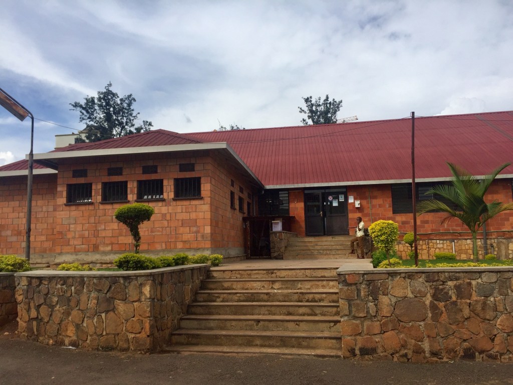
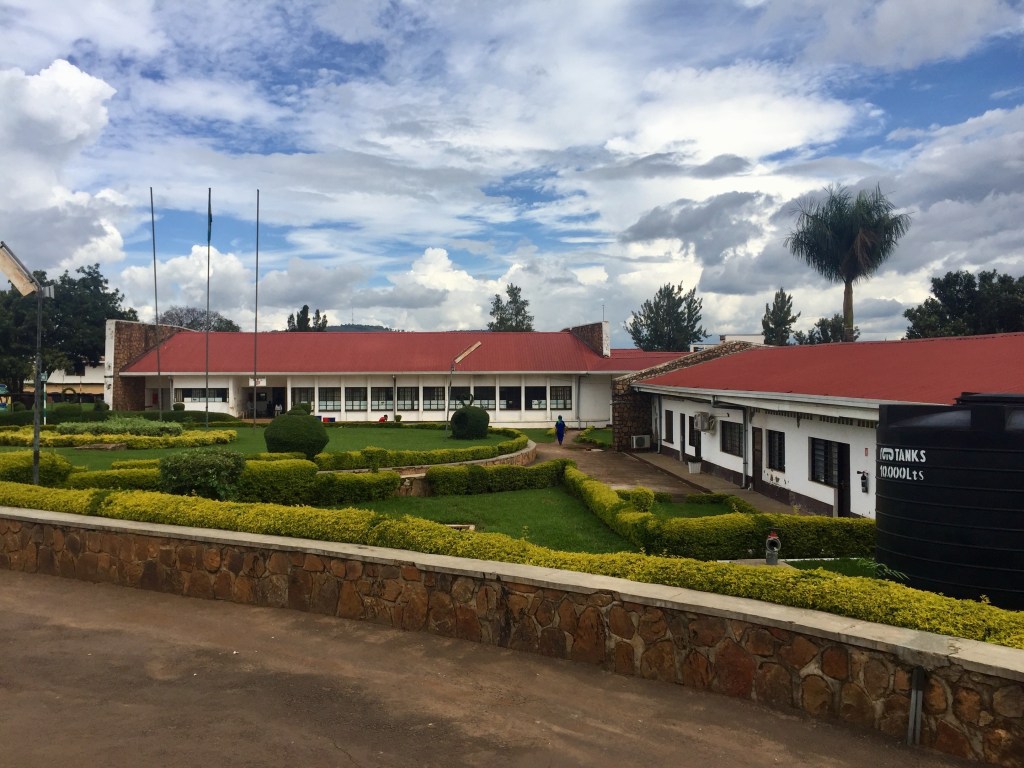
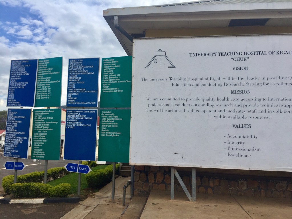
The University Teaching Hospital of Kigali (CHUK) is the largest hospital in its District of Nyarugenge and the biggest national referral hospital in the country of Rwanda, with a 565 hospital bed capacity and 6 operating theaters. It is located in the heart of the capital of the country, Kigali, contributing to its easy accessibility by patients. Rwanda is a country of over 12.5 million people, with an estimated 70.2% of the population living in a rural setting. Per the World Bank, there is an estimated 1 physician per 10,000 people in-country. The government of Rwanda is focused on elevating the country from a low-income developing nation to a middle-income country with a robust health sector capable of ensuring a healthy people with adequate healthcare access. It provides universal healthcare, at a small cost, to all Rwandan citizens who aren’t provided health insurance through employment. In Rwanda there are a total of 14 practicing pathologists, which equates to approximately 1.1 pathologists per million people in the country. In contrast, within the United States there are an estimated 60 pathologists per million people. CHUK offers an array of outpatient, inpatient, surgical, and diagnostic medical services. Inpatient and outpatient services include surgery, accident & emergency, internal medicine, mental health, anesthesiology & critical care, gynecology, pediatrics, maternal & neonatology, ear/nose/throat, ophthalmology, neurosurgery, pediatric surgery, urology, nephrology, dialysis, oncology, and dermatology. Surgical services include general surgery, general pediatric surgery, neurosurgery, orthopaedics, ophthalmology, ear/nose/throat, and obstetrics/gynecology. Diagnostic services include ultrasound, digital x-ray, CT scan, and anatomic and clinical pathology services. In its current state, the hospital has a total of 18 divisions.
There are two facets to the pathology laboratory at CHUK: the Anatomic Pathology (AP) and the Clinical Pathology (CP) laboratories. Within the AP laboratory, also known as the histopathology laboratory, all surgical specimens are grossly examined by a pathology resident and/or pathologist, prepared by a pathology resident for processing, and processed by laboratory technicians into formalin-fixed paraffin-embedded tissue placed onto glass slides. These glass slides are then reviewed by both the pathology residents and the pathologists in order to render a diagnosis, which is communicated to the clinician in order to help direct appropriate patient management. Specimens reviewed at CHUK are predominantly “in-house” specimens generated by the surgeons and clinicians functioning within the walls of the institution. “Referral” specimens are a rarity and generally consist of small biopsies. Cytopathology specimens are also processed within the AP laboratory and include a mixture of fine needle aspiration (FNA) specimens, obtained by pathology residents via superficial FNA, as well as exfoliative cytology specimens such as effusions and urines collected by “in-house” clinicians. Cervical screening conventional pap smears are a rarity. Within the AP laboratory, Diff-Quik, Papanicolaou, and hematoxylin & eosin (H&E) staining was available for slides, as well as a limited panel of special stains: PAS-D, auramine, and a modified acid-fast stain. No immunohistochemistry was available on-site, though cases could be sent for free to nearby Butaro Hospital for IHC or consultation via digital slide scanning.
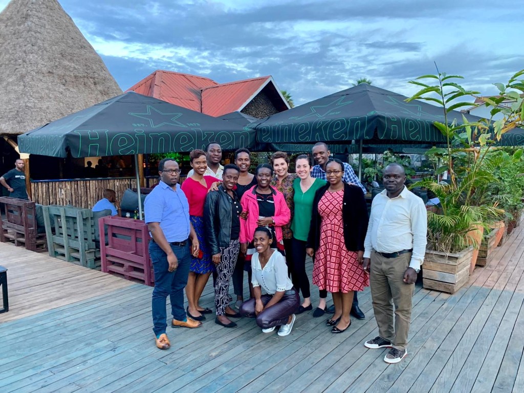
Regarding my experience at CHUK, I departed the United States on a Saturday evening and reached Kigali, Rwanda by 1AM the following Monday morning. On my first day at CHUK, I was introduced to the 5 anatomic pathology staff, 9 anatomic pathology residents, and the single visiting pathologist serving as a laboratory inspector conducting a mock inspection/assessment. I was given a tour of the pathology facilities as well as the entire hospital system.
There were two aspects to my primary job at CHUK: teaching the residents cytopathology and microscopic review of all live cytopathology cases received in the laboratory. Regarding resident education, there were four ways in which I interacted with the residents during my time to facilitate cytopathology education: lectures, multi-headed microscope unknown slide sessions (unknown case conference where I provided the residents with cases they had never seen before), multi-headed microscope “stump the chump” unknown slide sessions (where the residents presented me with unknown cases I had never seen before), and interactive practicals where we performed various hands-on aspects of cytopathology and general pathology practice.
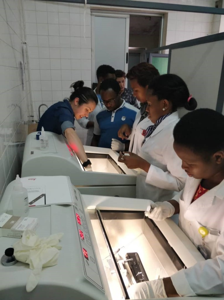
In respect to lectures, I delivered a total of eight 1.5 hour powerpoint-based lectures covering the following topics: breast cytology, thyroid cytology, lymph node cytology, salivary gland cytology, urine cytology, effusion cytology, peritoneal washing cytology, and frozen section pathology (frozen section lecture presented as a combined effort with Dr. Raina Flores). For unknown slide sessions in which I presented cases to the residents, we had 6 sessions covering the following topics: breast, thyroid, salivary gland, urine, conventional pap, and cerebrospinal fluid. We completed a total of 5 “stump the chump” sessions, where residents gave me slides that I had never seen before and we discussed each case and its work-up as well as its associated differential diagnosis or final pathologic diagnosis at the multi-headed microscope. Topics covered included: breast, thyroid, salivary gland, lymph node, and effusions. Finally, with the assistance of “in-house” pathologists, I helped conduct 2 hands-on practicals with the residents: the first regarding fine needle aspiration technique and slide smearing technique (with Dr. Claire Nadyisaba) and the second regarding performance of frozen section intraoperative consultations using Leica CM1850 cryostats and cow liver (with Dr. Raina Flores).
The second of my duties, live cytopathology case review, was also performed at the multi-headed microscope with the residents each afternoon. On a given day, we would typically receive somewhere between 1 and 4 FNA consultations for which the residents would go to FNA clinic and perform the procedure. The laboratory also received various aspirated and exfoliative cytology specimens, such as pleural effusion and ascites fluids, from clinicians within the hospital system. In total, we reviewed 51 cytopathology cases together at the microscope. 27.5% were neoplastic, with 7.8% being malignant and 2% being lymphoma. 56.8% of cases were negative for malignancy, with 21.5% being inflammatory/infectious. In total, 9.8% of cases were interpreted as “atypical” and 5.9% of cases were non-diagnostic. Of the 51 cases, 21 (41.2%) were FNA consultations that I attended and the resident performed.
On my final day of work, I provided the residents with a 41-page cytology knowledge assessment (in PDF format) to complete at their leisure. This test covered the following topics: cervical and vaginal cytology (19 questions), urine and bladder cytology (11 questions), effusion cytology and peritoneal washings (13 questions), cerebrospinal fluid cytology (12 questions), breast cytology (8 questions), thyroid cytology (17 questions), salivary gland cytology (13 questions), and lymph node cytology (11 questions). Within the document, an answer key with associated detailed explanations was provided so it could serve as a learning aid/study guide for the trainees. On my last workday, the residents were asked to evaluate their experience with the Cytopathology Module/Course. A total of 7 of 9 residents completed the evaluation. Regarding preparation and organization of different topics, all residents found the quality of the powerpoints to be “very good” or “excellent”. The quality of the practical sessions was rated as “good,” “very good” or “excellent by all residents and the entire module was given an overall rating of “very good” or “excellent” by all of the residents. The majority of residents felt their time was used effectively during this module and that the venues for theoretical and practical learning were appropriate. In the free-text areas for additional comments, suggestions for improvement included a longer duration (at least 4 weeks) of the module, more hands-on practical time, the opportunity for residents to present information, and more microscopy sessions. For additional topics to be covered, respiratory cytology was suggested. In overarching comments regarding their module experience, the residents felt the module was well-prepared, the teaching sessions were well-organized, and that the course was interesting and helpful.
Finally, though not within the confines of my assigned “duties”, I also spent a portion of each day acting as “consultant” to the on-site pathologists for challenging surgical pathology cases, offering opinions as able for various lesions that were challenging to classify on H&E morphology alone. I also served as a “second reviewer” for new malignant diagnoses being rendered in the laboratory, offering my name to be included in the report as a board certified pathologist who has laid eyes on the case and agrees with the interpretation. Examples of some interesting surgical pathology cases I saw in “consultation” included Wilms tumor (nephroblastoma), cystic partially differentiated nephroblastoma (CPDN), pleomorphic xanthoastrocytoma (PXA), sinonasal undifferentiated carcinoma, basaloid moderately-differentiated carcinoma of the uterine cervix, high-grade large cell lymphoma of the cervical lymph node, high-grade squamous intraepithelial lesion of the vulva arising within a condyloma acuminatum, and low-grade papillary urothelial carcinoma of the bladder. I also attend a single Tumor Board Multidisciplinary Conference with two residents and 1 staff pathologist in which a resident presented a case of mucinous moderately-differentiated adenocarcinoma of the colon transmurally invading adjacent ileum. It was interesting to hear the clinicians, pathologists, and radiologists interact in addressing quality of care, efficiency of care, and clinical decision-making. The time of initial presentation to the time of surgery was greater than 1 year for this patient.
My time spent at CHUK in Kigali, Rwanda was an invaluable experience. The work setting granted me the opportunity to expand my role as an academic educator. I was offered the opportunity to present as many lectures as possible to the resident trainees, participate as the leader of multi-headed microscope slide sessions, serve as a spearheading physician in laboratory services expansion efforts, and work as an ‘attending’ physician overseeing trainees’ performance of FNAs. It was an experience that demanded personal growth, via the assumption of roles that I am not privy to as a post-graduate medical education trainee in the United States. Additionally, I was exposed to a cytopathology and surgical pathology workload for a patient population quite dissimilar from the community I am used to serving. With limited ancillary testing capabilities, I returned to a more “pure” form of rendering pathologic diagnoses, based on H&E morphology alone rather than on the synthesis of cyto- and/or histomorphologic appearance coupled with various ancillary diagnostic testing data points. In conclusion, this was an experience that expanded my understanding of the ways in which I can be useful as a board certified anatomic and clinical pathologist interested in incorporating medical mission work into my clinical practice. Beyond arriving in countries without expansive pathology laboratory systems and simply doing the work, I can also pursue opportunities where I can help educate and shape burgeoning in-country pathologists who will then go on to have productive, hopefully decades-long careers in their country, serving their countrymen. This trip certainly expanded my understanding of the role of a “visiting” pathologist. This experience was made possible by the ASCP Trainee Global Health Fellowship Award. Thank you so much to the ASCP, Dr. Dan Milner, Alpa Pandya, and the CHUK pathology department for helping to facilitate this opportunity!
-Kelsey McHugh, MD is a board certified anatomic and clinical pathologist, with cytopathology subspecialty certification, who is currently completing gastrointestinal, hepatic, and pancreatobiliary pathology subspecialty training. She anticipates graduating from the Cleveland Clinic Gastrointestinal, Hepatic, and Pancreatobiliary Pathology Fellowship in June 2020, after which she will remain at the Cleveland Clinic as a staff pathologist beginning July 2020.
