Microbiology Case Study: A 56 Year Old Man with Bacteremia

Case History

A 56 year old man with a history of hypertension, asthma and COPD, bladder stones, congenital bladder abnormality, and bladder exstrophy presented to the ED with fatigue, generalized weakness, poor appetite, fever, and nausea. He had intermittent right flank pain and generalized abdominal pain. The patient also reported he had passed bladder stones in the past year.

In the ED he had an elevated peripheral white blood cell count of 12.9 x10³/μl with a differential of 81% neutrophils, 11% lymphocytes, and 6% monocytes. His creatinine was elevated at 5.4 mg/dl. CT imaging of the urogenital tract showed hydronephrosis with evidence of infection.

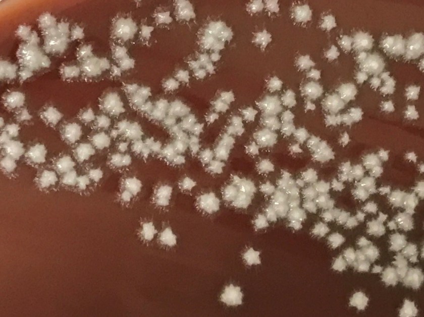
Blood and urine specimens were sent for culture. The urine culture grew 10-50,000 cfu/ml Aerococcus urinae. After 30 hours of incubation, the anaerobic blood culture was positive for small, coccoid-looking gram positive rods (Image 1). The following bacteria grew on solid media under anaerobic conditions after 48 hours of incubation (Image 2).

actscha1
Image 1. Gram stain showing small, coccoid-looking Gram-positive rods
actscha2
Image 2. Tiny colonies growing on non-selective anaerobic media after 48 hours.

Discussion

The organism growing in the blood culture was identified as Actinotignum schaalii. A. schaalii is a facultative aerobe that prefers enriched media and grows best in anaerobic or 5% CO2 environments. It is a non-hemolytic, non-motile, and non-spore forming Gram-positive rod. A. schaalii is negative for catalase, oxidase, CAMP factor, and nitrate reduction, but is hippurate positive. It is also negative for lactose, mannitol, raffinose, sorbitol, and trehalose acid production.

Actinotignum schaalii was formerly named Actinobaculum schaalii. The Actinotignum genus now consists of A. urinale, A. sanguinis, and A. schaalii. A. schaalii is part of urogenital microbiota in healthy adults and can cause urinary tract infections, especially as it thrives in the humid atmosphere of diapered children or elderly adults. Isolation of this organism is associated with urinary incontinence, bladder cancer, catherization, neurogenic bladder, renal failure, prostate cancer, and chronic renal failure. The presence of A. schaalii in the urogenital tract can progress to bacteremia and rarely it can also cause groin abscesses.

Actinotignum schaalii is an under recognized uropathogen due to its fastidious nature—it is slowly growing and prefers enriched media grown under anaerobic conditions. None of these conditions are met as part of a routine urine culture as urine cultures are often finaled in < 48 hours, they may not use enriched media, and they are incubated under aerobic rather than anaerobic conditions. Because of these factors, A. schaalii rarely grows in urine culture despite it being a known colonizer of the urogenital tract and occasional uropathogen. Instead it is most commonly recovered from blood cultures as a result of urosepsis. When colonies do grow from blood culture, they are very difficult to identify by biochemical methods. A. schaalii is often misidentified by biochemical panels as Arcanobacterium spp. or Gardnerella vaginalis (API Coryne system, bioMerieux), Actinomyces meyeri (Rapid ID32A system, bioMerieux), or Actinomyces israelii (Rapid ANA II system, Remel). In our lab Bruker Biotyper MALDI-TOF MS was able to identify A. schaalii with high confidence. Based on the most current literature, Vitek MS misidentifies A. schaalii as G. vaginalis or A. meyeri. This data is from 2015, so it’s possible the Vitek MS spectra database has been updated and now identifies A. schaalii.

A. schaalii is routinely susceptible to all β-lactams but requires an extended duration of antimicrobial treatment compared to other uropathogens. It is resistant to trimethoprim/sulfamethoxazole and quinolones, both of which are commonly prescribed for urinary tract infection. Therefore, patients with A. schaalii are at risk for recurrent urinary tract infections due to inappropriate or inadequate antibiotic treatment.

Our patient was prescribed ciprofloxacin for presumed urinary tract infection prior to his bacteremia. He was switched to ceftriaxone upon hospital admission. His bacteremia was cleared and the patient was discharged 5 days later on an oral β-lactam antibiotic. 

References

  1. Lotte, R., L. Lotte, and R. Ruimy. “Actinotignum schaalii (formerly Actinobaculum schaalii): a newly recognized pathogen—review of the literature.” Clinical Microbiology and Infection1 (2016): 28-36. Le
  2. Le Brun, Cécile, et al. “Urinary tract infection caused by Actinobaculum schaalii: a urosepsis pathogen that should not be underestimated.” JMM Case Reports2 (2015).
  3. Manual of Clinical Microbiology, 11th edition


-Erin McElvania, PhD, D(ABMM), is the Director of Clinical Microbiology NorthShore University Health System in Evanston, Illinois.

Essential Diagnostics List – Ready, Set, Go!

In a recent post I introduced the WHO’s draft of the Essential Diagnostics List (the EDL). The EDL is a catalog of in vitro diagnostics (IVD) designed to complement the Essential Medicines List. The EDL is not necessary meant to be a global list, but something to be adopted and adapted by each country, and tests added, subtracted, or prioritized based on each country’s disease burden. While the draft is still in development, at least one country is already on track to have adopted a country specific EDL by the end of the year!

India is working towards being the first country to have an EDL by the end of 2018. It so happens that the draft of the EDL was announced while India was in the process of rolling out a Free Diagnostics Initiative (FDI) in 29 of its states. The goal of the FDI is to “ensure the availability of a minimum set of diagnostics appropriate to the level of care to reduce out of pocket expenditure on diagnostics and to encourage appropriate treatment of based on accurate diagnosis”. Similar to the EDL, the FDI plans for different IVDs at different levels of laboratories, from community healthcare centers to reference laboratories. The development of an EDL seems like a natural product of India’s FDI. Talk about perfect timing!

The Indian Council on Medical Research (ICMR), comprised of clinicians, microbiologists, and medical device industry leaders, has convened to adapt the EDL to India’s infection patterns and diseases. They plan to have their national EDL ready to present by the beginning of 2019. The ICMR intends that an Indian EDL will optimize utilization of the Indian EML. “The objective is to test and treat rather than treat and test” states Dr. Kamina Walia of the ICMR. The ICMR also realizes that in order for diagnostics to be affordable, the country’s laboratory infrastructure will need to be strengthened, including building laboratory capacity where none currently exists.

It is so exciting to see the EDL already under consideration by a nation. It’s even more exciting to hear medical experts speak about how laboratory infrastructure should be strengthened, and to know that medical device industry leaders are coming to the table. It’s going to be fun to watch this develop over the next decade.

Do you want to be involved in the EDL project? There is time! The WHO is accepting applications for IVDs to be added to the second edition of the EDL, which will be released in 2019. The deadline for submissions is September 15, 2019. Instructions can be found here.


Sarah Brown Headshot_small

Sarah Riley, PhD, DABCC, is an Assistant Professor of Pediatrics and Pathology and Immunology at Washington University in St. Louis School of Medicine. She is passionate about bringing the lab out of the basement and into the forefront of global health.  

Microbiology Case Study: A 62 Year Old Male with Productive Cough

Case History

62 year old male from Louisiana with a medical history of COPD presents with fever, productive cough, weight loss, and a red nodule on the left hand. Chest x-ray shows interstitial and lobar infiltrates. Patient reports no recent travel history. Mycobacterial culture of the sputum is positive for an organism. MALDI-ToF of the sputum culture confirms the result.

Lab Identification

mycokans1
Image 1. Kinyoun stained long bacilli with a banded, ladder like pattern isolated from sputum culture.
mycokans2
Image 2. Bright lemon yellow colonies growing on LJ slant after exposure to light on the right compared with unpigmented control colonies on the left.
mycokans3
Image 3. Bright yellow colonies growing on 7H11 media after exposure to light on the right compared with unpigmented control colonies on the left.

Mycobacterium kansasii grows on 7H11 media and Lowenstein-Jensen (LJ) slants. The colonies appear smooth or rough and unpigmented when isolated in the dark. Upon exposure to light, colonies turn bright lemon yellow due to enhanced b-carotene production, which makes M. kansasii a photochromogen. M. kansasii stains positive with acid-fast stains. On Kinyoun stain, it shows long bacilli with a banded, ladder like pattern. M. kansasii is positive for nitrate reduction, catalase, urease, and tween hydrolysis. It is negative for niacin and pyrazinamindase. The identification of M. kansasii is confirmed by MALDI or DNA probe of an isolate.

Discussion

Mycobacterium kansasii is a slow growing photochromogen, an organism that grows unpigmented colonies in the dark but produces a bright lemon yellow pigment upon exposed to light3. It is also an acid-fast positive long bacillus that causes TB-like chronic pneumonia, which is the second most common non-TB mycobacterial infection after MAC in the AIDS population. There are five genotypes of Mycobacterium kansasii. Genotypes I and II infect humans, with I being the most prevalent. Because environmental sources of M. kansasii are rarely identified, isolation of M. kansasii from a culture is never considered a contaminant.

M. kansasii is usually found in the tap water in cities endemic for the infection. In the U.S., it is most prevalent in the central and southern states including Louisiana, Illinois, Texas, and Florida1. Internationally, it is most prevalent in Israel, Korea, France, Japan, Portugal, with the highest incidence in England, Wales, and among South American gold miners.

The majority of patients with M. kansasii infection have an underlying pulmonary disease such as COPD, bronchiectasis, or TB infection3. The clinical manifestation of M. kansasii infection includes a primarily unilateral cavitary infiltrate in the lungs without pleural effusions. Patients are generally older compared with those infected with TB. They present with productive cough, weight loss, fever, night sweats, and dyspnea. Symptoms are usually less severe but more chronic compared with those of TB pneumonia. M. kansasii can also present as cutaneous lesions similar to sporothrichosis. Lesions include nodules, pustules, red plaques, and ulcers1.

M. kansasii infection is diagnosed by chest X-ray or chest CT, positive respiratory culture, and clinical exclusion of other diagnoses2. The criteria for a positive culture include either two consecutive positive sputum cultures, one positive culture from bronchoscopy specimens, or one positive culture with compatible clinical symptoms2. The treatment of M. kansasii infection depends on the resistance of the organism to rifampin. Rifampin-sensitive organisms are treated with at least three drugs including rifampin, ethambutol, isoniazid, and pyridoxine1. Rifampin-resistant organisms are treated with three drugs including clarithromycin or azithromycin, moxifloxacin, ethambutol, sulfamethoxazole, or streptomycin1. Due to drug interaction, rifampin is contra-indicated among HIV patients taking protease inhibitors and nonnucleoside reverse transcriptase inhibitors. Because rifampin increases the metabolism of these HIV medications, it can lead to HIV drug resistance among these patients.


References

  1. Akram SM, Bhimji SS. Mycobacterium Kansasii. StatPearls. 2018. https://www.ncbi.nlm.nih.gov/books/NBK430906/
  2. Johnston JC, Chiang L, Elwood K. Mycobacterium Microbiol Spectrum. 2017;5(1):TNMI7-0011-2016. doi:10.1128/microbiolspec.TNMI7-0011-2016.
  3. Meraz A, Raheem S. Pulmonary Mycobacterium Kansasii Infection –A Tale of a Right Upper Lobe Cavitary Lesion [abstract]. Journal of Hospital Medicine. 2015; 10 (suppl 2). https://www.shmabstracts.com/abstract/pulmonary-mycobacterium-kansasii-infection-a-tale-of-a-right-upper-lobe-cavitary-lesion/. Accessed March 12, 2018.


-Ting Chen, MD is a 1st year anatomic and clinical pathology resident at the University of Vermont Medical Center.

Wojewoda-small

-Christi Wojewoda, MD, is the Director of Clinical Microbiology at the University of Vermont Medical Center and an Associate Professor at the University of Vermont.

Microbiology Case Study: A 50 Year Old Man with Altered Mental Status

Case Histor

A 50 year old man presented to the emergency room with altered mental status, fever, and hypotension. His medical history was significant for quadriplegia, chronic indwelling urinary catheter with frequent urinary tract infections, and diabetes mellitus. The patient was intubated and started on vasopressors for presumed septic shock. CT scan of the abdomen showed dilated loops of bowel with possible ileus and pneumoperitoneum around the sigmoid colon.

Laboratory Identification

  • Clostridium difficile Fecal PCR: Negative
  • Fecal Bacterial Pathogen PCR: Negative for Salmonella spp., Shigella or Enteroinvasive E. coli spp., Campylobacter spp., or Shiga toxin producing genes
  • Urine culture: 2 strains of Pseudomonas aeruginosa and Klebsiella pneumoniae

Blood culture: At 41 hours, two of four blood culture bottles became positive (one aerobic bottle in each set). Gram stain showed budding yeast with pseudohyphae (Image 1). Growth of white, smooth colonies with foot-like projections was observed on agar (Image 2). MALDI-TOF analysis confirmed identification of the organism as Candida albicans.

candalbi1
Image 1: Gram stain of blood culture bottle demonstrating yeast form (left) and pseudohyphae (right).
candalbi2
Image 2: Colony morphology on chocolate agar demonstrating smooth white colonies with surrounding “feet.”

Discussion

Candida spp. are the most common yeast cultured from clinical specimens. Candida albicans is characterized macroscopically by growth of smooth, white colonies with surrounding “feet” which represent projections of pseudohyphae. The pseudohyphae are distinguished microscopically from true hyphae by constriction of the cells where they meet (with true hyphae, the cell walls will remain parallel). When incubated at 37 C for 2 hours C. albicans will produce germ tubes (extensions from the yeast cell representing an attempt at forming true hyphae).

Candida spp. are normal flora found in the gastrointestinal tract, mucous membranes, and skin. Invasive candidiasis is typically an opportunistic infection with the patient’s own, endogenous flora (although nosocomial spread also occurs). Although it can be normal skin flora, the growth of Candida spp. from a blood culture should be presumed to be pathogenic; treatment should be initiated, and attempts made to identify the source. Typically, invasive candidiasis arises from one of three sources: 1) colonization and biofilm formation on an indwelling intravenous catheter 2) dissemination from a deep nidus of infection (often urinary tract) or 3) translocation from the gastrointestinal tract. Patients in intensive care, those who are immunocompromised, at extremes of age, those who have been on broad spectrum antibiotics, and those with GI tract perforation or anastamotic leaks post-operatively are at greatest risk for developing invasive candidiasis.

C. albicans has historically been the most common species of yeast causing invasive disease. However, non-albicans species (C. glabrata, C. parapsilosis, C. tropicalis and C. krusei) now cause almost 50% of invasive candidiasis. This changing epidemiology is relevant because the different species demonstrate different susceptibility profiles to antifungal agents, including azoles and echinocandins. C. auris is another recently emerging species that shows resistance to multiple antifungal agents, and has been described as causing outbreaks of healthcare-associated infections.

Although the patient’s chronic indwelling urinary catheter raised suspicion for a urinary source of the candidemia, the urine culture failed to support that theory. Given the findings of the CT abdomen in this patient, translocation from the GI tract is favored as the source of the candidemia. The patient’s history of treatment with antibiotics for repeated urinary tract infections, however, may have placed him at greater risk for developing invasive candidiasis.

-Alison Krywanczyk, MD is a 4th year anatomic and clinical pathology resident at the University of Vermont Medical Center.

Wojewoda-small

-Christi Wojewoda, MD, is the Director of Clinical Microbiology at the University of Vermont Medical Center and an Associate Professor at the University of Vermont.